
 metals · microbes · host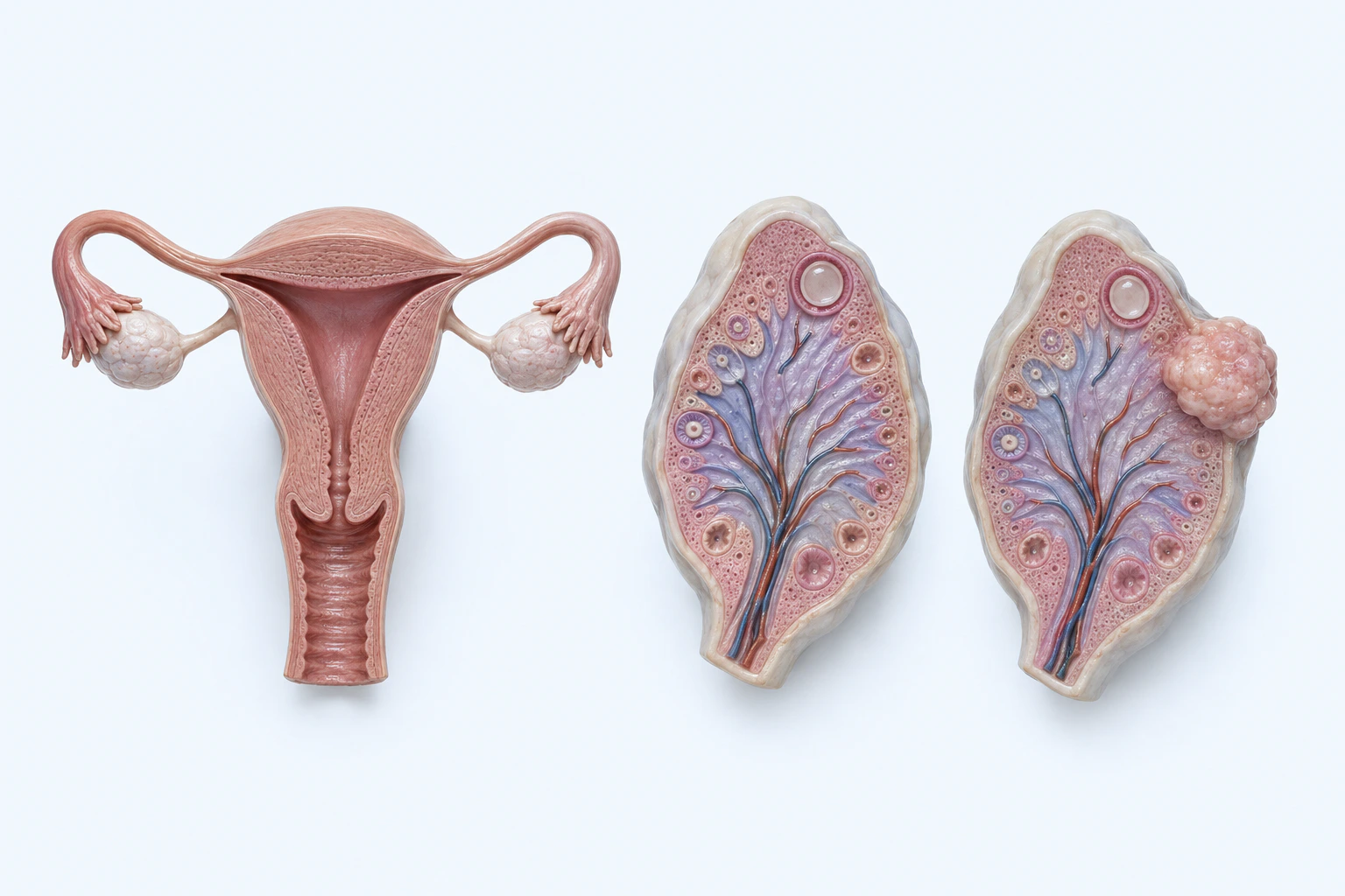
Representative ovarian-neoplasm orientation with one bounded nonspecific growth. Appearance does not establish benignity, malignancy, histologic subtype, origin, grade, stage, spread, prognosis, or diagnosis.
Scientific media record1 verified identifier
- Subject
- Ovarian Neoplasmscondition
- Identifiers
- MeSH:D010051
- Review
- Editorial review completeIdentifiers authority-verified · Accessibility validated · · ovarian-cancer|ovarian-cancer-pathology-v1.webp
- Digital source
- Trained-algorithmic mediaCreated with a trained generative algorithm and reviewed by WikiBiome for subject identity, scientific framing, identifiers, provenance, and accessibility.
- Scientific basis
- Ovarian Neoplasms — MeSHOvarian Epithelial, Fallopian Tube, and Primary Peritoneal Cancers
- License
- CC BY-SA 4.0Created
Ovarian cancer is the most lethal gynecological malignancy, with approximately 314,000 new cases and 207,000 deaths annually worldwide. Its poor prognosis stems largely from late-stage diagnosis (>70% at stage III/IV) and the development of chemoresistance.
From a metallomics perspective, ovarian cancer sits at the intersection of metalloestrogen biology, iron-driven cell death (ferroptosis), and an emerging understanding of the peritoneal and tumor-associated microbiome. The metallomic dimension offers both mechanistic insight and potential therapeutic targets, particularly through ferroptosis-inducing strategies.
Evidence map29 cited passagesInspect provenance +
Glutathione depleted/dysregulated: Central to platinum resistance. Glutathione synthesis is the most significantly altered pathway in platinum-resistant OC. Platinum-resistant cells upregulate glutathione, resisting ferroptosis.
Taurine depleted: Antioxidant defense compromised in OC tissue.
| Source | Metals | Relevance | |--------|--------|-----------| | Smoking | Cadmium (primary) | 35-50% higher Cd body burden in smokers | | Diet | Cd, As, Ni | Contaminated soils, rice, shellfish, leafy greens | | Occupational | Cd, Ni, As | Battery production, smelting, electronics | | Endometriosis | Iron | Repeated retrograde menstruation deposits iron in
| Marker | Direction | Evidence | |--------|-----------|---------| | Tumor-associated macrophages | Elevated | Infiltrate inflammatory tumor microenvironment | | Inflammatory cytokines | Elevated; mediate gut-OC axis | Genetically predicted causal mediation | | LPS | Present in tumor tissue | Confirmed by immunohistochemistry | | B-cell/IgA response | Suppre
Tumor Tissue: | Taxon | Direction | Evidence | |-------|-----------|---------| | Proteobacteria (phylum) | Enriched | Proteobacteria/Firmicutes ratio increased (, n=137) | | Propionibacterium acnes | Enriched | Inflammatory potential | | Firmicutes (phylum) | Depleted | Relative decrease | | Crenarchaeota (archaea) | Depleted | Archaeal diversity reduced |
Gut: | Taxon | Direction | Evidence | |-------|-----------|---------| | Escherichia-Shigella | Enriched | Markedly higher in OC (, n=382) | | Dialister | Enriched (causal) | Two independent MR studies (, ) | | Coprococcus | Depleted | Butyrate producer | | Fusicatenibacter | Depleted | SCFA producer | | Butyricicoccus | Depleted | SCFA producer | | Oscilliba
Cervicovaginal: | Taxon | Direction | Evidence | |-------|-----------|---------| | Lactobacillus dominance | Lost | Non-Lactobacillus community type O: OR 2.80 for OC in women <50 (, Lancet Oncology, n=580) |
Peritoneal: 18 microbial features specific to OC pathology identified; combined with CA-125 and HE4, improved diagnostic accuracy.
Oxidative stress tolerance enzymes: Enriched in tumor tissue bacteria (KEGG pathway analysis, )
Keap1-Nrf2-GPX4 axis: Ferroptosis resistance pathway upregulated in platinum-resistant OC; targetable by Tripterygium + Lactobacillus
Ascending infection model: Vaginal pathogens (Neisseria gonorrhoeae, Chlamydia trachomatis) increase OC risk. Lactobacillus depletion in cervicovaginal compartment enables pathogen ascension to upper reproductive tract. Tubal ligation disrupts this pathway.
IgA-coated bacteria in ascites: Unique microbial-immune interaction at the tumor site; commensal microbiota required for B-cell-mediated antitumor immunity.
Simultaneous dysbiosis across gut, cervicovaginal, peritoneal, and tumor tissue. Shannon index significantly decreased in OC tumor tissue (P=0.0215, ). Gut-based random forest classifier achieves AUC = 0.86 for OC detection.
Systematic loss of butyrate-producing genera (Coprococcus, Fusicatenibacter, Butyricicoccus, Oscillibacter) in gut. Butyric acid metabolites identified as protective factors by Mendelian randomization.
Antibiotic-induced dysbiosis increases tumor growth AND cisplatin resistance. Gut microbiome is required for immune surveillance of cancer stem cells. IPA and indoxyl sulfate are key protective metabolites suppressed by antibiotics, restored by FMT.
Warburg-like shift in OC tissue: carnitine elevated 1.75-fold, butyrylcarnitine 3.62-fold, taurine depleted. Stage IV OC ascites metabolome converges with GI cancers, suggesting gut barrier breakdown in advanced disease.
High-fat and ketogenic diets accelerate EOC tumor growth via microbiome disruption and polyamine biosynthesis upregulation. Mediterranean diet shows survival benefit (HR=0.59, ).
Endometriosis is a direct risk factor for OC (RR 1.265, ), with iron deposits from retrograde menstruation contributing to carcinogenesis in clear cell and endometrioid subtypes.
Tripterygium glycosides + L. paracasei—induces ferroptosis via GPX4 inhibition, microbiome-dependent (, animal model)
Post-surgical probiotics—restores diversity, activates omega-oxidation
E. coli Nissle 1917—reduces tumor via TLR-4/IL-23, but only in unstressed mice
| Metal | Direction | Key Evidence | |-------|-----------|-------------| | cadmium | Elevated | Primary metalloestrogen. Binds ERalpha with Kd ~4.5 x 10^-10 M (nearly equivalent to estradiol). Activates ER-dependent transcription in OC cells at 1 uM. Also signals through GPR30/GPER in ER-negative cells at 50-500 nM. Half-life 12-30 years creates cumulative o
| Source | Metals | Relevance | |--------|--------|-----------| | Smoking | Cadmium (primary) | 35-50% higher Cd body burden in smokers | | Diet | Cd, As, Ni | Contaminated soils, rice, shellfish, leafy greens | | Occupational | Cd, Ni, As | Battery production, smelting, electronics | | Talc | As, trace metals | Historical concern for perineal talc use | | W
The peritoneal cavity, long assumed sterile, harbors a low-biomass microbiome altered in ovarian cancer. Ascitic fluid from OC patients contains distinct bacterial communities compared to benign conditions.
Showing 24 of 29 evidence-bearing passages. Every remaining citation is still indexed in the reference record below.
One disease. Five evidence layers.
A generated systems view of the metals, organisms, host sequestration signals, ecological conditions, and microbial functions indexed for Ovarian Cancer.
Evidence layer
Taxonomic signature
Organisms reported as enriched or depleted, with their indexed functional context kept beside the name.Gut enrichment; LPS production; siderophore-mediated iron scavenging in iron-accumulating tumor microenvironment
Phylum-level increase in tumor tissue; high inflammatory potential; Proteobacteria/Firmicutes ratio elevated
P. acnes enriched in tumor tissue; inflammatory potential
Identified in ovarian tumor tissue; beta-glucan immune modulation via Dectin-1; iron-dependent virulence
Causal risk factor identified by two independent Mendelian randomization studies
Enriched in ovarian tumor tissue; FadA-mediated E-cadherin/beta-catenin disruption; NF-kB inflammation
Cervicovaginal Lactobacillus dominance lost; protective acid barrier compromised; ascending infection enabled
Butyrate producer depleted in gut (n=382)
MR-identified protective factor; SCFA producer; heavy metal binder
Butyrate producer; depleted at baseline, restored by probiotic therapy
SCFA producer depleted in OC gut
SCFA producer depleted in OC gut
Archaeal/fungal diversity reduced in tumor tissue (Crenarchaeota depleted)
Butyrate producer depleted in gut; loss removes anti-inflammatory protection
Evidence layer
Nutritional immunity
Host metal-withholding, inflammatory, antioxidant, and microbial-metabolite signals indexed in the signature.Elevated host signals
6Depleted protective signals
7Evidence layer
Ecological state
The environmental conditions that connect the organism-level observations into a system.Evidence layer
Virulence functions
Microbial structures, enzymes, and acquisition systems implicated by the linked evidence.The disease record, in full.
The original WikiBiome disease narrative remains intact beneath the generated signature atlas.
Metalloestrogen Connections#
Cadmium as Metalloestrogen#
Cadmium is the most established metalloestrogen relevant to ovarian cancer. cadmium (Cd) binds estrogen receptor alpha (ERa) with a dissociation constant (Kd) of approximately 4.5 x 10^-10 M—nearly equivalent to estradiol. cadmium activates ER-dependent gene transcription in ovarian cancer cell lines at concentrations as low as 1 uM.
cadmium also signals through the membrane-bound estrogen receptor GPR30/GPER, inducing proliferative responses in ER-negative cells at 50-500 nM. Epidemiological studies have linked urinary and blood cadmium levels with increased ovarian cancer risk, though results are inconsistent across populations.
cadmium's half-life in the body is 12-30 years, meaning chronic low-level exposure produces cumulative ovarian tissue burden. Smoking is the primary non-occupational cadmium exposure source; dietary cadmium from contaminated soils (phosphate fertilizers) adds chronic background exposure. See Cadmium and Metalloestrogens for broader mechanisms.
Nickel and Arsenic#
Nickel binds ERa noncompetitively with estradiol and induces cell growth in hormone-sensitive cell lines. nickel (Ni) also drives epigenetic changes—global loss of histone acetylation, H3K9 methylation—that may promote ovarian carcinogenesis independently of estrogenic effects.
Arsenic exposure is associated with increased ovarian cancer risk through Oxidative Stress, DNA damage, and interference with DNA repair pathways. cobalt (Co)-exposure to multiple metals (cadmium (Cd) + nickel + arsenic (As)) may produce synergistic carcinogenic effects through converging estrogenic and epigenetic mechanisms.
Iron and Ferroptosis#
Iron Dysregulation in Ovarian Cancer#
Ovarian cancer cells exhibit altered iron metabolism with upregulation of transferrin receptor 1 (TfR1) and downregulation of ferroportin, creating an iron-accumulating phenotype. Elevated intracellular iron drives Fenton chemistry, generating reactive oxygen species (ROS) that promote genomic instability.
Endometriosis-associated ovarian cancers (clear cell and endometrioid subtypes) develop in an iron-rich environment from repeated retrograde menstruation and hemoglobin breakdown.
Ferroptosis as Therapeutic Target#
Ferroptosis—iron-dependent regulated cell death driven by lipid peroxidation—has emerged as a promising therapeutic strategy.
Ovarian cancer cells with high iron content are particularly vulnerable to ferroptosis induction. Erastin and RSL3 (GPX4 inhibitors) trigger ferroptosis in cisplatin-resistant ovarian cancer cells. Combination of ferroptosis inducers with conventional chemotherapy may overcome Platinum resistance.
The cystine/glutamate antiporter (system Xc-) is a key target; its inhibition depletes glutathione and sensitizes cells to ferroptosis. Iron chelation paradoxically reduces ferroptosis susceptibility, confirming iron's central role. See Ferroptosis for detailed pathway mechanisms.
Tumor and Peritoneal Microbiome#
Distinct Tumor-Associated Microbiome#
Ovarian cancer tissues harbor a distinct microbiome compared to normal ovarian tissue and adjacent peritoneum. Fusobacterium enrichment has been documented in ovarian cancer tissue, paralleling its well-established role in colorectal cancer. Fusobacterium nucleatum promotes tumor progression through FadA adhesin binding to E-cadherin, activating beta-catenin signaling and NF-kB-mediated Metal-Driven Inflammation.
See Fusobacterium for detailed mechanisms.
Peritoneal Microbiome#
The peritoneal cavity, long assumed sterile, harbors a low-biomass microbiome that is altered in ovarian cancer. Ascitic fluid from ovarian cancer patients contains distinct bacterial communities compared to benign conditions. Peritoneal microbiome composition may influence the tumor immune microenvironment and response to immunotherapy.
Mycobiome#
Fungal communities (mycobiome) in ovarian cancer are an emerging area of investigation. Candida and Malassezia species have been identified in ovarian tumor tissue. Fungal beta-glucans can activate complement and modulate anti-tumor immunity through Dectin-1 receptor signaling.
The mycobiome may interact with bacterial communities to shape the overall tumor microenvironment.
Environmental and Dietary Metal Exposure#
| Source | Metals | Relevance |
|---|---|---|
| Smoking | cadmium (Cd) (primary) | 35-50% higher cadmium body burden in smokers |
| Diet | cadmium, arsenic (As), nickel (Ni) | Contaminated soils, rice, shellfish, leafy greens |
| Occupational | cadmium, nickel, As | Battery production, smelting, electronics |
| Talc | arsenic, trace metals | Historical concern for perineal talc use |
| Water | As | Arsenic-contaminated groundwater in endemic areas |
Open Questions#
Unresolved questions identified by the current evidence record.
01Can ferroptosis-inducing agents overcome Platinum resistance in recurrent ovarian cancer?+
The current WikiBiome record identifies this as an unresolved evidence gap.
02Does the ovarian tumor microbiome composition predict chemotherapy response?+
The current WikiBiome record identifies this as an unresolved evidence gap.
03What is the relative contribution of cadmium (Cd) metalloestrogen signaling vs. genetic/hormonal risk factors?+
The current WikiBiome record identifies this as an unresolved evidence gap.
04Can peritoneal microbiome profiling improve early detection through liquid biopsy of ascitic fluid?+
The current WikiBiome record identifies this as an unresolved evidence gap.
05Do mycobiome-bacteria interactions in the peritoneal cavity influence ovarian cancer progression?+
The current WikiBiome record identifies this as an unresolved evidence gap.
Connections#
- Metalloestrogens—cadmium (Cd) and nickel (Ni) as ERa-binding metals driving ovarian cell proliferation
- Cadmium—Primary metalloestrogen; mammary and ovarian accumulation; smoking as exposure source
- Nickel—Epigenetic carcinogenesis via histone modification; noncompetitive ERa binding
- Iron—Iron accumulation in endometriosis-associated subtypes; Fenton chemistry
- Ferroptosis—Iron-dependent cell death as therapeutic target; GPX4 inhibition in cisplatin-resistant cells
- Fusobacterium—Enriched in ovarian tumor tissue; FadA-mediated E-cadherin/beta-catenin activation
References 33
Numbered by first appearance in the article, then reconciled with its declared source list.
- 1
AlHilli MM, Sangwan N, Myers A et al. (2025). The effects of dietary fat on gut microbial composition and function in a mouse model of ovarian cancer. Journal of Ovarian Research.
- 2
Al-Natsheh M (2022). Al-Natsheh 2022 — Stress and Probiotics in Ovarian Cancer. University of Brighton PhD Thesis.
- 3
Qin X, Zhou J, Wang Z et al. (2022). Qin 2022 — Metagenomic Analysis of Upper Reproductive Tract Microbiome in Ovarian Cancer. EPMA Journal.
- 4
Chen YH, Bao RH, Liu JC et al. (2024). Association between pre-diagnosis and post-diagnosis Alternate Mediterranean Diet and ovarian cancer survival. Journal of Translational Medicine.
- 5
Reid BM, Permuth JB, Sellers TA (2017). Reid 2017 — Epidemiology of Ovarian Cancer: A Review. Cancer Biology and Medicine.
- 6
Yammine SG, Huybrechts I, Biessy C et al. (2020). Dietary and circulating fatty acids and ovarian cancer risk in the European Prospective Investigation into Cancer and Nutrition. Cancer Epidemiology Biomarkers and Prevention.
- 7
Lori M. Poisson, Adnan Munkarah, Hala Madi et al. (2015). Poisson 2015 — A Metabolomic Approach to Identifying Platinum Resistance in Ovarian Cancer. Journal of Ovarian Research.
- 8
Fong MY, McDunn J, Kakar SS (2011). Fong 2011 — Identification of Metabolites in the Normal Ovary and Their Transformation in Primary and Metastatic Ovarian Cancer. PLoS ONE.
- 9
Kim HS, Kim TH, Chung HH et al. (2014). Risk and prognosis of ovarian cancer in women with endometriosis: a meta-analysis. British Journal of Cancer.
- 10
Min Zhang, Jiao Mo, Wei Huang et al. (2024). Zhang 2024 — The Ovarian Cancer-Associated Microbiome Contributes to the Tumor's Inflammatory Microenvironment. Frontiers in Cellular and Infection Microbiology.
- 11
Li Chuan, Bingke Yao, Hui Zhang et al. (2026). Chuan 2026 — Genetically Predicted Inflammatory Cytokines Mediate the Associations Between the Gut Microbiota and Ovarian Cancer. Journal of Ovarian Research.
- 12
Wang Q, Zhao L, Han L et al. (2020). The differential distribution of bacteria between cancerous and noncancerous ovarian tissues in situ. Journal of Ovarian Research.
- 13
Jonathan Zorea, Yair Motro, Rami D. Mazor et al. (2023). Zorea 2023 — TRAF3 Suppression Encourages B Cell Recruitment and Prolongs Survival of Microbiome-Intact Mice with Ovarian Cancer. Journal of Experimental and Clinical Cancer Research.
- 14
Hawkins SM, Nephew KP (2022). Hawkins 2022 — Unintended Consequences of Antibiotic Therapy on the Microbiome Delivers a Gut Punch in Ovarian Cancer. Cancer Research.
- 15
Chao Geng, Qi Cao, Zhiyu Chen et al. (2026). Geng 2026 — The Protective Effect of Probiotic Therapy on Gut Microbiota and the Activation of Omega-Oxidation After Ovarian Cancer Surgery. Journal of Cancer Research and Clinical Oncology.
- 16
Zhou B, Sun C, Huang J et al. (2019). The biodiversity Composition of Microbiome in Ovarian Carcinoma Patients. Scientific Reports.
- 17
Gong W, Jin G, Bao Y et al. (2025). Characteristics and potential diagnostic value of gut microbiota in ovarian tumor patients. Scientific Reports.
- 18
Jia Guo, Cheng Wang, Hui Li et al. (2025). Guo 2025 — Exploring the Causal Associations of the Gut Microbiota and Plasma Metabolites with Ovarian Cancer. Journal of Ovarian Research.
- 19
Li Zhang, Ting Cao, Kunhong Liu et al. (2024). Zhang 2024 — Genetically Predicted Blood Metabolites Mediate Relationships Between Gut Microbiota and Ovarian Cancer. Frontiers in Cellular and Infection Microbiology.
- 20
Nene NR, Reisel D, Leimbach A et al. (2019). Association between the cervicovaginal microbiome, BRCA1 mutation status, and risk of ovarian cancer: a case-control study. The Lancet Oncology.
- 21
Miao R, Badger TC, Groesch K et al. (2020). Assessment of peritoneal microbial features and tumor marker levels as potential diagnostic tools for ovarian cancer. PLoS ONE.
- 22
Tian M, Zeng X, Zhong Y et al. (2026). Tian et al. 2026 — Tripterygium Glycosides and Lactobacillus paracasei Sensitize EOC to Cisplatin via Keap1-Nrf2-GPX4 Pathway. Cellular and Molecular Biology Letters.
- 23
Shutian Deng, Woojin Kim, Kai Cheng et al. (2025). Deng 2025 — Identification and Impact of Microbiota-Derived Metabolites in Ascites of Ovarian and Gastrointestinal Cancer. Cancer and Metabolism.
- 24
★Monisha Jaishankar, Tenzin Tseten, Naresh Anbalagan et al. (2014). Toxicity, Mechanism and Health Effects of Some Heavy Metals. Interdisciplinary Toxicology.
- 25
Yan Zhang, Jie He, Jiao Jin et al. (2022). Recent advances in the application of metallomics in diagnosis and prognosis of human cancer. Metallomics.
- 26
Sipos A, Ujlaki G, Miko E et al. (2021). The role of the microbiome in ovarian cancer: mechanistic insights into oncobiosis and to bacterial metabolite signaling. Molecular Medicine.
- 27
Lee SR, Lee JC, Kim SH et al. (2021). Altered Composition of Microbiota in Women with Ovarian Endometrioma: Microbiome Analyses of Extracellular Vesicles in the Peritoneal Fluid. International Journal of Molecular Sciences.
- 28
Englert-Golon M, Sajdak S, Plagens-Rotman KM et al. (2025). Englert-Golon 2025 — Potential Role of Microbiota in Ovarian Cancer Treatment. Archives of Medical Science.
- 29
Montoya VK (2013). Metagenomic Analyses of Two Female Genital Tract Diseases: Bacterial Vaginosis and Ovarian Cancer. University of British Columbia MSc Thesis.
- 30
Dash R, Hosen SMZ, Karim MR et al. (2015). In silico analysis of indole-3-carbinol and its metabolite DIM as EGFR tyrosine kinase inhibitors in platinum resistant ovarian cancer. Journal of Applied Pharmaceutical Science.
- 31
Taylor-Harding B, Agadjanian H, Nassanian H et al. (2012). Indole-3-carbinol synergistically sensitises ovarian cancer cells to bortezomib treatment. British Journal of Cancer.
- 32
Bin Zheng, Hong Shen, Huan Han et al. (2018). Zheng 2018 — Dietary Fiber Intake and Reduced Risk of Ovarian Cancer: A Meta-Analysis. Nutrition Journal.
- 33
Zhu F, Li F, Zhang R et al. (2026). Indole-3-carbinol alleviates cisplatin-induced ovarian damage by inhibiting ovarian fibrosis through the TGF-beta1/Smad pathway. Journal of Ovarian Research.
Article network
Mentioned here 10
Pages linking here 14
Connect the evidence
Publicly readable discussion by ORCID-authenticated researchers. Questions, interpretation, methods, corrections, and new evidence stay attached to this record.
No discussion yet. Start with a precise question or a source-backed observation.
Activity and accepted changes
Accepted researcher context, editorial status, public discussion, and upstream Git revisions are shown together. Pending, declined, and withdrawn proposals remain private.
- published revision
Backfill oxidative stress concept links
Karen Pendergrass · +1 −1
Inspect exact Git diff ↗ - published revision
Backfill inflammation concept links
Karen Pendergrass · +1 −1
Inspect exact Git diff ↗ - published revision
massive wiki expansion: 149 stubs fixed, 100+ new pages, Rule 15 scan, keystone papers
WikiBiome Deploy Bot · +6 −6
Inspect exact Git diff ↗ - published revision
nightly maintenance: 94 stub demotions, 181 source_count fixes, 22 auto-discovered stubs, 5 adversarial audits, 3 boundary fixes, 3 evidence-level corrections
WikiBiome Deploy Bot · +1 −0
Inspect exact Git diff ↗ - published revision
cycle 1: health check + lint fixes + 8 ingests + 2 stubs + gestational-diabetes signature
WikiBiome Deploy Bot · +1 −0
Inspect exact Git diff ↗ - published revision
pre-overnight checkpoint 2026-04-18
WikiBiome Deploy Bot · +2 −2
Inspect exact Git diff ↗ - published revision
Batch: fix 1025 broken wikilinks, wire 13 STOP pages, deepen PPD/GERD/T1D/8 microbes
WikiBiome Deploy Bot · +8 −8
Inspect exact Git diff ↗ - published revision
Deepen metal/concept entities + 8 new sources for T1D/schizophrenia
WikiBiome Deploy Bot · +251 −0
Inspect exact Git diff ↗ - published revision
v2 migration Priority 2: All 29 disease entity pages upgraded with associated_conditions, seo_target, wikipedia_differentiation
WikiBiome Deploy Bot · +6 −0
Inspect exact Git diff ↗ - published revision
WikiBiome v2 migration: signature pages + safety fixes + gap analysis
WikiBiome Deploy Bot · +10 −1
Inspect exact Git diff ↗ - published revision
WikiBiome update — 2026-04-11 22:49
WikiBiome Deploy Bot · +99 −0
Inspect exact Git diff ↗