
 metals · microbes · host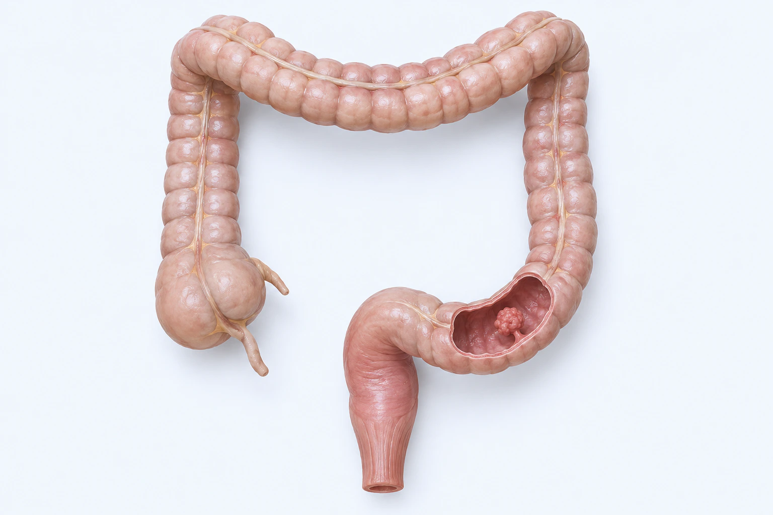
Representative colorectal anatomy with one localized raised mucosal lesion. The open lumen does not communicate stage, spread, obstruction, procedure, or diagnosis.
Scientific media record1 verified identifier
- Subject
- Colorectal Neoplasmscondition
- Identifiers
- MeSH:D015179
- Review
- Editorial review completeIdentifiers authority-verified · Accessibility validated · · colorectal-cancer|colorectal-cancer-pathology-v1.webp
- Digital source
- Trained-algorithmic mediaCreated with a trained generative algorithm and reviewed by WikiBiome for subject identity, scientific framing, identifiers, provenance, and accessibility.
- License
- CC BY-SA 4.0Created
Colorectal cancer (CRC) is the third most common cancer worldwide and the second leading cause of cancer death. Over 90% of cases are sporadic, driven by a complex interplay between genetic predisposition, dietary habits, Gut Microbiome composition, and environmental exposures.
The metallomic and microbiome dimensions of CRC are deeply intertwined: metal dyshomeostasis shapes the microbial community, while microbial metabolites (bile acids, SCFAs, genotoxins) drive or suppress carcinogenesis at the colonic epithelium.
CRC rates vary dramatically by geography and diet—common in Western populations (65 per 100,000 in African Americans) and rare in rural Africa (<5 per 100,000)—underscoring the dominance of environmental and dietary factors over genetics alone.
Evidence map59 cited passagesInspect provenance +
The metallomic profile of CRC from and supporting literature:
demonstrated that Cr(VI) ingestion causes DNA damage and metastasis through hub genes VEGFA, EGFR, APP, SGK1, JUN, and TLR2—directly relevant to colorectal carcinogenesis. Cadmium and arsenic converge on similar pathways of oxidative DNA damage, epigenetic modification, and DNA repair disruption. Heavy metal exposure also depletes SCFA-producing bacteria,
Fusobacterium nucleatum is the most consistently enriched bacterium in CRC across global cohorts. Its pro-tumorigenic mechanisms are multifaceted:
FadA adhesin disrupts E-cadherin, activating beta-catenin/Wnt signaling and promoting proliferation
Fap2 protein binds TIGIT receptor on NK cells, inhibiting anti-tumor immunity, and binds Gal-GalNAc on tumor cells for selective colonization
F. nucleatum abundance increases progressively through the adenoma-carcinoma sequence, validated by qPCR in cohorts exceeding 400 patients.
Enterotoxigenic B. fragilis (ETBF) is found in the mucosa of 80% of CRC patients. The BFT metalloprotease toxin (three isoforms with 93% sequence identity) cleaves E-cadherin, disrupting cell-cell junctions and activating Wnt/beta-catenin and NF-kB pathways. identified chenodeoxycholic acid (CDCA), a primary bile acid and FXR agonist, as a natural BFT inhibi
From Table 1 and supporting studies:
pks+ E. coli: produces colibactin genotoxin causing double-strand DNA breaks and single-base substitutions
S. gallolyticus: classic CRC-associated bacterium
Peptostreptococcus anaerobius: enriched in tumor microenvironment
Bacteria consistently depleted in CRC include Bifidobacterium, Lactobacillus, Roseburia, Faecalibacterium prausnitzii, and Ruminococcus—the core SCFA-producing community.
demonstrated that GPR43 is markedly reduced or lost in ~80% of colorectal adenocarcinomas and 8 of 9 colon cancer cell lines. Loss occurs through promoter hypermethylation. Restoration of GPR43 sensitizes cells to propionate/butyrate-induced cell cycle arrest and apoptosis via caspase activation, p21 upregulation, and Bcl-2/Survivin downregulation.
found that the CRC fecal virome demonstrates increased network connectivity compared to non-cancer individuals. Bacteriophages constitute 90% of the gut virome and modulate bacterial community composition through selective lysis. Key findings:
Dietary fiber: metabolized by gut microbiome into SCFAs (butyrate, propionate, acetate); maintains microbial diversity; 20-30g daily recommended
Mediterranean diet: multi-component protection via n-3 fatty acids (COX-2/TNF-alpha suppression), fiber/butyrate (NF-kB inhibition), olive oil (COX-2/iNOS/STAT3 reduction), and polyphenols (IL-1beta/IL-6/TNF-alpha reduction)
Polyphenols: function as prebiotics increasing Bifidobacterium, Lactobacillus, and Akkermansia; curcumin increases Lactobacillales and decreases Coriobacteriales in CRC models
Consistent oral pathogen enrichment across young-onset and old-onset CRC, CMS subtypes, and geographic populations
Stage-specific microbial markers: adenoma biomarkers are distinct from CRC biomarkers, and both are distinct from healthy controls
Iron-dependent dysbiosis: fermentative anaerobes exploit iron-rich tumor microenvironment; SCFA producers are competitively excluded
Interkingdom ecological reshaping: Candida biofilms and bacteria co-evolve in hypoxic, acidic CRC microenvironment
Bile acid-driven cancer stem cell generation: secondary bile acids (DCA, LCA) directly induce stemness and drug resistance via M3R/Wnt pathways
Iron (Fe) is the dominant selective pressure in CRC:
This is NOT due to low dietary fiber alone—it is competitive exclusion by pathogenic taxa exploiting iron-rich, hypoxic, acidic conditions
Showing 24 of 59 evidence-bearing passages. Every remaining citation is still indexed in the reference record below.
One disease. Five evidence layers.
A generated systems view of the metals, organisms, host sequestration signals, ecological conditions, and microbial functions indexed for Colorectal Cancer.
Evidence layer
Taxonomic signature
Organisms reported as enriched or depleted, with their indexed functional context kept beside the name.Primary pathobiont — FadA adhesin disrupts E-cadherin/beta-catenin, Fap2 inhibits NK cells via TIGIT, LPS-TLR4 drives NF-kB and chemoresistance; progressively increases through adenoma-carcinoma sequence
Early adenoma pathogen — enriched in ~80% of CRA; appears before F. nucleatum in adenoma-carcinoma sequence
ETBF BFT zinc-metalloprotease cleaves E-cadherin, activates Wnt/beta-catenin and NF-kB; found in mucosa of >80% of CRC patients
Oral pathogen, consistent cross-population biomarker — enriched in CRC across all age groups
Oral pathogen — consistently enriched in advanced stages; pks+ colibactin producer
pks+ strains synthesize colibactin — induces double-strand DNA breaks and mutations; siderophore competition in iron-rich environment
CRC-enriched driver taxon — consistently elevated in both young- and old-onset CRC; bile acid metabolism
Oral pathogen — enriched in CMS1 immune subtype; periodontal-gut-CRC link
Paradoxical role — elevated 4x in CRC stool vs healthy; maintains glycerophospholipid metabolism for PD-1 escape; SCFA paradox
Interkingdom interaction — biofilm formation, oxygen depletion, functional shielding with bacteria, reduced inflammatory detection
Enriched in tumor microenvironment; implicated in carcinogenesis
Produces extracellular superoxide and hydroxyl radicals causing oxidative DNA damage
Classic CRC-associated bacterium; bloodstream presence signals colonic neoplasia
Fungal abundance increases in CRC vs healthy controls; part of mycobiome dysbiosis
SCFA producers (butyrate, propionate) — protective; lost in CRC microenvironment; competitive exclusion by iron-rich pathogens
Major butyrate producer — loss reduces HDAC inhibition of tumor cells and anti-inflammatory IL-10 signaling
SCFA producer — butyrate generators depleted in established CRC but may be recruited as 'passengers' in tumor microenvironment
Protective in healthy microbiota — 40-fold depleted in CRC; associated with fiber fermentation and SCFA production
SCFA producer — 10x depleted in CRC; fiber fermentation and short-chain fatty acid production capacity lost
Protective species (Dialister invisus 48.7x depleted) — SCFA production and immune tolerance functions lost
Depleted in CRC; loss compromises colonization resistance, IgA responses, and tight junction integrity
Depleted in CRC; loss of lactic acid production and competitive exclusion of pathogens
Fiber-fermenting SCFA producer depleted in CRC
Evidence layer
Nutritional immunity
Host metal-withholding, inflammatory, antioxidant, and microbial-metabolite signals indexed in the signature.Elevated host signals
7Depleted protective signals
5Evidence layer
Ecological state
The environmental conditions that connect the organism-level observations into a system.Evidence layer
Virulence functions
Microbial structures, enzymes, and acquisition systems implicated by the linked evidence.The disease record, in full.
The original WikiBiome disease narrative remains intact beneath the generated signature atlas.
Metallomic Signature#
The metallomic profile of CRC from[1]Recent advances in the application of metallomics in diagnosis and prognosis of human cancerYan Zhang, Jie He, Jiao Jin et al. · 2022Open reference 1 ↓ and supporting literature:
| Metal | Direction | Key Evidence | |
|---|---|---|---|
| [[copper | copper (Cu)]] | Elevated | Increased in serum/plasma across multiple studies; copper/zinc (Zn) ratio first proposed as a CRC marker |
| [[zinc | zinc]] | Depleted | European study of 58,221 participants confirmed significant association; zinc deficiency is a general cancer feature across multiple meta-analyses |
| [[selenium | selenium (Se)]] | Depleted | Decreased across cancer types; impairs glutathione peroxidase defense |
| [[iron | iron (Fe)]] | Elevated (tissue) | Heme iron from red meat drives N-nitroso compound formation and oxidative DNA damage; iron dysregulation via ferroptosis pathways |
| [[cadmium | cadmium (Cd)]] | Exposure risk | IARC Group 1 carcinogen; disrupts DNA repair and epigenetic modification |
| [[arsenic | As]] | Exposure risk | IARC Group 1 carcinogen; environmental contamination source |
| [[chromium | chromium (Cr)]] | Exposure risk (chromium VI) | Ingestion route associated with CRC and GI tract diseases; hub genes include VEGFA and EGFR |
| [[nickel | nickel (Ni)]] | Elevated (inconsistent) | Increased in some studies; IARC Group 1 carcinogen alongside chromium and cadmium |
The Cu/Zn Ratio#
The copper/zinc ratio was first proposed as a CRC biomarker and captures the two most consistent metallomic changes—copper elevation and zinc depletion. Elevated copper displaces zinc from metallothionein due to higher binding affinity, simultaneously impairing copper/zinc superoxide dismutase (Cu/Zn-SOD) antioxidant defense and creating a pro-oxidant environment favorable to carcinogenesis.
This ratio is now documented as elevated across breast, prostate, lung, and thyroid cancers as well.
Metal-Carcinogenesis Mechanisms#
[2]Adverse Human Health Effects of Chromium by Exposure Route: A Comprehensive Review Based on Toxicogenomic ApproachDong Yeop Shin, Sang Min Lee, Yujin Jang et al. · 2023Open reference 2 ↓ demonstrated that chromium (Cr)(VI) ingestion causes DNA damage and metastasis through hub genes VEGFA, EGFR, APP, SGK1, JUN, and TLR2—directly relevant to colorectal carcinogenesis. Cadmium and arsenic converge on similar pathways of oxidative DNA damage, epigenetic modification, and DNA repair disruption.
Heavy metal exposure also depletes SCFA-producing bacteria, removing a key protective mechanism against carcinogenesis.
Microbiome Signatures#
Fusobacterium nucleatum as Key Pathobiont#
Fusobacterium nucleatum is the most consistently enriched bacterium in CRC across global cohorts.[3]Towards the Human Colorectal Cancer MicrobiomeMarchesi JR, Dutilh BE, Hall N et al. · 2011Open reference 3 ↓[4]Microbiota disbiosis is associated with colorectal cancerZhiguang Gao, Bomin Guo, Renyuan Gao et al. · 2015Open reference 4 ↓ Its pro-tumorigenic mechanisms are multifaceted. FadA adhesin disrupts E-cadherin, activating beta-catenin/Wnt signaling and promoting proliferation.[5]Immune System, Microbiota, and Microbial Metabolites: The Unresolved Triad in Colorectal Cancer MicroenvironmentHanus M, Parada-Venegas D, Landskron G et al. · 2021Open reference 5 ↓
Fap2 protein binds TIGIT receptor on NK cells, inhibiting anti-tumor immunity, and binds Gal-GalNAc on tumor cells for selective colonization.[5]Immune System, Microbiota, and Microbial Metabolites: The Unresolved Triad in Colorectal Cancer MicroenvironmentHanus M, Parada-Venegas D, Landskron G et al. · 2021Open reference 5 ↓
LPS–TLR4 interaction activates NF-kB signaling, autophagy, and chemotherapy resistance (5-FU). miR21/TLR4/NF-kB axis and ULK1/ATG7 autophagy pathway activation.
F. nucleatum abundance increases progressively through the adenoma-carcinoma sequence, validated by qPCR in cohorts exceeding 400 patients.[6]Features of combined gut bacteria and fungi from a Chinese cohort of colorectal cancer, colorectal adenoma, and post-operative patientsXiaopeng Li, Jiahui Feng, Zhanggui Wang et al. · 2023Open reference 6 ↓
Bacteroides fragilis BFT Toxin#
Enterotoxigenic B. fragilis (ETBF) is found in the mucosa of >80% of CRC patients.[7]Drug Discovery and Repurposing Inhibits a Major Gut Pathogen-Derived Oncogenic ToxinPaul Metz, Martijn J. H. Tjan, Shaoguang Wu et al. · 2019Open reference 7 ↓
The BFT metalloprotease toxin (three isoforms with >93% sequence identity) cleaves E-cadherin, disrupting cell-cell junctions and activating Wnt/beta-catenin and NF-kB pathways.[7]Drug Discovery and Repurposing Inhibits a Major Gut Pathogen-Derived Oncogenic ToxinPaul Metz, Martijn J. H. Tjan, Shaoguang Wu et al. · 2019Open reference 7 ↓[7]Drug Discovery and Repurposing Inhibits a Major Gut Pathogen-Derived Oncogenic ToxinPaul Metz, Martijn J. H. Tjan, Shaoguang Wu et al. · 2019Open reference 7 ↓ identified chenodeoxycholic acid (CDCA), a primary bile acid and FXR agonist, as a natural BFT inhibitor—reducing IL-8 secretion by 67-69% at physiological concentrations, suggesting inter-individual bile acid variation may explain differential CRC susceptibility.
Other CRC-Associated Bacteria#
From[5]Immune System, Microbiota, and Microbial Metabolites: The Unresolved Triad in Colorectal Cancer MicroenvironmentHanus M, Parada-Venegas D, Landskron G et al. · 2021Open reference 5 ↓ Table 1 and supporting studies:
- pks+ E. coli: produces colibactin genotoxin causing double-strand DNA breaks and single-base substitutions[5]Immune System, Microbiota, and Microbial Metabolites: The Unresolved Triad in Colorectal Cancer MicroenvironmentHanus M, Parada-Venegas D, Landskron G et al. · 2021Open reference 5 ↓
- S. gallolyticus: classic CRC-associated bacterium[5]Immune System, Microbiota, and Microbial Metabolites: The Unresolved Triad in Colorectal Cancer MicroenvironmentHanus M, Parada-Venegas D, Landskron G et al. · 2021Open reference 5 ↓
- Peptostreptococcus anaerobius: enriched in tumor microenvironment[4]Microbiota disbiosis is associated with colorectal cancerZhiguang Gao, Bomin Guo, Renyuan Gao et al. · 2015Open reference 4 ↓
- Enterococcus faecalis: produces extracellular superoxide and hydroxyl radicals
Bacteria consistently depleted in CRC include Bifidobacterium, Lactobacillus, Roseburia, Faecalibacterium prausnitzii, and Ruminococcus—the core SCFA-producing community.[4]Microbiota disbiosis is associated with colorectal cancerZhiguang Gao, Bomin Guo, Renyuan Gao et al. · 2015Open reference 4 ↓[5]Immune System, Microbiota, and Microbial Metabolites: The Unresolved Triad in Colorectal Cancer MicroenvironmentHanus M, Parada-Venegas D, Landskron G et al. · 2021Open reference 5 ↓
Bile Acid-Microbiome-Cancer Axis#
Secondary bile acids (deoxycholic acid/DCA and lithocholic acid/LCA), converted from primary bile acids by gut bacterial 7-alpha dehydroxylation, are potent CRC promoters. Their carcinogenic mechanisms include.
ROS/RNS generation via NAD(P)H oxidase activation and mitochondrial membrane disruption. NF-kB activation determining intestinal epithelial cell fate; persistent activation drives colitis-associated CRC. DNA damage through oxidative base modification and disruption of base excision repair.
Cancer stem cell induction: DCA/LCA induce CSC markers (CD44, CD166, ALDHA1) and pluripotency genes (KLF4, Nanog, OCT4, SOX2) through M3R/Wnt/beta-catenin signaling, with 12-15 fold increases in c-Myc.
EMT promotion: MMP upregulation (MMP1, MMP3, MMP10) and mesenchymal marker activation. Drug resistance: upregulation of ABCB1 and ABCG2 transporters in bile acid-exposed colonocytes.
High dietary fat and red meat consumption increase secondary bile acid levels. Western diet populations bear up to 60% of the global CRC burden. Vitamin D deficiency compounds the risk, as VDR functions as a secondary bile acid receptor (LCA) and activates detoxification pathways via PXR/SXR.
SCFAs as Protective Factors#
Short-chain fatty acids—acetate, propionate, and Butyrate constituting ~95% of total SCFAs at colonic concentrations of 70-100 mM—are the primary protective metabolites against CRC.
Butyrate and the Warburg Paradox#
Butyrate exhibits a dual role explained by the metabolic state of colonocytes: in normal cells, butyrate enters the Krebs cycle as an energy source promoting proliferation.
In tumor cells undergoing the Warburg effect (anaerobic glycolysis), butyrate accumulates in the nucleus where it acts as an HDAC inhibitor, upregulating p21/p27 tumor suppressors and inducing apoptosis.
GPR43/FFAR2 as Tumor Suppressor#
[8]G-protein-coupled receptor for short-chain fatty acids suppresses colon cancerYong Tang, Yakun Chen, Hongmei Jiang et al. · 2011Open reference 8 ↓ demonstrated that GPR43 is markedly reduced or lost in ~80% of colorectal adenocarcinomas and 8 of 9 colon cancer cell lines. Loss occurs through promoter hypermethylation.
Restoration of GPR43 sensitizes cells to propionate/butyrate-induced cell cycle arrest and apoptosis via caspase activation, p21 upregulation, and Bcl-2/Survivin downregulation.
SCFA Receptor Network#
Three GPCRs mediate SCFA tumor suppression. FFAR2/GPR43: promotes IL-18 secretion via NLRP3 inflammasome; enhances Treg differentiation; deficiency promotes tumorigenesis via Wnt/beta-catenin. FFAR3/GPR41: expressed on intestinal epithelial and immune cells.
HCAR2/GPR109A: butyrate-specific; suppresses carcinogenesis; knockdown accelerates colonic Metal-Driven Inflammation and cancer progression.
Fungal Microbiome (Mycobiome) in CRC#
Fungi represent ~0.1% of gut microorganisms but play important roles in CRC.
Candida albicans and Saccharomyces cerevisiae show significantly increasing abundance in CRC vs healthy controls, validated by qPCR (n=402). Basidiomycota/Ascomycota ratio increases with disease progression through the adenoma-carcinoma sequence. Bacterial-fungal antagonistic interactions (R = -0.17 to -0.32) suggest disruption of cross-kingdom balance contributes to CRC.
Fungal beta-glucans can be protective (upregulating tight junction proteins Claudin-1 and ZO-1), while C. albicans can be destructive (GelE-mediated E-cadherin degradation). Dectin-1 and TLR4 signaling are essential for anti-fungal immunity in the gut; C. albicans can inhibit NLRP3/NLRP6 inflammasomes.
Viral Microbiome (Virome) in CRC#
[9]Alterations in colorectal cancer virome and its persistence after surgerySi Xian Ho, Jia-Hao Law, Chin-Wen Png et al. · 2024Open reference 9 ↓ found that the CRC fecal virome demonstrates increased network connectivity compared to non-cancer individuals. Bacteriophages constitute 90% of the gut virome and modulate bacterial community composition through selective lysis. Key findings:
- cobalt (Co)-exclusion of healthy-associated viruses with CRC-enriched bacteria
- Enterobacteria phage P88 positively correlated with butyrate-producing Megasphaera
- Virome Dysbiosis partially persists after surgery, indicating stable alterations
- Trans-kingdom virus-bacteria networks are disrupted in CRC
Iron and Ferroptosis in CRC#
Iron intersects with CRC through multiple pathways.
Heme iron from red meat generates N-nitroso compounds (NOCs) causing KRAS mutations (G>A transitions at codons 12/13) and oxidative DNA damage. Ferroptosis (iron-dependent lipid peroxidation cell death) represents both a vulnerability and a defense mechanism in CRC cells; GPX4 is the key ferroptosis regulator.
Iron dysregulation parallels the mitochondrial membrane disruption caused by secondary bile acids. Metal-induced dysbiosis depletes butyrate-producing Firmicutes, reducing antioxidant defenses that normally protect against iron-mediated lipid peroxidation.
Diet Connections#
Protective Factors#
Dietary fiber: metabolized by gut microbiome into SCFAs (butyrate, propionate, acetate); maintains microbial diversity; 20-30g daily recommended.[10]Emerging Evidence on the Effects of Dietary Factors on the Gut Microbiome in Colorectal CancerAppunni S, Rubens M, Ramamoorthy V et al. · 2021Open reference 10 ↓[11]Diet, microbiota, and dysbiosis: a 'recipe' for colorectal cancerKishore Vipperla, Stephen J. O'Keefe · 2016Open reference 11 ↓
Mediterranean diet: multi-component protection via n-3 fatty acids (COX-2/TNF-alpha suppression), fiber/butyrate (NF-kB inhibition), olive oil (COX-2/iNOS/STAT3 reduction), and polyphenols (IL-1beta/IL-6/TNF-alpha reduction).[12]Mediterranean Diet: Prevention of Colorectal CancerMicah G. Donovan, Ornella I. Selmin, Tom C. Doetschman et al. · 2017Open reference 12 ↓[13]Design of the Building Research in CRC Prevention (BRIDGE-CRC) Trial: A 6-Month, Parallel Group Mediterranean Diet and Weight Loss Randomized Controlled Lifestyle Intervention Targeting the Bile Acid-Gut Microbiome Axis to Reduce Colorectal Cancer Risk Among African American/Black Adults with ObesityAndrew McLeod, Patricia Wolf, Robert S. Chapkin et al. · 2023Open reference 13 ↓
Polyphenols: function as prebiotics increasing Bifidobacterium, Lactobacillus, and Akkermansia; curcumin increases Lactobacillales and decreases Coriobacteriales in CRC models.[10]Emerging Evidence on the Effects of Dietary Factors on the Gut Microbiome in Colorectal CancerAppunni S, Rubens M, Ramamoorthy V et al. · 2021Open reference 10 ↓
Risk Factors#
Red and processed meat: generates NOCs, heterocyclic amines, and heme iron-mediated genotoxicity; gut microbiome metabolizes these into more harmful compounds. High-fat diet: increases secondary bile acid production via microbial 7-alpha dehydroxylation. Alcohol: metabolized to acetaldehyde (DNA-damaging intermediate) via gut microbiome pathways; increases Bacteroidetes and Ruminococcaceae.
Low-fiber Western diet: predisposes to CRC via reduced SCFA production and increased pathobiont abundance.
Geographic Diet Contrasts#
The starkest evidence comes from African American vs rural African comparisons: CRC rates of 65 vs <5 per 100,000, with Prevotella predominant in native Africans (high fiber) and Bacteroides predominant in African Americans (high fat/protein). This dietary selection of the microbiome mediates the majority of CRC risk.
Comorbidities#
Inflammatory Bowel Disease (IBD)—IBD is the strongest established risk factor for CRC; chronic colonic inflammation drives the inflammation-dysplasia-carcinoma sequence; shared Fusobacterium nucleatum enrichment and SCFA producer depletion.
Crohn's Disease—Crohn's colitis confers a 2-3x increased CRC risk; shared NF-kB/Wnt signaling crosstalk, barrier dysfunction, and Enterobacteriaceae bloom; the same metal-driven dysbiosis cascade underlies both.
Obesity—obesity increases CRC risk by 30%; shared high-fat diet, elevated secondary bile acids (DCA/LCA), and gut dysbiosis; adipokine dysregulation promotes tumor microenvironment inflammation.
Type 2 Diabetes—T2D increases CRC risk by 20-30%; shared insulin resistance, hyperinsulinemia (IGF-1 axis promotes cell proliferation), and SCFA depletion; metformin may be protective through AMPK activation and microbiome modulation.
Connections#
- Metal-Disease Matrix: A Cross-Source Synthesis
- Biomarkers—fecal occult blood, CEA, and microbial biomarkers (F. nucleatum qPCR) for CRC screening and monitoring
References 36
Numbered by first appearance in the article, then reconciled with its declared source list.
- 1
Yan Zhang, Jie He, Jiao Jin et al. (2022). Recent advances in the application of metallomics in diagnosis and prognosis of human cancer. Metallomics.
- 2
Dong Yeop Shin, Sang Min Lee, Yujin Jang et al. (2023). Adverse Human Health Effects of Chromium by Exposure Route: A Comprehensive Review Based on Toxicogenomic Approach. International Journal of Molecular Sciences.
- 3
Marchesi JR, Dutilh BE, Hall N et al. (2011). Towards the Human Colorectal Cancer Microbiome. PLOS ONE.
- 4
Zhiguang Gao, Bomin Guo, Renyuan Gao et al. (2015). Microbiota disbiosis is associated with colorectal cancer. Frontiers in Microbiology.
- 5
Hanus M, Parada-Venegas D, Landskron G et al. (2021). Immune System, Microbiota, and Microbial Metabolites: The Unresolved Triad in Colorectal Cancer Microenvironment. Frontiers in Immunology.
- 6
Xiaopeng Li, Jiahui Feng, Zhanggui Wang et al. (2023). Features of combined gut bacteria and fungi from a Chinese cohort of colorectal cancer, colorectal adenoma, and post-operative patients. Frontiers in Microbiology.
- 7
Paul Metz, Martijn J. H. Tjan, Shaoguang Wu et al. (2019). Drug Discovery and Repurposing Inhibits a Major Gut Pathogen-Derived Oncogenic Toxin. Frontiers in Cellular and Infection Microbiology.
- 8
Yong Tang, Yakun Chen, Hongmei Jiang et al. (2011). G-protein-coupled receptor for short-chain fatty acids suppresses colon cancer. International Journal of Cancer.
- 9
Si Xian Ho, Jia-Hao Law, Chin-Wen Png et al. (2024). Alterations in colorectal cancer virome and its persistence after surgery. Scientific Reports.
- 10
Appunni S, Rubens M, Ramamoorthy V et al. (2021). Emerging Evidence on the Effects of Dietary Factors on the Gut Microbiome in Colorectal Cancer. Frontiers in Nutrition.
- 11
Kishore Vipperla, Stephen J. O'Keefe (2016). Diet, microbiota, and dysbiosis: a 'recipe' for colorectal cancer. Food & Function.
- 12
Micah G. Donovan, Ornella I. Selmin, Tom C. Doetschman et al. (2017). Mediterranean Diet: Prevention of Colorectal Cancer. Frontiers in Nutrition.
- 13
Andrew McLeod, Patricia Wolf, Robert S. Chapkin et al. (2023). Design of the Building Research in CRC Prevention (BRIDGE-CRC) Trial: A 6-Month, Parallel Group Mediterranean Diet and Weight Loss Randomized Controlled Lifestyle Intervention Targeting the Bile Acid-Gut Microbiome Axis to Reduce Colorectal Cancer Risk Among African American/Black Adults with Obesity. Trials.
- 14
Youwen Qin, Xin Tong, Wei-Jian Mei et al. (2024). Consistent signatures in the human gut microbiome of old- and young-onset colorectal cancer. Nature Communications.
- 15
Wu Y, Jiao N, Zhu R et al. (2021). Identification of Microbial Markers across Populations in Early Detection of Colorectal Cancer. Nature Communications.
- 16
Loke MF, Chua EG, Gan HM et al. (2018). Metabolomics and 16S rRNA Sequencing of Human Colorectal Cancers and Adjacent Mucosa. PLOS ONE.
- 17
Lulu Farhana, Pratima Nangia-Makker, Evan Arbit et al. (2016). Bile acid: a potential inducer of colon cancer stem cells. Stem Cell Research & Therapy.
- 18
Xinjian Xu, Ji Lv, Fang Guo et al. (2020). Gut Microbiome Influences the Efficacy of PD-1 Antibody Immunotherapy on MSS-Type Colorectal Cancer via Metabolic Pathway. Frontiers in Microbiology.
- 19
Maria Daniella Carretta, John Quiroga, Rodrigo Lopez et al. (2021). Participation of short-chain fatty acids and their receptors in gut inflammation and colon cancer. Frontiers in Physiology.
- 20
Johanna Simin, Romina Fornes, Qing Liu et al. (2020). Antibiotic Use and Risk of Colorectal Cancer: A Systematic Review and Dose-Response Meta-Analysis. British Journal of Cancer.
- 21
Tung Hoang, Minjung Kim, Ji Won Park et al. (2023). Dysbiotic microbiome variation in colorectal cancer patients is linked to lifestyles and metabolic diseases. BMC Microbiology.
- 22
Xiaoliang Xie, Wenjing Wang, Haiming Zhang et al. (2025). Cholesterol-induced colorectal cancer progression and its mitigation through gut microbiota remodeling and simvastatin treatment. BMC Cancer.
- 23
Ma M, Zheng Z, Li J et al. (2024). Association between the Gut Microbiota, Inflammatory Factors, and Colorectal Cancer: Evidence from Mendelian Randomization Analysis. Frontiers in Microbiology.
- 24
Saito K, Koido S, Odamaki T et al. (2019). Metagenomic Analyses of the Gut Microbiota Associated with Colorectal Adenoma. PLOS ONE.
- 25
Bars-Cortina D, Ramon E, Rius-Sansalvador B et al. (2024). Comparison between 16S rRNA and Shotgun Sequencing in Colorectal Cancer, Advanced Colorectal Lesions, and Healthy Human Gut Microbiota. BMC Genomics.
- 26
Tiffany L Weir, Daniel K Manter, Amy M Sheflin et al. (2013). Stool Microbiome and Metabolome Differences between Colorectal Cancer Patients and Healthy Adults. PLoS ONE.
- 27
Rachel V. Purcell, Martina Visnovska, Patrick J. Biggs et al. (2017). Distinct gut microbiome patterns associate with consensus molecular subtypes of colorectal cancer. Scientific Reports.
- 28
Qinhan Gao, Yuwen Liu, Fayu Su et al. (2026). Ketogenic diet suppresses colorectal cancer through reshaping gut microbiota and modulating the intestinal FXR/NF-kB signaling pathway. Food Science and Human Wellness.
- 29
Lan Zhao, William C. Cho, Mark R. Nicolls (2021). Colorectal cancer-associated microbiome patterns and signatures. Frontiers in Genetics.
- 30
Hou X, Zheng Z, Wei J et al. (2022). Effects of Gut Microbiota on Immune Responses and Immunotherapy in Colorectal Cancer. Frontiers in Immunology.
- 31
Shaomin Zou, Chao Yang, Jieping Zhang et al. (2024). Multi-omic profiling reveals associations between the gut microbiome, host genome and transcriptome in patients with colorectal cancer. Journal of Translational Medicine.
- 32
Yu L, Zhao G, Wang L et al. (2022). A Systematic Review of Microbial Markers for Risk Prediction of Colorectal Neoplasia. British Journal of Cancer.
- 33
Liu Z, Zhou X, Kuang L et al. (2025). Novel Insights into Immune-Gut Microbiota Interactions in Colorectal Cancer: A Mendelian Randomization Study. Infectious Agents and Cancer.
- 34
Hana Ajouz, Deborah Mukherji, Ali Shamseddine (2014). Secondary bile acids: an underrecognized cause of colon cancer. World Journal of Surgical Oncology.
- 35
Rie Sugimoto, Lingaku Lee, Yuki Tanaka et al. (2024). Zinc Deficiency as a General Feature of Cancer: A Review of the Literature. Biological Trace Element Research.
- 36
Yilin Huang, Yang Wang, Xiaotian Huang et al. (2024). Unveiling the overlooked fungi: the vital of gut fungi in inflammatory bowel disease and colorectal cancer. Gut Pathogens.
Article network
Mentioned here 18
Pages linking here 102
Connect the evidence
Publicly readable discussion by ORCID-authenticated researchers. Questions, interpretation, methods, corrections, and new evidence stay attached to this record.
No discussion yet. Start with a precise question or a source-backed observation.
Activity and accepted changes
Accepted researcher context, editorial status, public discussion, and upstream Git revisions are shown together. Pending, declined, and withdrawn proposals remain private.
- published revision
Strengthen TLR4 and link high-leverage contexts
Karen Pendergrass · +1 −1
Inspect exact Git diff ↗ - published revision
Consolidate microbial metabolite knowledge
Karen Pendergrass · +1 −1
Inspect exact Git diff ↗ - published revision
Backfill gut microbiome concept links
Karen Pendergrass · +1 −1
Inspect exact Git diff ↗ - published revision
Backfill inflammation concept links
Karen Pendergrass · +1 −1
Inspect exact Git diff ↗ - published revision
Complete corpus-wide Dysbiosis linking
Karen Pendergrass · +1 −1
Inspect exact Git diff ↗ - published revision
Complete Tight junctions contextual coverage
Karen Pendergrass · +1 −1
Inspect exact Git diff ↗ - published revision
Add NLRP3 inflammasome concept and link batch
Karen Pendergrass · +3 −3
Inspect exact Git diff ↗ - published revision
massive wiki expansion: 149 stubs fixed, 100+ new pages, Rule 15 scan, keystone papers
WikiBiome Deploy Bot · +36 −36
Inspect exact Git diff ↗ - published revision
nightly maintenance: 94 stub demotions, 181 source_count fixes, 22 auto-discovered stubs, 5 adversarial audits, 3 boundary fixes, 3 evidence-level corrections
WikiBiome Deploy Bot · +1 −0
Inspect exact Git diff ↗ - published revision
cycle 1: health check + lint fixes + 8 ingests + 2 stubs + gestational-diabetes signature
WikiBiome Deploy Bot · +1 −0
Inspect exact Git diff ↗ - published revision
Batch: fix 1025 broken wikilinks, wire 13 STOP pages, deepen PPD/GERD/T1D/8 microbes
WikiBiome Deploy Bot · +2 −0
Inspect exact Git diff ↗ - published revision
Deep citation pass on 10 disease entities + expand 3 thin entities
WikiBiome Deploy Bot · +12 −12
Inspect exact Git diff ↗ - published revision
Deepen metal/concept entities + 8 new sources for T1D/schizophrenia
WikiBiome Deploy Bot · +378 −0
Inspect exact Git diff ↗ - published revision
WikiBiome update — 2026-04-15 17:23
WikiBiome Deploy Bot · +18 −18
Inspect exact Git diff ↗ - published revision
v2 migration Priority 2: All 29 disease entity pages upgraded with associated_conditions, seo_target, wikipedia_differentiation
WikiBiome Deploy Bot · +6 −0
Inspect exact Git diff ↗