

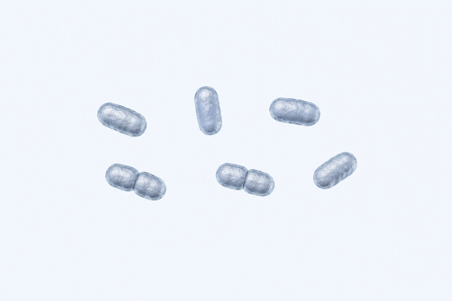
Type-strain-anchored Porphyromonas gingivalis reconstruction showing eight compact short rod-to-coccobacillary bodies in four single and two paired groupings. This species plate is representative, non-diagnostic, and not a micrograph.
Scientific media record1 verified identifier
- Subject
- Porphyromonas gingivalistaxon · species
- Identifiers
- NCBITaxon:837
- Review
- Editorial review completeIdentifiers authority-verified · Accessibility validated · · porphyromonas-gingivalis|porphyromonas-gingivalis-morphology-v1.webp
- Digital source
- Trained-algorithmic mediaCreated with a trained generative algorithm and reviewed by WikiBiome for subject identity, scientific framing, identifiers, provenance, and accessibility.
- Scientific basis
- Porphyromonas gingivalis — NCBI TaxonomyPorphyromonas gingivalis — LPSNProposal for reclassification in the new genus PorphyromonasPorphyromonas gingivalis type strain — BacDive
- License
- CC BY-SA 4.0Created
A Gram-negative obligate anaerobe and keystone pathogen of the oral microbiome that causes chronic periodontitis and is increasingly implicated in systemic diseases including Alzheimer's Disease and Cardiovascular Disease.
P. gingivalis virulence is fundamentally dependent on zinc, iron, and manganese, particularly for the expression and activity of its signature gingipains—virulence proteases that directly link the oral pathobiont to Neuroinflammation and atherosclerosis.
Evidence map7 cited passagesInspect provenance +
Immune evasion: Mixed biofilm attenuates macrophage cytokine/chemokine production; neutrophil elastase activity remains at baseline—the host cannot detect the bacterial invader.
Gingipain amplification: Rgp activity increases up to 10-fold under normoxic conditions in fungal co-culture, enhancing tissue destruction and immune evasion.
Chronic persistence: In vivo, C. albicans pre-infection reduced P. gingivalis mortality from 21% to 7% while bacterial persistence at 96 hours rose from 24% to 85%.
Co-aggregation: Stabilized by Als3 (fungal adhesin) binding to RgpA hemagglutinin domain, and PPAD-mediated citrullination of Candida surface proteins.
Cortisol upregulates T9SS-associated genes (mfa1, mfa5, sigP, porP, sprA) in a concentration-dependent manner.
Migration requires exogenous lactate or pyruvate—metabolic substrates from erythrocyte lysis (gingival bleeding) or oral streptococcal cross-feeding.
The fimbrial tip protein Mfa5 (containing a von Willebrand factor domain) is essential for cortisol-stimulated translocation.
Contents
1. Gingipains: Zinc-Dependent Virulence Proteases2. Iron and Heme Acquisition3. Manganese (Mn-SOD) and Oxidative Tolerance4. Systemic Disease Associations5. Biofilm Architecture and Periodontal Niche6. Oral Dysbiosis and Periodontitis7. Metal and Nutrient Context8. Ecological Modulators9. Functional Shielding by Candida albicans10. Cortisol-Stimulated Virulence11. ConnectionsGingipains: Zinc-Dependent Virulence Proteases#
The Three Gingipain Classes#
P. gingivalis secretes three major gingipain proteases (EC 3.4.21.-) that are essential virulence factors:
- RgpA and RgpB (Arg-specific gingipains): cleave proteins after arginine residues
- Kgp (Lys-specific gingipain): cleaves proteins after lysine residues
All three are zinc metallopeptidases with catalytic zinc in their active sites.
Zinc Requirement and Regulation#
Gingipain expression and activity are zinc-responsive: under high-zinc conditions, gingipain synthesis increases; under zinc limitation, expression is downregulated. This makes gingipain activity directly modulated by local zinc availability in periodontal lesions.
Host Calprotectin (S100A8/A9) and other nutritional immunity mechanisms that sequester zinc at Metal-Driven Inflammation sites may paradoxically reduce gingipain activity—a counterintuitive protective mechanism.
Gingipain Functions#
Gingipains perform multiple virulence roles. Degradation of barrier proteins: cleave collagen, fibronectin, and laminin in the gingival epithelium and periodontal ligament, deepening pocket formation. Immune evasion: cleave IgG, complement C3, and C4, disarming antibody-mediated and complement-based defense.
Activation of protease-activated receptors (PARs): gingipain-mediated cleavage of PAR1 and PAR4 on epithelial and immune cells triggers pro-inflammatory signaling (NF-kB, MAPK).
Amyloid-beta and tau generation: critically, gingipains can directly cleave amyloid precursor protein (APP) and tau protein, generating amyloidogenic fragments that persist and aggregate in the brain.
Iron and Heme Acquisition#
P. gingivalis is a hemin specialist: obtains iron primarily from heme in blood and broken-down hemoglobin from gingival bleeding. Expresses multiple hemin-binding proteins and heme-iron uptake transporters (HmuR, IhtA). Iron is essential for cytochrome c oxidase, catalase, and electron transport chain enzymes; zinc (Zn)-proteasome activity; and cofactors in numerous metabolic enzymes.
Periodontal bleeding directly feeds P. gingivalis by providing high-iron, high-heme substrate.
Manganese (Mn-SOD) and Oxidative Tolerance#
Expresses manganese-dependent superoxide dismutase (manganese (Mn)-SOD) for protection against neutrophil-generated reactive oxygen species (ROS). Elevated oral manganese (not well-studied but potentially elevated in periodontal lesions) may favor P. gingivalis persistence.
Systemic Disease Associations#
Alzheimer's Disease#
Mechanistic link: Gingipains (particularly Kgp and RgpA) are directly immunoreactive with antibodies against P. gingivalis found in Alzheimer's brains. Gingipain-cleaved amyloid-beta and tau peptides are more aggregation-prone and neurotoxic. Chronic periodontitis increases AD risk; oral P. gingivalis load correlates with cognitive decline and amyloid pathology.
Oral Dysbiosis with P. gingivalis enrichment may enable translocation of pathobiont-derived lipopolysaccharide (LPS) and gingipains across the intestinal barrier into systemic circulation, crossing the blood-brain barrier and perpetuating neuroinflammation.
Cardiovascular Disease#
P. gingivalis is enriched in subgingival plaques of periodontitis patients who have atherosclerosis. Gingipain-mediated cleavage of platelet aggregation inhibitors and endothelial barrier proteins promotes thrombosis and atherosclerotic plaque progression. P. gingivalis-derived LPS acts as a toll-like receptor 4 (TLR4) agonist, driving systemic inflammation.
Chronic bacteremia (transient seeding into bloodstream during chewing or dental procedures) exposes the endothelium to gingipain proteases and bacterial LPS.
Biofilm Architecture and Periodontal Niche#
P. gingivalis is a biofilm specialist that organizes complex polymicrobial communities in the subgingival pocket. Produces extracellular polysaccharides (mostly glucose polymers) that form the biofilm matrix, creating anaerobic microenvironments.
Exhibits synergistic virulence with Prevotella intermedia, Fusobacterium nucleatum, and other Gram-negative anaerobes: FadA and gingipains together degrade epithelial tight junctions more effectively than either alone.
Biofilm structure is resistant to both host immune attack and antimicrobial peptides/antibiotics.
Oral Dysbiosis and Periodontitis#
Keystone-Pathogen Concept#
P. gingivalis is the prototype of a keystone pathogen: despite representing <1% of oral biomass in periodontitis, its presence organizes the entire dysbiotic community around it.
Produces diffusible signaling molecules (e.g., via quorum sensing) that alter the behavior of commensals, transforming the biofilm into a virulent community. Non-toxigenic strains of P. gingivalis can persist as commensals in healthy gingiva; toxigenic (gingipain-producing) strains cause periodontitis.
Disease Progression#
Initiation: P. gingivalis and other Gram-negative anaerobes colonize the subgingival pocket. Amplification: Bleeding from early inflammation provides iron/heme substrate, fueling P. gingivalis proliferation and gingipain expression. Persistence: Biofilm + gingipain-mediated immune evasion = chronic pocket deepening and bone loss.
Systemic spillover: Chronic bacteremia and endotoxemia drive systemic inflammation; gingipains reach distant tissues.
Metal and Nutrient Context#
Zinc limitation in healthy gingiva (controlled by Calprotectin (S100A8/A9) and other nutritional immunity factors) restricts gingipain production. Zinc elevation in inflamed pockets (from tissue breakdown and transudation) enhances gingipain synthesis and activity.
Iron availability from bleeding is the major source; heme-dependent pathways are essential for P. gingivalis competitiveness in the bloodfilled pocket.
Ecological Modulators#
Mechanical plaque removal disrupts biofilm architecture, eliminating the polymicrobial community structure that sustains P. gingivalis. Chlorhexidine and other antimicrobials reduce P. gingivalis load but may select for resistant strains, illustrating the limitation of direct-kill approaches.
Zinc sequestration: Enhancing nutritional immunity mechanisms that restrict zinc in the periodontal pocket suppresses gingipain production by limiting cofactor availability—the organism's primary virulence dependency.
Iron/heme competition: Lactoferrin competes with P. gingivalis heme acquisition systems, reducing the iron pool that drives pathogen competitiveness in the blood-filled pocket.
Functional Shielding by Candida albicans#
P. gingivalis forms mixed-species biofilms with Candida albicans that dramatically enhance bacterial persistence and virulence while suppressing host immune recognition. Immune evasion: Mixed biofilm attenuates macrophage cytokine/chemokine production; neutrophil elastase activity remains at baseline—the host cannot detect the bacterial invader.[1]Bartnicka et al. 2020 — Candida albicans Shields the Periodontal Killer Porphyromonas gingivalis from Recognition by the Host Immune System and Supports the Bacterial Infection of Gingival TissueDominika Bartnicka, Miriam Gonzalez-Gonzalez, Joanna Sykut et al. · 2020Open reference 1 ↓
Gingipain amplification: Rgp activity increases up to 10-fold under normoxic conditions in fungal co-culture, enhancing tissue destruction and immune evasion.[1]Bartnicka et al. 2020 — Candida albicans Shields the Periodontal Killer Porphyromonas gingivalis from Recognition by the Host Immune System and Supports the Bacterial Infection of Gingival TissueDominika Bartnicka, Miriam Gonzalez-Gonzalez, Joanna Sykut et al. · 2020Open reference 1 ↓
Chronic persistence: In vivo, C. albicans pre-infection reduced P. gingivalis mortality from 21% to 7% while bacterial persistence at 96 hours rose from 24% to 85%.[1]Bartnicka et al. 2020 — Candida albicans Shields the Periodontal Killer Porphyromonas gingivalis from Recognition by the Host Immune System and Supports the Bacterial Infection of Gingival TissueDominika Bartnicka, Miriam Gonzalez-Gonzalez, Joanna Sykut et al. · 2020Open reference 1 ↓
cobalt (Co)-aggregation: Stabilized by Als3 (fungal adhesin) binding to RgpA hemagglutinin domain, and PPAD-mediated citrullination of Candida surface proteins.[1]Bartnicka et al. 2020 — Candida albicans Shields the Periodontal Killer Porphyromonas gingivalis from Recognition by the Host Immune System and Supports the Bacterial Infection of Gingival TissueDominika Bartnicka, Miriam Gonzalez-Gonzalez, Joanna Sykut et al. · 2020Open reference 1 ↓
See Functional Shielding for the full interkingdom framework.
Cortisol-Stimulated Virulence#
The stress hormone cortisol directly promotes P. gingivalis surface translocation and virulence. Cortisol upregulates T9SS-associated genes (mfa1, mfa5, sigP, porP, sprA) in a concentration-dependent manner.[2]Kim et al. 2022 — Cortisol Promotes Surface Translocation of Porphyromonas gingivalisHey-Min Kim, Christina Magda Rothenberger, Mary Ellen Davey · 2022Open reference 2 ↓
Migration requires exogenous lactate or pyruvate—metabolic substrates from erythrocyte lysis (gingival bleeding) or oral streptococcal cross-feeding.[2]Kim et al. 2022 — Cortisol Promotes Surface Translocation of Porphyromonas gingivalisHey-Min Kim, Christina Magda Rothenberger, Mary Ellen Davey · 2022Open reference 2 ↓
The fimbrial tip protein Mfa5 (containing a von Willebrand factor domain) is essential for cortisol-stimulated translocation.[2]Kim et al. 2022 — Cortisol Promotes Surface Translocation of Porphyromonas gingivalisHey-Min Kim, Christina Magda Rothenberger, Mary Ellen Davey · 2022Open reference 2 ↓
Provides a direct mechanism for stress-induced periodontitis progression: elevated salivary cortisol (>0.15 µM in periodontitis, >1.24 µM in acute stress) activates P. gingivalis migration and tissue invasion.
Connections#
- Zinc—gingipain cofactor; zinc availability directly modulates virulence
- Functional Shielding—C. albicans shields P. gingivalis from immune recognition
- Iron—hemin-dependent; bleeding provides substrate; high-iron selects for P. gingivalis
- Manganese—manganese (Mn)-SOD for ROS defense in inflammatory niche
- Alzheimer's Disease—gingipains directly generate amyloid-beta and tau cleavage products; oral dysbiosis link
- Cardiovascular Disease—gingipain-mediated endothelial dysfunction; atherosclerotic plaque involvement
- Biofilm—keystone pathogen organizing polymicrobial communities
- Nutritional Immunity (Metal Sequestration)—calprotectin and lactoferrin suppress gingipain activity via metal sequestration
- barrier-disruption—gingipains cleave tight junction proteins and ECM
- periodontal P. gingivalis drives endotoxemia and neuroinflammation
- Amyloid-Beta—direct proteolytic generation by gingipains
References 15
Numbered by first appearance in the article, then reconciled with its declared source list.
- 1
★Dominika Bartnicka, Miriam Gonzalez-Gonzalez, Joanna Sykut et al. (2020). Bartnicka et al. 2020 — Candida albicans Shields the Periodontal Killer Porphyromonas gingivalis from Recognition by the Host Immune System and Supports the Bacterial Infection of Gingival Tissue. International Journal of Molecular Sciences.
- 2
Hey-Min Kim, Christina Magda Rothenberger, Mary Ellen Davey (2022). Kim et al. 2022 — Cortisol Promotes Surface Translocation of Porphyromonas gingivalis. Pathogens.
- 3
Diego F. Gualtero, Gloria Ines Lafaurie, Diana Marcela Buitrago et al. (2023). Gualtero 2023 — Oral Microbiome Mediated Inflammation, a Potential Inductor of Vascular Diseases: A Comprehensive Review. Frontiers in Cardiovascular Medicine.
- 4
Various (2021). Periodontitis and T2D — Endogenous and Microbial Biomarkers for Periodontitis and Type 2 Diabetes Mellitus. Various.
- 5
Max Foroughi, Keykavous Parang (2026). Periodontal Biomarkers in Cardiovascular Disease: Mechanisms, Diagnostics, and Clinical Implications. Infection.
- 6
Pedro A B Pereira, Velma T E Aho, Filip Scheperjans (2020). Pereira 2020 -- The Oral Microbiome of Early Stage Parkinson's Disease and Its Relationship with Functional Measures of Motor and Non-Motor Symptoms. BMC Biology.
- 7
Kausar Sadia Fakhruddin, Iman Kamal, Tatia Maglaperidze et al. (2025). Fakhruddin 2025 — Autism and the Oral Microbiome: Systematic Review. International Dental Journal.
- 8
Kawar N, Park SG, Schwartz JL et al. (2021). Salivary microbiome with gastroesophageal reflux disease and treatment. Scientific Reports.
- 9
Yixuan Meng, Feng Wu, Soyoung Kwak et al. (2025). Oral bacterial and fungal microbiome and subsequent risk for pancreatic cancer. JAMA Oncology.
- 10
Sungyang Jo, Wooyoung Jang, Eungseok Oh (2022). Jo 2022 -- Association Between Oral Dysbiosis and Parkinson's Disease: A Systematic Review. Journal of Movement Disorders.
- 11
Rosemeire Arai Yoshida, Tiago Bertola Lobato, Renata Gorjão et al. (2023). Yoshida 2023 — Detection and Quantification of Pathogens in Saliva of Adolescents With Cerebral Palsy: A Cross-Sectional Study. Frontiers in Dental Medicine.
- 12
Boyu Tang, Caixia Yan, Xin Shen et al. (2022). Tang 2022 — The Bidirectional Biological Interplay between Microbiome and Viruses in Periodontitis and Type-2 Diabetes Mellitus. Frontiers in Immunology.
- 13
Kudra A, Muszynski D, Sobocki BK et al. (2023). Insights into Oral Microbiome and Colorectal Cancer - On the Way of Searching New Perspectives. Frontiers in Cellular and Infection Microbiology.
- 14
Rachel L. Fitzjerrells, Leeann Aguilar Meza, Meeta Yadav et al. (2025). Multiple Sclerosis Patients Exhibit Oral Dysbiosis with Decreased Early Colonizers and Lower Hypotaurine Level. npj Biofilms and Microbiomes.
- 15
Asmaa Ibrahim, Saravanan Subramanian, Vijaya Anand et al. (2023). Ibrahim 2023 -- Association Between Oral Dysbiosis and Parkinson's Disease: A Systematic Review. Journal of Oral Microbiology.
Article network
Mentioned here 18
Pages linking here 22
Connect the evidence
Publicly readable discussion by ORCID-authenticated researchers. Questions, interpretation, methods, corrections, and new evidence stay attached to this record.
No discussion yet. Start with a precise question or a source-backed observation.
Activity and accepted changes
Accepted researcher context, editorial status, public discussion, and upstream Git revisions are shown together. Pending, declined, and withdrawn proposals remain private.
- published revision
Add reviewed neuroinflammation coverage batch
Karen Pendergrass · +1 −1
Inspect exact Git diff ↗ - published revision
Backfill inflammation concept links
Karen Pendergrass · +1 −1
Inspect exact Git diff ↗ - published revision
Complete corpus-wide Dysbiosis linking
Karen Pendergrass · +1 −1
Inspect exact Git diff ↗ - published revision
Complete Tight junctions contextual coverage
Karen Pendergrass · +1 −1
Inspect exact Git diff ↗ - published revision
massive wiki expansion: 149 stubs fixed, 100+ new pages, Rule 15 scan, keystone papers
WikiBiome Deploy Bot · +61 −27
Inspect exact Git diff ↗ - published revision
nightly maintenance: 94 stub demotions, 181 source_count fixes, 22 auto-discovered stubs, 5 adversarial audits, 3 boundary fixes, 3 evidence-level corrections
WikiBiome Deploy Bot · +3 −0
Inspect exact Git diff ↗ - published revision
pre-overnight checkpoint 2026-04-18
WikiBiome Deploy Bot · +1 −1
Inspect exact Git diff ↗ - published revision
Adversarial audit fixes: confidence downgrades, disclaimers, STOP safety, entity cleanup
WikiBiome Deploy Bot · +5 −5
Inspect exact Git diff ↗ - published revision
WikiBiome update — 2026-04-15 17:23
WikiBiome Deploy Bot · +1 −1
Inspect exact Git diff ↗ - published revision
wiki: bulk entity upgrades, new article pages, and site regeneration
WikiBiome Deploy Bot · +6 −0
Inspect exact Git diff ↗ - published revision
WikiBiome update — integrity fixes, metallomic diet pages, cross-condition analyses
WikiBiome Deploy Bot · +3 −1
Inspect exact Git diff ↗ - published revision
WikiBiome update — 2026-04-10 17:16
WikiBiome Deploy Bot · +119 −0
Inspect exact Git diff ↗