
 metals · microbes · host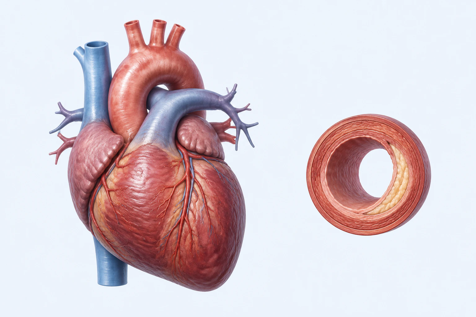
Cardiovascular-system orientation with one representative arterial-wall process. Cardiovascular diseases are a broad category spanning heart, blood-vessel, and pericardial conditions; the plaque inset does not define the category and this reconstruction is not a scan or diagnostic image.
Scientific media record1 verified identifier
- Subject
- Cardiovascular Diseasescondition
- Identifiers
- MeSH:D002318
- Review
- Editorial review completeIdentifiers authority-verified · Accessibility validated · · cardiovascular-disease|cardiovascular-disease-pathology-v1.webp
- Digital source
- Trained-algorithmic mediaCreated with a trained generative algorithm and reviewed by WikiBiome for subject identity, scientific framing, identifiers, provenance, and accessibility.
- Scientific basis
- Cardiovascular Diseases — MeSHHow the Heart Works — Anatomy
- License
- CC BY-SA 4.0Created
Cardiovascular disease (CVD)—encompassing atherosclerosis, coronary artery disease, hypertension, myocardial infarction, heart failure, and stroke—is the leading cause of death globally. The gut-heart axis has emerged as a central paradigm linking microbial metabolites (TMAO, SCFAs, bile acids, tryptophan derivatives) to CVD pathogenesis.
Metallomic profiling reveals a consistent signature of copper elevation and selenium depletion in acute events, while chronic exposures to lead and cadmium drive CVD risk through both direct vascular toxicity and microbiome-mediated pathways.
The convergence of metal dyshomeostasis, microbial Dysbiosis, and metabolite disruption creates a multi-layered framework for understanding CVD etiology.
Evidence map73 cited passagesInspect provenance +
provided the most detailed metallomic profiling of CVD using ICP-MS/MS in 101 AMI patients and 66 controls:
demonstrated functional enrichment of TMA lyase genes (CutC/D, YeaW/X) in the ACVD gut microbiome, directly linking microbial TMAO production capacity to disease state.
characterized 1,241 individuals across the cardiometabolic disease spectrum (healthy to heart failure) using integrated metagenomics and metabolomics:
detailed how microbiota-derived tryptophan metabolites drive vascular disease:
: Malassezia enrichment increases from normotensive to pre-hypertension to hypertension, positively correlated with immunoglobulin light chains; fungal dysbiosis detected already at the pre-hypertension stage
: HTN+CKD patients show distinct mycobiome with increased Apiotrichum, Cystobasidium, Saccharomyces and decreased Candida; cytokine associations with fungal genera
established five mechanisms linking oral dysbiosis to CVD:
The metallomic signature in CVD is characterized by elevated nickel, lead, cadmium, and iron, with depleted selenium and glutathione,,.
Nickel elevation in CVD patients is robust. NHANES 2017-2018 cross-sectional analysis (n=2702) found elevated urinary nickel (UNi) in CVD patients with an inverted L-shaped dose-response relationship. Early clinical observations documented endogenous nickel release post-myocardial infarction, suggesting that cardiac tissue damage itself liberates nickel, whi
Metal interaction studies show plasma nickel positively correlated with zinc, vanadium, and chromium but negatively correlated with copper, indicating dysregulated metal homeostasis in CVD. Experimental evidence in Swiss mice fed 1,100-1,600 ppm dietary nickel showed reduced cardiac and renal enzyme activity and dose-dependent inhibition of cardiac contracti
These metals drive mis-metallation events (Primitive 3). They enter cells via calcium channels and displace correct metal cofactors. The combination of lead and cadmium produces synergistic oxidative stress beyond either metal alone. Their presence in the GI microenvironment selects for metal-tolerant taxa while eliminating metal-sensitive commensals, contri
Elevated iron in CVD creates a selective pressure favoring siderophore-dependent Enterobacteriaceae like E. coli and Bacteroides. The iron elevation may reflect both environmental exposure and functional anemia (elevated hepcidin) as a host defense mechanism against pathogenic iron-scavenging bacteria. However, in the CVD context, the iron-enriched environme
Selenium is a cofactor for selenoproteins including glutathione peroxidase (GPx), the primary antioxidant defense against TMAO-induced and metal-induced ROS. Its depletion impairs the host's ability to counteract oxidative damage from dysbiotic TMAO producers and metal-dependent pathogens.
The dietary iron content in red meat is particularly relevant because it drives both siderophore-dependent Enterobacteriaceae overgrowth AND promotes heme-iron absorption, creating dual mechanisms of CVD risk.
The host is actively withholding metals from pathogens, not failing nutritionally,:
The hallmark of CVD dysbiosis is massive enrichment of Enterobacteriaceae family species, particularly E. coli and Klebsiella, alongside elevated Streptococcus spp.. This consortium is unified by three pathogenic functions: TMAO production, LPS synthesis, and iron piracy.
Role: The primary TMAO producer in CVD. Encodes TMA lyase enzymes (CutC/D and CntA/B) that metabolize dietary choline and carnitine to trimethylamine (TMA), which is absorbed into portal circulation and hepatically oxidized to TMAO by FMO3,.
E. coli additionally produces LPS (lipopolysaccharide), which translocates through the disrupted gut barrier (via butyrate loss) and activates TLR4 on endothelial cells and macrophages, driving systemic inflammation.
Role in gut: Enriched in CVD gut microbiome; TMAO-producer. Encoded in the metagenomic classifiers that distinguish ACVD patients from healthy controls with AUC 0.86.
Role in oral cavity: Streptococcus spp., particularly viridans group streptococci (VGS), colonize the periodontal pocket in periodontitis,. VGS express:
Oral VGS bacteremia seeding atherosclerotic plaques is confirmed by bacterial DNA recovery from plaques.
Role: Strict anaerobe—indicator of local hypoxia. Produces fragilisin (BFT), a zinc-dependent metalloproteolytic toxin that disrupts intestinal tight junctions (destroys claudins and occludin), increasing permeability and enabling translocation of intact bacterial cells and LPS.
Role in periodontitis: The keystone periodontal pathogen. Expressed gingipains degrade host proteins and matrix metalloproteinases, creating the periodontal pocket. Produces LPS and HSP60 (molecular mimicry target). When periodontal disease is active, P. gingivalis cells and LPS breach the inflamed periodontal vasculature and enter systemic circulation, inoc
The CVD microbiome is characterized by severe depletion of SCFA-producing bacteria,:
Showing 24 of 73 evidence-bearing passages. Every remaining citation is still indexed in the reference record below.
One disease. Five evidence layers.
A generated systems view of the metals, organisms, host sequestration signals, ecological conditions, and microbial functions indexed for Cardiovascular Disease.
Evidence layer
Taxonomic signature
Organisms reported as enriched or depleted, with their indexed functional context kept beside the name.TMA lyase (CutC/D) carriers — TMAO production driving atherosclerosis; LPS biosynthesis; siderophore-mediated iron piracy
TMA/TMAO production; LPS source; enriched virulence factors in ACVD; copper homeostasis systems conferring survival advantage
Oral-to-gut translocation; enriched in ACVD and infective endocarditis; molecular mimicry (M protein in rheumatic heart disease)
LPS-producer; beta-glucuronidase activity; strict anaerobe indicator of hypoxia
Periodontal pathogen; molecular mimicry via HSP60; bacteremia in CVD
Enriched in ACVD metagenome; associated with drug metabolism confounding
Enriched in ACVD; pro-inflammatory mucin degrader
Mycobiome: enriched in atherosclerosis and heart failure; barrier disruption via epithelial damage
Mycobiome: progressive enrichment from normotensive to pre-hypertension to hypertension; correlated with immunoglobulin light chains
Butyrate producer — lost in ACVD; depletion reduces SCFA-mediated blood pressure regulation and anti-inflammatory signaling
Major butyrate producer — depleted in ACVD; loss reduces gut barrier integrity and anti-inflammatory IL-10 production
SCFA-producing family — reduced capacity for butyrate synthesis in ACVD
Reported as protective in one hypertension MR analysis; not species-resolved and not established as an SCFA producer or metal-sensitive taxon
Indole metabolite producer (IPA, IAA); anti-inflammatory; lost in ACVD
Butyrate producers and protective IPA (indole-3-propionic acid) producers — depleted in untreated ACVD
Mycobiome: M. racemosus decreased in CHD; potentially protective fungal taxon
Evidence layer
Nutritional immunity
Host metal-withholding, inflammatory, antioxidant, and microbial-metabolite signals indexed in the signature.Elevated host signals
6Depleted protective signals
7Evidence layer
Ecological state
The environmental conditions that connect the organism-level observations into a system.Evidence layer
Virulence functions
Microbial structures, enzymes, and acquisition systems implicated by the linked evidence.The disease record, in full.
The original WikiBiome disease narrative remains intact beneath the generated signature atlas.
Metallomic Signature#
[1]Plasma metallomics reveals potential biomarkers and insights into the ambivalent associations of elements with acute myocardial infarctionSi Ying Lim, Hiranya Dayal, Song Jie Seah et al. · 2023Open reference 1 ↓ provided the most detailed metallomic profiling of CVD using ICP-MS/MS in 101 AMI patients and 66 controls:
| Metal | Direction | Key Evidence | |
|---|---|---|---|
| [[copper | copper (Cu)]] | Elevated | 0.85 vs 0.73 ug/mL in AMI (p<0.01); remained elevated 1 month post-PCI; ceruloplasmin-mediated acute phase response |
| [[selenium | selenium (Se)]] | Depleted | 90.31 vs 99.98 ng/mL in AMI (p<0.01); persistent depression at 6 months; impairs selenoprotein antioxidant defense |
| [[iron | iron (Fe)]] | Decreased | 0.95 vs 1.17 ug/mL in AMI; iron/copper ratio significantly decreased |
| [[lead | lead (Pb)]] | CVD risk factor | Chronic low-level exposure drives hypertension and atherosclerosis |
| [[cadmium | cadmium (Cd)]] | CVD risk factor | Smoking-associated; vascular toxicity and microbiome disruption |
| [[arsenic | As]] | Decreased in AMI | 1.32 vs 2.12 ng/mL (p<0.001); paradoxically lower in acute events |
| [[nickel | nickel (Ni)]] | Cardiometabolic risk | NHANES epidemiological data linking nickel exposure to metabolic syndrome and CVD |
Cu/Se Ratio as Biomarker#
The copper (Cu)/selenium (Se) ratio captures both copper elevation and selenium depletion in a single metric and is the most discriminating element-pair ratio for AMI. A random forest classifier incorporating copper/selenium and iron (Fe)/copper ratios alongside traditional risk factors achieved an AUC of 0.942 (95% CI 0.889-0.970).
Essential elements exhibit ambivalent (U- or J-shaped) relationships with AMI risk, meaning both deficiency and excess are harmful.
Pb and Cd as CVD Risk Factors#
Lead and cadmium exert cardiovascular toxicity through both direct and microbiome-mediated mechanisms. Smoking status is the predominant determinant of non-essential/toxic element plasma levels (aluminum (Al), cadmium (Cd), Rb, strontium (Sr), U, vanadium (V)). Cadmium drives vascular damage through Oxidative Stress, endothelial dysfunction, and disruption of the Gut Microbiome toward pro-inflammatory configurations.
Lead exposure contributes to hypertension and atherosclerosis progression.
Gut Microbiome-CVD Axis#
TMAO as Microbiome-Derived CVD Risk Factor#
Trimethylamine N-oxide (TMAO) is the most established microbiome-derived cardiovascular risk metabolite. Produced from dietary choline, phosphatidylcholine, and L-carnitine by gut bacteria via the CutC/CutD enzyme complex, TMA is oxidized to TMAO in the liver.
High TMAO levels increase atherosclerosis risk via cholesterol deposition, platelet activation, and endothelial damage through the MAPK pathway.
[2]The gut microbiome in atherosclerotic cardiovascular diseaseZhuye Jie, Huihua Xia, Shi-Long Zhong et al. · 2017Open reference 2 ↓ demonstrated functional enrichment of TMA lyase genes (CutC/D, YeaW/X) in the ACVD gut microbiome, directly linking microbial TMAO production capacity to disease state.
Jie 2017 ACVD Microbiome Signature#
The landmark metagenome-wide association study of 218 ACVD patients and 187 controls established. ACVD enriched: Enterobacteriaceae, Streptococcus spp., E. lenta, R. gnavus, L. salivarius, E. coli. ACVD depleted: Butyrate-producing Roseburia spp., F. prausnitzii, Clostridiales.
Functional enrichment: TMA lyase genes, LPS biosynthesis, virulence factors, simple sugar transport.
Functional depletion: butyrate synthesis capacity, vitamin biosynthesis. Classifier: 47 MLGs achieved AUC of 86% (83-90%). Critical caveat: drug use (fondaparinux, acarbose, metoprolol, atorvastatin) was the major confounding factor.
MetaCardis Multi-Omic Study#
[3]Microbiome and metabolome features of the cardiometabolic disease spectrumSebastien Fromentin, Sofia K. Forslund, Kanta Chechi et al. · 2022Open reference 3 ↓ characterized 1,241 individuals across the cardiometabolic disease spectrum (healthy to heart failure) using integrated metagenomics and metabolomics:
- Identified 767 dysmetabolism features and 283 IHD-specific features
- Uremic toxins (4-cresol, phenylacetylglutamine, indoxyl sulfate) showed escalation from dysmetabolism to IHD
- Gene richness significantly reduced in ACS, CIHD, and HF
- Microbiome classifier AUC >0.8 for healthy vs IHD
- The dysmetabolism-to-IHD trajectory provides a framework for understanding progressive CVD development
SCFAs as Protective Factors#
SCFAs protect against CVD through multiple mechanisms. Blood pressure regulation: butyrate suppresses intrarenal renin-angiotensin system via PRR; propionate modulates renin release via Olfr78 receptor; GPR41 and Olfr78 play opposing roles creating a buffering system.
Anti-atherosclerotic: butyrate inhibits HDAC3, induces FGF21, promotes lipid oxidation; SCFAs suppress NF-kB and NLRP3 inflammasome in macrophages; reduce VCAM-1 and MCP-1 expression. Post-MI protection: SCFAs promote M2 macrophage polarization; butyrate downregulates TNF-alpha and IL-1beta while upregulating IL-10.
Gut barrier maintenance: butyrate induces tight junction proteins and mucin production, preventing LPS translocation—a key pathway in CVD pathogenesis. Appetite and metabolism: colonic SCFAs increase fat oxidation and energy expenditure; propionate stimulates PYY and GLP-1 release.
Metal-induced depletion of SCFA-producing bacteria (Roseburia, F. prausnitzii, Clostridia) directly reduces these cardioprotective effects, creating a vicious cycle of barrier disruption, endotoxemia, and vascular Metal-Driven Inflammation.
Bile Acids as CVD Mediators#
Primary bile acids (CA, CDCA) are converted to secondary bile acids (DCA, LCA) by gut bacteria via 7-alpha dehydroxylation. In CVD:
- BAs act through FXR and TGR5 receptors to affect fatty acid oxidation, triglyceride accumulation, and NF-kB signaling
- FXR activation reduces triglycerides; TGR5 activation promotes energy expenditure
- Elevated secondary-to-primary BA ratio is associated with CVD
- Iron and copper homeostasis affects bile acid metabolism; metal-induced bile acid shifts may promote atherosclerosis via FXR/TGR5 dysregulation
Tryptophan Metabolites and Vascular Inflammation#
[4]Microbiota-derived tryptophan metabolites in vascular inflammation and cardiovascular diseaseNadja Paeslack, Maximilian Mimmler, Stefanie Becker et al. · 2022Open reference 4 ↓ detailed how microbiota-derived tryptophan metabolites drive vascular disease.
Indoxyl sulfate (pro-atherogenic): promotes vascular inflammation, procoagulant state, endothelial dysfunction; increases tissue factor and inhibits wound healing. Indole-3-propionic acid (IPA) (protective): negatively associated with atherosclerosis despite cardiovascular effects. Indole-3-acetic acid and indole-3-aldehyde (anti-inflammatory): promote IL-10 production via AHR activation.
Serotonin: microbiota-modulated; affects blood pressure and thromboinflammation.
Cadmium exposure upregulates indoxyl sulfate production, directly connecting metal exposure to the pro-atherogenic tryptophan metabolite pathway. Metal-induced depletion of Clostridium and Lactobacillus reduces protective IPA and IAld production.
Mycobiome in CVD#
The fungal microbiome is an underexplored but increasingly recognized contributor to CVD:
Hypertension#
:[5]Gut mycobiome dysbiosis contributes to the development of hypertension and its response to immunoglobulin light chainsYeqing Zou, Anxing Ge, Brako Lydia et al. · 2022Open reference 5 ↓ Malassezia enrichment increases from normotensive to pre-hypertension to hypertension, positively correlated with immunoglobulin light chains; fungal dysbiosis detected already at the pre-hypertension stage.
:[6]Exploring the gut mycobiome: differential composition and clinical associations in hypertension, chronic kidney disease, and their comorbidityJuan Qiu, Longyou Zhao, Yiwen Cheng et al. · 2023Open reference 6 ↓ HTN+CKD patients show distinct mycobiome with increased Apiotrichum, Cystobasidium, Saccharomyces and decreased Candida; cytokine associations with fungal genera.
Atherosclerosis#
- C. albicans, Exophiala, Malassezia, Penicillium, Wallemia increased in ACVD
- Mucor racemosus and Fusarium decreased in CHD (potentially protective)
Heart Failure#
- Candida proliferation in CHF patients with elevated intestinal permeability
- S. boulardii supplementation failed to improve cardiac function in the GutHeart trial
Oral Microbiome-CVD Connection#
[7]The oral microbiome in the pathophysiology of cardiovascular diseaseAndrea Tonelli, Evelyn N. Lumngwena, Ntobeko A. B. Ntusi · 2023Open reference 7 ↓ established five mechanisms linking oral dysbiosis to CVD:
- Transmigration: oral bacteria seed systemic circulation through inflamed periodontium, inoculating atherosclerotic plaques
- Cytokine release: chronic oral inflammation causes endothelial dysfunction and oxidative stress
- Molecular mimicry: oral bacterial antigens activate autoimmune B cells targeting atherosclerotic plaques
- Toxin release: bacterial toxins increase lipid peroxidation and LDL/HDL modification
- Abnormal lipid profile: LPS-induced lipoprotein metabolism abnormalities
Viridans group streptococci and S. aureus are the most common causes of infective endocarditis. Streptococcus M protein in Group A beta-hemolytic streptococci triggers molecular mimicry in rheumatic heart disease. Multi-site dysbiosis (oral + gut) may synergistically amplify CVD risk.
Diet and CVD-Microbiome Interactions#
Mediterranean Diet#
The Mediterranean diet offers multi-pathway CVD protection by modulating gut microbiome composition toward anti-inflammatory profiles, increasing SCFA production, and reducing TMAO precursor availability.
Fiber#
Dietary fiber increases SCFA production, maintaining gut barrier integrity and reducing LPS translocation. Inulin supplementation in hypertensives showed correlation between gut permeability and systolic blood pressure.
Polyamines#
Bacterially synthesized polyamines (cadaverine, putrescine, spermidine) have cardioprotective effects. Spermidine reduces cardiac hypertrophy, improves echocardiographic parameters, and modifies intestinal microbiota toward anti-inflammatory composition.
Comorbidities#
Type 2 Diabetes—shared insulin resistance, TMAO pathway, and SCFA depletion; T2D doubles CVD risk; the MetaCardis study documented a continuous dysmetabolism-to-IHD trajectory spanning both conditions.
Obesity—shared metabolic inflammation, gut barrier dysfunction, and dysbiosis; adipose tissue is a source of pro-inflammatory cytokines (TNF-alpha, IL-6) that drive atherosclerosis.
Hypertension—the most direct CVD comorbidity; shared mycobiome dysbiosis (Malassezia enrichment tracks from pre-hypertension to hypertension); SCFAs regulate blood pressure via Olfr78 and GPR41.
Chronic Kidney Disease—shared uremic toxin accumulation (indoxyl sulfate, p-cresol sulfate); CKD accelerates atherosclerosis; gut mycobiome alterations overlap in HTN+CKD patients.
Depression—CVD patients have 2-3x higher depression rates; shared gut-brain axis disruption, tryptophan pathway shifts toward pro-inflammatory kynurenine, and SCFA depletion; depression independently increases CVD mortality.
Connections#
- Metal-Disease Matrix: A Cross-Source Synthesis
- Atherosclerosis—lead and cadmium exposure accelerate atherosclerotic plaque formation via endothelial damage
- Trimethylamine N-Oxide (TMAO)—microbiome-derived TMAO promotes atherosclerosis and thrombosis; elevated in CVD patients
- Biomarkers—copper (Cu)/zinc (Zn) ratio, iron (Fe)/copper ratio, selenium (Se) levels, and CRP as metallomic and inflammatory CVD biomarkers
- microbiome-derived metabolites—TMAO, SCFAs, and bile acids as microbiome-CVD mediators
- Environmental Metal Exposure—occupational and dietary lead (Pb)/cadmium (Cd) exposure as modifiable CVD risk factors
References 25
Numbered by first appearance in the article, then reconciled with its declared source list.
- 1
Si Ying Lim, Hiranya Dayal, Song Jie Seah et al. (2023). Plasma metallomics reveals potential biomarkers and insights into the ambivalent associations of elements with acute myocardial infarction. Journal of Trace Elements in Medicine and Biology.
- 2
Zhuye Jie, Huihua Xia, Shi-Long Zhong et al. (2017). The gut microbiome in atherosclerotic cardiovascular disease. Nature Communications.
- 3
Sebastien Fromentin, Sofia K. Forslund, Kanta Chechi et al. (2022). Microbiome and metabolome features of the cardiometabolic disease spectrum. Nature Medicine.
- 4
Nadja Paeslack, Maximilian Mimmler, Stefanie Becker et al. (2022). Microbiota-derived tryptophan metabolites in vascular inflammation and cardiovascular disease. Amino Acids.
- 5
Yeqing Zou, Anxing Ge, Brako Lydia et al. (2022). Gut mycobiome dysbiosis contributes to the development of hypertension and its response to immunoglobulin light chains. Frontiers in Immunology.
- 6
Juan Qiu, Longyou Zhao, Yiwen Cheng et al. (2023). Exploring the gut mycobiome: differential composition and clinical associations in hypertension, chronic kidney disease, and their comorbidity. Frontiers in Immunology.
- 7
Andrea Tonelli, Evelyn N. Lumngwena, Ntobeko A. B. Ntusi (2023). The oral microbiome in the pathophysiology of cardiovascular disease. Nature Reviews Cardiology.
- 8
★Yucheng Liu, Xiaomin Luo, Yongde Peng et al. (2025). Cardio-Metabolic Effects of Nickel: A Narrative Review. Cardiovascular Toxicology.
- 9
Edward S. Chambers, Tom Preston, Gary Frost et al. (2018). Role of Gut Microbiota-Generated Short-Chain Fatty Acids in Metabolic and Cardiovascular Health. Current Nutrition Reports.
- 10
Hilde Herrema, Max Nieuwdorp, Albert K. Groen (2020). Microbiome and Cardiovascular Disease. Handbook of Experimental Pharmacology (Prevention and Treatment of Atherosclerosis).
- 11
Jing Zhen, Zhou Zhou, Meng He et al. (2023). The gut microbial metabolite trimethylamine N-oxide and cardiovascular diseases. Frontiers in Endocrinology.
- 12
Max Foroughi, Keykavous Parang (2026). Periodontal Biomarkers in Cardiovascular Disease: Mechanisms, Diagnostics, and Clinical Implications. Infection.
- 13
Yihui Li, Ru Fu, Ruixuan Li et al. (2023). Causality of gut microbiome and hypertension: A bidirectional mendelian randomization study. Frontiers in Cardiovascular Medicine.
- 14
Xiao-Ce Dai, Yi Yu, Si-Yu Zhou et al. (2024). Assessment of the Causal Relationship between Gut Microbiota and Cardiovascular Diseases: A Bidirectional Mendelian Randomization Analysis. BioData Mining.
- 15
Qiang Luo, Yilan Hu, Xin Chen et al. (2022). Effects of Gut Microbiota and Metabolites on Heart Failure and Its Risk Factors: A Two-Sample Mendelian Randomization Study. Frontiers in Nutrition.
- 16
Rebecca L. Walker, Hera Vlamakis, Jonathan Wei Jie Lee et al. (2021). Population Study of the Gut Microbiome: Associations with Diet, Lifestyle, and Cardiometabolic Disease. Genome Medicine.
- 17
Dorothea Katharina Hoffelner, Tim Hendrikx (2025). Emerging therapy targets to modulate microbiome-mediated effects evident in cardiovascular disease. Frontiers in Cardiovascular Medicine.
- 18
★Sweta Ghosh, Syam P. Nukavarpu, Venkatakrishna Rao Jala (2024). Effects of Heavy Metals on Gut Barrier Integrity and Gut Microbiota. Microbiota and Host.
- 19
★Puthiyavalappil Rasin, Ashwathi A V, Sabeel M Basheer et al. (2025). Exposure to Cadmium and Its Impacts on Human Health: A Short Review. Journal of Hazardous Materials Advances.
- 20
Xiaoyu Wei, Zixin Guo, Jingyang Wang et al. (2025). Gut mycobiome in cardiometabolic disease progression: current evidence and future directions. Frontiers in Microbiology.
- 21
Yingdong Lu, Yang Zhang, Xin Zhao et al. (2022). Microbiota-derived short-chain fatty acids: Implications for cardiovascular and metabolic disease. Frontiers in Cardiovascular Medicine.
- 22
★Karen Pendergrass (2026). Heavy Metals, Microbial Metallomics, and the US Obesity Epidemic: A Mechanistic Examination of a Population-Level Metabolic Disruption. Zenodo Preprint.
- 23
Lu Wang, Shiqi Wang, Qing Zhang et al. (2022). The role of the gut microbiota in health and cardiovascular diseases. Molecular Biomedicine.
- 24
Catia Almeida, J. Guilherme Goncalves-Nobre, Diogo Alpuim Costa et al. (2023). The potential links between human gut microbiota and cardiovascular health and disease - is there a gut-cardiovascular axis?. Frontiers in Gastroenterology.
- 25
★Qinheng Zhu, Boyan Chen, Fu Zhang et al. (2024). Toxic and Essential Metals: Metabolic Interactions with the Gut Microbiota and Health Implications. Frontiers in Nutrition.
Article network
Mentioned here 18
Pages linking here 85
Connect the evidence
Publicly readable discussion by ORCID-authenticated researchers. Questions, interpretation, methods, corrections, and new evidence stay attached to this record.
No discussion yet. Start with a precise question or a source-backed observation.
Activity and accepted changes
Accepted researcher context, editorial status, public discussion, and upstream Git revisions are shown together. Pending, declined, and withdrawn proposals remain private.
- published revision
Consolidate microbial metabolite knowledge
Karen Pendergrass · +2 −2
Inspect exact Git diff ↗ - published revision
Backfill oxidative stress concept links
Karen Pendergrass · +1 −1
Inspect exact Git diff ↗ - published revision
Backfill gut microbiome concept links
Karen Pendergrass · +1 −1
Inspect exact Git diff ↗ - published revision
Backfill inflammation concept links
Karen Pendergrass · +1 −1
Inspect exact Git diff ↗ - published revision
Complete corpus-wide Dysbiosis linking
Karen Pendergrass · +1 −1
Inspect exact Git diff ↗ - published revision
Complete Tight junctions contextual coverage
Karen Pendergrass · +1 −1
Inspect exact Git diff ↗ - published revision
Add NLRP3 inflammasome concept and link batch
Karen Pendergrass · +3 −3
Inspect exact Git diff ↗ - published revision
Reconcile current taxonomic identities
Karen Pendergrass · +6 −6
Inspect exact Git diff ↗ - published revision
massive wiki expansion: 149 stubs fixed, 100+ new pages, Rule 15 scan, keystone papers
WikiBiome Deploy Bot · +27 −27
Inspect exact Git diff ↗ - published revision
nightly maintenance: 94 stub demotions, 181 source_count fixes, 22 auto-discovered stubs, 5 adversarial audits, 3 boundary fixes, 3 evidence-level corrections
WikiBiome Deploy Bot · +1 −0
Inspect exact Git diff ↗ - published revision
cycle 1: health check + lint fixes + 8 ingests + 2 stubs + gestational-diabetes signature
WikiBiome Deploy Bot · +1 −0
Inspect exact Git diff ↗ - published revision
Batch: fix 1025 broken wikilinks, wire 13 STOP pages, deepen PPD/GERD/T1D/8 microbes
WikiBiome Deploy Bot · +1 −1
Inspect exact Git diff ↗ - published revision
Deepen metal/concept entities + 8 new sources for T1D/schizophrenia
WikiBiome Deploy Bot · +427 −0
Inspect exact Git diff ↗ - published revision
WikiBiome update — 2026-04-15 17:23
WikiBiome Deploy Bot · +14 −14
Inspect exact Git diff ↗ - published revision
v2 migration Priority 2: All 29 disease entity pages upgraded with associated_conditions, seo_target, wikipedia_differentiation
WikiBiome Deploy Bot · +6 −0
Inspect exact Git diff ↗