

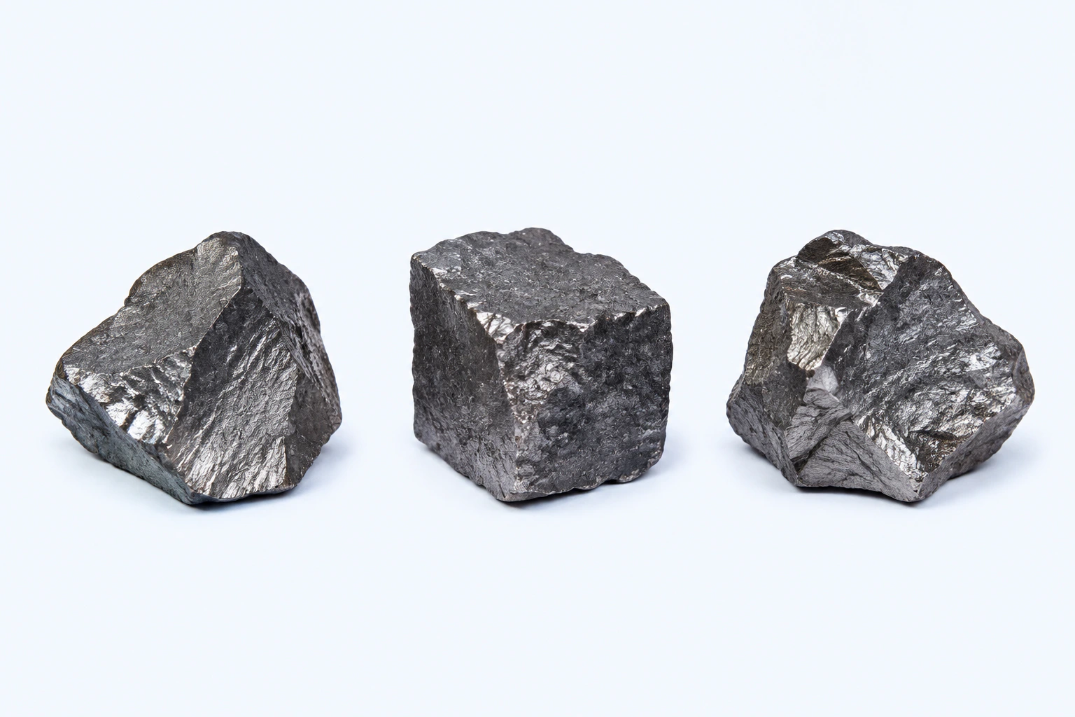
Elemental iron (Fe), shown as three representative dark silver-gray solid specimens. Form and surface vary with purity, processing, and oxidation; this is not ore, iron oxide, analytical reference material, or a photograph.
Scientific media record2 verified identifiers
- Subject
- Ironelement
- Identifiers
- Atomic number 26PubChem CID:23925
- Review
- Editorial review completeIdentifiers authority-verified · Accessibility validated · · iron|iron-technical-specimen-v1.webp
- Digital source
- Trained-algorithmic mediaCreated with a trained generative algorithm and reviewed by WikiBiome for subject identity, scientific framing, identifiers, provenance, and accessibility.
- Scientific basis
- Iron — PubChem ElementIron, PubChem CID 23925
- License
- CC BY-SA 4.0Created
The most abundant transition metal in the human body and arguably the most consequential metal in biology.
Iron sits at the center of a paradox that drives pathology across virtually every disease domain: organisms need it for oxygen transport, energy metabolism, DNA synthesis, and immune defense, yet its redox activity makes it dangerous when uncontrolled.
This tension between necessity and toxicity shapes the ecology of the Gut Microbiome, the outcome of infections, and the trajectory of neurodegeneration, cancer, and cardiovascular disease.
What sets iron apart from every other metal in this wiki is its role as the primary currency of the war between hosts and pathogens. The host sequesters iron to starve invaders. Pathogens evolve siderophores, hemolysins, and heme receptors to steal it back.
The microbiome's composition at any given moment is, in large part, a reflection of who is winning the iron war.
Evidence map79 cited passagesInspect provenance +
Iron's centrality to biology is not accidental—it reflects the chemistry of the planet where life originated. Analysis of 3.33-billion-year-old carbonaceous material from the Barberton greenstone belt reveals that iron was among the key bio-functional elements in Earth's earliest ecosystems, enriched in biogenic material alongside vanadium, nickel, and co
Fenton reaction: Fe2+ + H2O2 - Fe3+ + OH- + OH generates hydroxyl radicals, the most reactive oxygen species. Iron, copper, vanadium, chromium, and cobalt all participate in Fenton-type chemistry, but iron is the most biologically abundant.
Iron is one of the two most abundant intracellular transition metals (alongside zinc), reaching tens of millimolar total concentration in typical cells.
Fe enzymes come in three structural forms: nonheme iron, iron-sulfur (Fe-S) cluster-containing, and heme enzymes—each with distinct vulnerability profiles.
In the Irving-Williams series (Mg < Mn < Fe < Co < Ni < Cu Zn), iron occupies the middle ground—strong enough to be a versatile catalytic cofactor, but weak enough to be displaced by copper or zinc when homeostasis fails.
Regulated by the hepcidin-ferroportin axis: hepcidin controls iron absorption from dietary sources, macrophages, and body stores.
Key transport proteins: DMT1 (SLC11A2) for import, ferroportin (SLC40A1) for export, transferrin for plasma transport, ferritin for storage.
Iron regulatory proteins (IRP1/2) sense cellular iron and post-transcriptionally regulate transferrin receptor and ferritin expression.
Nickel can oxidize iron in iron-sulfur clusters and iron-containing hydroxylases, disrupting IRP-1/IRP-2, transferrin receptor, and ferritin levels.
A landmark finding from the Imlay lab demonstrates that iron was the ancestral metal cofactor for mononuclear enzymes, later replaced by manganese or zinc in organisms that colonized oxidizing environments. The enzyme ribulose-5-phosphate 3-epimerase (Rpe) illustrates this: E. coli and Bacteroides metalate Rpe with iron, while B. subtilis and Lactococcus use
E. coli can conditionally switch Rpe from iron to manganese under peroxide stress, mediated by OxyR-induced MntH manganese import and Dps miniferritin sequestration of free iron. This conditional metal switching is a key survival strategy that connects iron biology to the oxidative stress response.
Approximately 25% of all metalloenzymes use iron as a cofactor, and 33-40% of a typical proteome consists of metalloproteins,. Iron's three enzyme families serve distinct functions:
Iron is in high demand for Fe-S clusters and heme cofactors that manganese cannot substitute for, making these enzyme families irreplaceable even when organisms switch other enzymes to manganese.
Copper destroys Fe-S clusters in isopropylmalate isomerase (IPMI), fumarase A, and glutamate synthase (GOGAT), causing branched-chain amino acid auxotrophy and glutamate starvation.
Nickel and copper together produce synergistic Fe-S cluster damage at environmentally relevant concentrations where neither metal alone causes toxicity. Combined Ni/Cu exposure in E. coli triggers upregulation of ISC Fe-S cluster assembly genes and produces 70% of differentially expressed genes unique to the combination.
Reactive nitrogen species from macrophage iNOS also damage Fe-S clusters, adding a third axis of vulnerability alongside copper and nickel.
In a murine wound healing time course, iron shows a distinctive temporal profile: elevated at days 7-14 during the proliferation and remodeling phases, linked to oxidoreductase activity, heme-binding, steroid hydroxylase, and PPAR signaling pathways. Metal-linked genes constitute 16% of wound-responsive genes with nearly 2-fold overrepresentation, establishi
Iron detected in 100% of tampon samples at measurable concentrations.
Essential elements follow a characteristic dose-response curve: deficiency causes harm at low concentrations, optimal performance at moderate levels, and toxicity at high concentrations. For iron this curve is unusually narrow—the margin between deficiency (anemia, impaired immunity) and excess (oxidative damage, pathogen feeding) is clinically treacherou
Maternal trace element profiles shape the infant gut microbiome. In a prospective cohort of 146 mother-infant pairs, prenatal metal exposure patterns measurably affected gut microbial diversity and composition at 3, 6, and 12 months of life. Iron supplementation in infants increases Enterobacteriaceae and decreases Lactobacillus; in African children, iron su
Iron is a co-exposure in welding fumes alongside manganese and nickel.
Urinary iron levels showed statistically significant differences across infant feeding groups in an aerodigestive clinic cohort, though levels did not exceed toxicity thresholds.
Iron availability in the gut lumen is one of the primary determinants of microbial community structure. Iron deficiency reduces commensal beneficial bacteria (Lactobacillus, Bacillota) while iron excess increases harmful bacteria (Bacteroides, E. coli, Enterobacteriaceae). Iron deficiency anemia (IDA) specifically reduced Bacillota abundance while increasing
The mechanism is competitive: siderophore-producing Enterobacteriaceae outcompete commensals under high-iron conditions because commensals generally lack the aggressive iron acquisition systems that pathogens deploy. Iron, manganese, and nickel availability in the gut lumen determines competitive outcomes between commensals and pathogens.
Showing 24 of 79 evidence-bearing passages. Every remaining citation is still indexed in the reference record below.
Contents
1. Overview2. Biological Roles3. Dietary and Environmental Sources4. Microbiome Interactions5. Nutritional Immunity6. Conditions Associated7. Key Studies8. Biomarkers9. Interactions with Other Metals10. Open Questions11. Cross-ReferencesOverview#
Evolutionary Origins#
Iron's centrality to biology is not accidental—it reflects the chemistry of the planet where life originated.
Analysis of 3.33-billion-year-old carbonaceous material from the Barberton greenstone belt reveals that iron was among the key bio-functional elements in Earth's earliest ecosystems, enriched in biogenic material alongside vanadium, nickel, and cobalt.[1]Metallomics in deep time and the influence of ocean chemistry on the metabolic landscapes of Earth's earliest ecosystemsHickman-Lewis K, Cavalazzi B, Sorieul S et al. · 2020Open reference 1 ↓
The pre-Great Oxygenation Event world was an iron-rich, anoxic environment where life evolved to depend on iron's redox versatility. Metals that became biologically important only after oxygenation—zinc and molybdenum—are absent or below detection in these ancient specimens.[1]Metallomics in deep time and the influence of ocean chemistry on the metabolic landscapes of Earth's earliest ecosystemsHickman-Lewis K, Cavalazzi B, Sorieul S et al. · 2020Open reference 1 ↓
The modern biological dependency on iron is therefore a 3.3-billion-year inheritance from the geochemistry of early Earth.
Chemical Properties#
Transition metal cycling between iron(II) (Fe2+) (ferrous) and iron(III) (ferric) states—this redox cycling is both its biological utility and its danger.
Fenton reaction: iron(II) + H2O2 -> iron(III) + OH- + OH* generates hydroxyl radicals, the most reactive oxygen species.[2]Common and Trace Metals in Alzheimer's and Parkinson's DiseasesDoroszkiewicz J, Farhan JA, Mroczko J et al. · 2023Open reference 2 ↓
Iron, copper, vanadium, chromium, and cobalt all participate in Fenton-type chemistry, but iron is the most biologically abundant.[3]Heavy Metal Pollution in the Environment and Their Toxicological Effects on HumansBriffa J, Sinagra E, Blundell R · 2020Open reference 3 ↓
Iron is one of the two most abundant intracellular transition metals (alongside zinc), reaching tens of millimolar total concentration in typical cells.[4]Capdevila 2024 — Bacterial Metallostasis: Metal Sensing, Metalloproteome Remodeling, and Metal TraffickingDaiana A. Capdevila, Johnma J. Rondon, Katherine A. Edmonds et al. · 2024Open reference 4 ↓
iron enzymes come in three structural forms: nonheme iron, iron-sulfur (iron-S) cluster-containing, and heme enzymes—each with distinct vulnerability profiles.[4]Capdevila 2024 — Bacterial Metallostasis: Metal Sensing, Metalloproteome Remodeling, and Metal TraffickingDaiana A. Capdevila, Johnma J. Rondon, Katherine A. Edmonds et al. · 2024Open reference 4 ↓
In the Irving-Williams series (magnesium (Mg) < manganese (Mn) < iron < cobalt (Co) < nickel (Ni) < copper (Cu) > zinc (Zn)), iron occupies the middle ground—strong enough to be a versatile catalytic cofactor, but weak enough to be displaced by copper or zinc when homeostasis fails.[5]Robinson & Glasfeld 2020 — Metalation and Mis-metalation: Nature's Challenge in Metal CoordinationNigel J. Robinson, Andrea Glasfeld · 2020Open reference 5 ↓
Homeostatic Regulation#
Regulated by the hepcidin-ferroportin axis: hepcidin controls iron absorption from dietary sources, macrophages, and body stores.[6]Mechanism of Iron Ion Homeostasis in Intestinal Immunity and Gut Microbiota RemodelingHonghong Bao, Yi Wang, Hanlin Xiong et al. · 2024Open reference 6 ↓
Key transport proteins: DMT1 (SLC11A2) for import, ferroportin (SLC40A1) for export, transferrin for plasma transport, ferritin for storage.[6]Mechanism of Iron Ion Homeostasis in Intestinal Immunity and Gut Microbiota RemodelingHonghong Bao, Yi Wang, Hanlin Xiong et al. · 2024Open reference 6 ↓
Iron regulatory proteins (IRP1/2) sense cellular iron and post-transcriptionally regulate transferrin receptor and ferritin expression.[6]Mechanism of Iron Ion Homeostasis in Intestinal Immunity and Gut Microbiota RemodelingHonghong Bao, Yi Wang, Hanlin Xiong et al. · 2024Open reference 6 ↓ Nickel can oxidize iron in iron-sulfur clusters and iron-containing hydroxylases, disrupting IRP-1/IRP-2, transferrin receptor, and ferritin levels.[7]Genetic and Epigenetic Mechanisms in Metal Carcinogenesis and Cocarcinogenesis: Nickel, Arsenic, and ChromiumKonstantin Salnikov, Anatoly Zhitkovich · 2008Open reference 7 ↓
Biological Roles#
Iron as the Ancestral Enzyme Cofactor#
A landmark finding from the Imlay lab demonstrates that iron was the ancestral metal cofactor for mononuclear enzymes, later replaced by manganese or zinc in organisms that colonized oxidizing environments.
The enzyme ribulose-5-phosphate 3-epimerase (Rpe) illustrates this: E. coli and Bacteroides metalate Rpe with iron, while B. subtilis and Lactococcus use manganese, and S. cerevisiae uses zinc—yet the metal-coordinating residues are identical across all organisms.[8]Rohaun 2024 — Microbes Vary Strategically in Their Metalation of Mononuclear EnzymesSanjay Kumar Rohaun, Ramakrishnan Sethu, James A Imlay · 2024Open reference 8 ↓
The metal pool, not the protein, determines which cofactor is incorporated. Iron-charged Rpe has the highest catalytic turnover but is instantly inactivated by 0.1 mM hydrogen peroxide via Fenton chemistry at the active site; manganese-charged Rpe is fully resistant.[8]Rohaun 2024 — Microbes Vary Strategically in Their Metalation of Mononuclear EnzymesSanjay Kumar Rohaun, Ramakrishnan Sethu, James A Imlay · 2024Open reference 8 ↓
This reveals a fundamental evolutionary trade-off: organisms sacrifice catalytic efficiency for Oxidative Stress resistance when they switch from iron to manganese cofactors.
E. coli can conditionally switch Rpe from iron to manganese under peroxide stress, mediated by OxyR-induced MntH manganese import and Dps miniferritin sequestration of free iron.[8]Rohaun 2024 — Microbes Vary Strategically in Their Metalation of Mononuclear EnzymesSanjay Kumar Rohaun, Ramakrishnan Sethu, James A Imlay · 2024Open reference 8 ↓
This conditional metal switching is a key survival strategy that connects iron biology to the oxidative stress response.
Metalloproteome Architecture#
Approximately 25% of all metalloenzymes use iron as a cofactor, and 33-40% of a typical proteome consists of metalloproteins.[4]Capdevila 2024 — Bacterial Metallostasis: Metal Sensing, Metalloproteome Remodeling, and Metal TraffickingDaiana A. Capdevila, Johnma J. Rondon, Katherine A. Edmonds et al. · 2024Open reference 4 ↓[9]McEwan 2024 — Metalloproteome Plasticity: A Factor in Bacterial Pathogen Adaptive Responses?Alastair G. McEwan · 2024Open reference 9 ↓ Iron's three enzyme families serve distinct functions.
Nonheme iron enzymes: Include ribonucleotide reductase (essential for DNA synthesis) and lipoxygenases (lipid signaling). Iron-sulfur cluster enzymes: Aconitase, respiratory complexes I-III, nitrogenase—these are exquisitely vulnerable to disruption by copper, nickel, and oxidative stress. Heme enzymes: Cytochromes, Catalase, peroxidases, hemoglobin, myoglobin.
Iron is in high demand for iron (Fe)-S clusters and heme cofactors that manganese cannot substitute for, making these enzyme families irreplaceable even when organisms switch other enzymes to manganese.[9]McEwan 2024 — Metalloproteome Plasticity: A Factor in Bacterial Pathogen Adaptive Responses?Alastair G. McEwan · 2024Open reference 9 ↓
Iron-Sulfur Clusters as the Critical Vulnerability#
Iron-sulfur clusters are the single most important target connecting iron biology to disease, toxicity, and immune defense. iron (Fe)-S clusters are solvent-accessible in many enzymes and thermodynamically vulnerable to displacement by stronger-binding metals.
Copper destroys iron-S clusters in isopropylmalate isomerase (IPMI), fumarase A, and glutamate synthase (GOGAT), causing branched-chain amino acid auxotrophy and glutamate starvation.[10]Sullivan 2024 — Resisting Death by Metal: Metabolism and Cu/Zn Homeostasis in BacteriaMatthew J. Sullivan, Ignacio Teran, Kelvin GK Goh et al. · 2024Open reference 10 ↓
Nickel and copper together produce synergistic iron-S cluster damage at environmentally relevant concentrations where neither metal alone causes toxicity.
Combined nickel (Ni)/copper (Cu) exposure in E. coli triggers upregulation of ISC iron-S cluster assembly genes and produces 70% of differentially expressed genes unique to the combination.[11]Darwiche 2025 — The Molecular Basis of the Synergistic Toxicity of Nickel and Copper, Common Environmental Co-ContaminantsLinda Darwiche, Carlos A Rodriguez-Bornot, Rebecca A Ingrassia et al. · 2025Open reference 11 ↓
Reactive nitrogen species from macrophage iNOS also damage iron-S clusters, adding a third axis of vulnerability alongside copper and nickel.[12]Goh 2024 — An Opportunistic Pathogen Under Stress: How Group B Streptococcus Responds to Cytotoxic Reactive Species and Conditions of Metal Ion Imbalance to SurviveKelvin G K Goh, Devika Desai, Ruby Thapa et al. · 2024Open reference 12 ↓
The vulnerability of iron-S clusters means that iron biology cannot be understood in isolation—it is the target where copper toxicity, nickel toxicity, and oxidative/nitrosative stress all converge.
Role in Wound Healing#
In a murine wound healing time course, iron shows a distinctive temporal profile: elevated at days 7-14 during the proliferation and remodeling phases, linked to oxidoreductase activity, heme-binding, steroid hydroxylase, and PPAR signaling pathways.[13]Combined Metallomics/Transcriptomics Profiling Reveals a Major Role for Metals in Wound RepairWilkinson HN, Guinn BA, Hardman MJ · 2021Open reference 13 ↓
Metal-linked genes constitute 16% of wound-responsive genes with nearly 2-fold overrepresentation, establishing metals as active orchestrators of tissue repair rather than passive bystanders.[13]Combined Metallomics/Transcriptomics Profiling Reveals a Major Role for Metals in Wound RepairWilkinson HN, Guinn BA, Hardman MJ · 2021Open reference 13 ↓
Iron-binding gene keywords in the wound transcriptome cluster with oxidoreductase, heme binding, aromatase, and metabolic processes.[13]Combined Metallomics/Transcriptomics Profiling Reveals a Major Role for Metals in Wound RepairWilkinson HN, Guinn BA, Hardman MJ · 2021Open reference 13 ↓
Dietary and Environmental Sources#
Dietary#
Heme iron (meat, fish) is more bioavailable than non-heme iron (plants, fortified foods). Iron-fortified infant cereals and formulas are significant sources for infants. Iron detected in 100% of tampon samples at measurable concentrations.[14]Tampons as a Source of Exposure to Metal(loid)sJenni A. Shearston, Kristen Upson, Milo Gordon et al. · 2024Open reference 14 ↓
Iron and the Dose-Response Paradox#
Essential elements follow a characteristic dose-response curve: deficiency causes harm at low concentrations, optimal performance at moderate levels, and toxicity at high concentrations.[3]Heavy Metal Pollution in the Environment and Their Toxicological Effects on HumansBriffa J, Sinagra E, Blundell R · 2020Open reference 3 ↓
For iron this curve is unusually narrow—the margin between deficiency (anemia, impaired immunity) and excess (oxidative damage, pathogen feeding) is clinically treacherous, which is why iron status is the most frequently disrupted metal parameter across disease states.
Prenatal and Infant Exposure#
Maternal trace element profiles shape the infant gut microbiome. In a prospective cohort of 146 mother-infant pairs, prenatal metal exposure patterns measurably affected gut microbial diversity and composition at 3, 6, and 12 months of life.[15]Xiong 2025 — Prenatal Exposure to Trace Elements Impacts Mother-Infant Gut Microbiome, Metabolome and Resistome During the First Year of LifeShimao Xiong, Bing Xie, Naiyi Yin et al. · 2025Open reference 15 ↓
Iron supplementation in infants increases Enterobacteriaceae and decreases Lactobacillus; in African children, iron supplementation increased Bacteroidetes.[6]Mechanism of Iron Ion Homeostasis in Intestinal Immunity and Gut Microbiota RemodelingHonghong Bao, Yi Wang, Hanlin Xiong et al. · 2024Open reference 6 ↓ The developmental window of iron-microbiome interaction is a critical period whose long-term consequences remain poorly understood.
Environmental and Occupational#
Iron is a co-exposure in welding fumes alongside manganese and nickel.[16]Dose-Dependent Progression of Parkinsonism in Manganese-Exposed WeldersBrad A. Racette, Susan Searles Nielsen, Susan R. Criswell et al. · 2017Open reference 16 ↓
Urinary iron levels showed statistically significant differences across infant feeding groups in an aerodigestive clinic cohort, though levels did not exceed toxicity thresholds.[17]Heavy metal exposures in aerodigestive clinic cohort of infants with reflux or dysphagiaDu N, Du M, Punshon T et al. · 2025Open reference 17 ↓
Microbiome Interactions#
Iron Shapes the Commensal-Pathogen Balance#
Iron availability in the gut lumen is one of the primary determinants of microbial community structure. Iron deficiency reduces commensal beneficial bacteria (Lactobacillus, Bacillota) while iron excess increases harmful bacteria (Bacteroides, E. coli, Enterobacteriaceae).[6]Mechanism of Iron Ion Homeostasis in Intestinal Immunity and Gut Microbiota RemodelingHonghong Bao, Yi Wang, Hanlin Xiong et al. · 2024Open reference 6 ↓
Iron deficiency anemia (IDA) specifically reduced Bacillota abundance while increasing Bacteroidota, Pseudomonadota, and Patescibacteria.[6]Mechanism of Iron Ion Homeostasis in Intestinal Immunity and Gut Microbiota RemodelingHonghong Bao, Yi Wang, Hanlin Xiong et al. · 2024Open reference 6 ↓
The mechanism is competitive: siderophore-producing Enterobacteriaceae outcompete commensals under high-iron conditions because commensals generally lack the aggressive iron acquisition systems that pathogens deploy.[18]Microbial Metallomics and Parkinson's Disease: A Unified Metal-Driven Framework Linking Ferroptosis, Dysbiosis, and alpha-Synuclein PathologyKaren Pendergrass · 2025Open reference 18 ↓
Iron, manganese, and nickel availability in the gut lumen determines competitive outcomes between commensals and pathogens.[18]Microbial Metallomics and Parkinson's Disease: A Unified Metal-Driven Framework Linking Ferroptosis, Dysbiosis, and alpha-Synuclein PathologyKaren Pendergrass · 2025Open reference 18 ↓
Strategic Metal Cofactor Selection#
Different gut organisms have adopted fundamentally different iron strategies. Anaerobes and facultative anaerobes (E. coli, Bacteroides thetaiotaomicron) use iron as the default enzyme cofactor, exploiting its superior catalytic properties in the low-oxygen gut environment.[8]Rohaun 2024 — Microbes Vary Strategically in Their Metalation of Mononuclear EnzymesSanjay Kumar Rohaun, Ramakrishnan Sethu, James A Imlay · 2024Open reference 8 ↓
Aerotolerant organisms like Lactobacillus and Lactococcus constitutively use manganese instead, sacrificing catalytic efficiency for protection against the oxidative damage that iron cofactors invite.[8]Rohaun 2024 — Microbes Vary Strategically in Their Metalation of Mononuclear EnzymesSanjay Kumar Rohaun, Ramakrishnan Sethu, James A Imlay · 2024Open reference 8 ↓ This metal strategy difference is one reason why iron supplementation selectively favors iron-dependent pathobionts over manganese-dependent commensals.
Metalloproteome Plasticity During Infection#
Under immune-imposed metal restriction, bacteria can deliberately switch the metal cofactors in their enzymes. Salmonella in macrophages increases manganese(II) (Mn2+) uptake via MntH and SitABC transporters while simultaneously reducing iron(II) (Fe2+) use—a purposeful metalloproteome remodeling that trades catalytic efficiency for oxidative stress resistance.[9]McEwan 2024 — Metalloproteome Plasticity: A Factor in Bacterial Pathogen Adaptive Responses?Alastair G. McEwan · 2024Open reference 9 ↓
Cambialistic enzymes like S. aureus SodM can function with either iron or manganese, giving pathogens metabolic flexibility that strict iron-dependent organisms lack.[9]McEwan 2024 — Metalloproteome Plasticity: A Factor in Bacterial Pathogen Adaptive Responses?Alastair G. McEwan · 2024Open reference 9 ↓
The Microbiome as Intermediary in Neurodegeneration#
A unifying framework proposes that metal dyshomeostasis initiates vulnerability, and the microbiome amplifies and operationalizes it.
Altered host iron handling increases labile metal pools, creating selection pressure for microbes capable of surviving and exploiting metal-rich, inflammatory environments along the gut-brain axis.[19]Pendergrass 2025 — From Dysbiosis to Dyshomeostasis: Why Parkinson's Requires a Metallomic–Microbiome LensKaren Pendergrass · 2025Open reference 19 ↓ Dysbiosis in this model reflects functional enrichment for metal resistance and virulence—not a nonspecific imbalance.[19]Pendergrass 2025 — From Dysbiosis to Dyshomeostasis: Why Parkinson's Requires a Metallomic–Microbiome LensKaren Pendergrass · 2025Open reference 19 ↓
Gut microbiota influence the host via metabolites: SCFAs (Butyrate enhances epithelial barrier), indole derivatives, bile acids, and neurotransmitters (serotonin, dopamine, GABA).[6]Mechanism of Iron Ion Homeostasis in Intestinal Immunity and Gut Microbiota RemodelingHonghong Bao, Yi Wang, Hanlin Xiong et al. · 2024Open reference 6 ↓
Metal-Microbiome-Behavior Link#
Heavy metal load, microbiome-associated metabolites, and catecholamine ratios together account for 32% of variance in social behaviors in children.
The gut microbiome alters the metabolic outcomes of Heavy Metals, and heavy metals influence microbiome viability and metabolism—creating a bidirectional interaction where iron, zinc, and copper displacement by toxic metals disrupts microbial enzyme function.[20]Krajewski 2025 -- Heavy metals, noradrenaline/adrenaline ratio, and microbiome-associated hormone precursor metabolites: biomarkers for social behaviour, ADHD symptoms, and executive function in childrenKristin Krajewski · 2025Open reference 20 ↓
Nutritional Immunity#
Iron is the centerpiece of Nutritional Immunity (Metal Sequestration)—the host strategy of sequestering metals to starve invading pathogens. But nutritional immunity is not just about withholding iron.
It is a coordinated two-front war: the host simultaneously starves pathogens of iron in the extracellular space and floods them with toxic copper and zinc inside phagosomes.
Host Iron Sequestration#
Transferrin and lactoferrin bind extracellular iron; hemopexin and haptoglobin scavenge heme from lysed red blood cells.[21]Metal Ion Acquisition in Staphylococcus aureus: Overcoming Nutritional ImmunityJames E. Cassat, Eric P. Skaar · 2012Open reference 21 ↓
Lactoferrin concentrations increase 6.6-fold with bacterial vaginosis and 11.5-fold with Trichomonas vaginalis infection; lactoferrin is positively associated with serum hepcidin and ferritin.[22]Mucosal lactoferrin response to genital tract infections is associated with iron and nutritional biomarkers in young Burkinabe womenRoberts SA, Brabin L, Diallo S et al. · 2019Open reference 22 ↓
Iron-deficient women had lower vaginal lactoferrin and were more susceptible to genital infections—a vicious cycle where iron deficiency weakens the very defense mechanism designed to restrict iron from pathogens.[22]Mucosal lactoferrin response to genital tract infections is associated with iron and nutritional biomarkers in young Burkinabe womenRoberts SA, Brabin L, Diallo S et al. · 2019Open reference 22 ↓
Hepcidin dysfunction is proposed as a primary trigger for iron dysregulation in Parkinson's disease; SARS-CoV-2 proteins interact with the TMPRSS6/hepcidin pathway, suggesting viral infection could trigger neurodegeneration via iron dysregulation.[23]Riederer 2021 — Iron as Concert Master in Parkinson's DiseaseRiederer P, Monoranu C, Strobel S et al. · 2021Open reference 23 ↓
Metal Weaponization: The Other Side of Immunity#
While iron is withheld, the immune system simultaneously floods pathogens with toxic zinc and copper. Host peptidoglycan recognition proteins (PGRPs) induce 60-100x increases in intracellular zinc(II) (Zn2+) and copper (Cu)+ in target bacteria.[24]Kashyap et al. 2014 — Peptidoglycan Recognition Proteins Kill Bacteria by Inducing Oxidative, Thiol, and Metal StressDipika R. Kashyap, Minhui Wang, Li-Hung Liu et al. · 2014Open reference 24 ↓
This metal weaponization is not optional—chelation of either zinc or copper completely abolished PGRP bactericidal activity, demonstrating that metal intoxication is a required component of immune killing.[24]Kashyap et al. 2014 — Peptidoglycan Recognition Proteins Kill Bacteria by Inducing Oxidative, Thiol, and Metal StressDipika R. Kashyap, Minhui Wang, Li-Hung Liu et al. · 2014Open reference 24 ↓
The iron connection is direct: copper toxicity kills bacteria primarily by destroying iron-sulfur clusters in metabolic enzymes,[10]Sullivan 2024 — Resisting Death by Metal: Metabolism and Cu/Zn Homeostasis in BacteriaMatthew J. Sullivan, Ignacio Teran, Kelvin GK Goh et al. · 2024Open reference 10 ↓ and zinc toxicity kills by displacing manganese from enzymes that bacteria switched to precisely to avoid iron-related oxidative damage.[25]Chandrangsu & Helmann 2016 — Intracellular Zn Intoxication Mis-metalates PerR, Causing Heme Toxicity and Oxidative DeathPete Chandrangsu, John D. Helmann · 2016Open reference 25 ↓
Every axis of metal-mediated immune killing ultimately converges on disrupting the iron-dependent or iron-replacement enzyme systems.
Pathogen Iron Acquisition#
Pathogens have evolved elaborate systems to overcome host iron restriction. Staphylococcus aureus: Produces staphyloferrin A and B siderophores; uses the Isd heme acquisition system; hemolysins lyse red blood cells to release hemoglobin. Heme is the preferred iron source during infection.[21]Metal Ion Acquisition in Staphylococcus aureus: Overcoming Nutritional ImmunityJames E. Cassat, Eric P. Skaar · 2012Open reference 21 ↓
Streptococcus pneumoniae: Requires iron for viability; uses hemoglobin and heme (NOT transferrin or lactoferrin) as sole iron sources via 22 and 37 kDa membrane proteins.[26]Streptococcus pneumoniae Requires Iron for Its Viability and Expresses Two Membrane Proteins That Bind Haemoglobin and HaemMaria Elena Romero-Espejel, Marco A. Gonzalez-Lopez, Jose de Jesus Olivares-Trejo · 2013Open reference 26 ↓
The pneumococcal cell wall itself serves as a metal reservoir—peptidoglycan and teichoic acids bind divalent cations that buffer against host-imposed metal restriction.[27]Paterson 2025 — Enhanced Resistance of Metal Sequestering Agents by Reconfiguration of the Staphylococcus aureus Cell WallJoy R Paterson, Joshua M Wadsworth, Rebecca J Lee et al. · 2025Open reference 27 ↓
Group B Streptococcus: A manganese (Mn)-centric organism that relies on manganese for SOD activity; host manganese restriction via calprotectin is therefore a potent anti-GBS strategy. GBS has evolved copper efflux (CopA) and zinc efflux systems specifically to counter phagosomal metal flooding.[12]Goh 2024 — An Opportunistic Pathogen Under Stress: How Group B Streptococcus Responds to Cytotoxic Reactive Species and Conditions of Metal Ion Imbalance to SurviveKelvin G K Goh, Devika Desai, Ruby Thapa et al. · 2024Open reference 12 ↓
Siderophores are the most diagnostically important metallophores; detection via mass spectrometry can identify pathogens faster than culture.[28]Infection metallomics for critical care in the post-COVID eraPatil RH, Luptakova D, Havlicek V · 2021Open reference 28 ↓
The Siderophore Ecology#
Siderophores are small molecules evolved by bacteria to scavenge iron from the host environment. They represent the pathogen's answer to nutritional immunity, and their diversity reveals the intensity of the evolutionary arms race for iron.
Siderophore-antibiotic conjugates (Trojan horses) exploit the pathogen's own iron hunger against it. By attaching antibiotics to siderophore structures, drugs are actively imported through the pathogen's iron uptake machinery.
This achieves MICs 100-fold lower than passive diffusion because active transport concentrates the drug precisely where it acts.[29]de Carvalho & Fernandes 2014 — Siderophores as Trojan Horses Against MDR Pathogensde Carvalho, C.C.C.R., Fernandes et al. · 2014Open reference 29 ↓
Salmochelin-beta-lactam conjugates achieve 100-1000x enhanced activity against uropathogenic E. coli while sparing commensal Lactobacillus, because the IroN salmochelin receptor is encoded on a virulence-associated genomic island absent from commensals—the first demonstration of pathogen-selective antibiotic action.[30]Chairatana et al. 2015 — Salmochelin Conjugates for Pathogen-Selective KillingChairatana, P., et al. · 2015Open reference 30 ↓
Competitive iron deprivation takes a different approach: natural pyoverdine siderophores from Pseudomonas species can outcompete target pathogens' own siderophores for iron. A systematic screen of 320 pyoverdine variants identified specific structural variants that potently inhibit Acinetobacter baumannii, Klebsiella pneumoniae, and Staphylococcus aureus.[31]Vollenweider et al. 2024 — Pyoverdines as Iron-Depriving AntimicrobialsVollenweider, V., et al. · 2024Open reference 31 ↓
Resistance evolution against pyoverdines is slower than against conventional antibiotics, because the target—iron acquisition—is a fundamental metabolic requirement rather than a single enzyme.[31]Vollenweider et al. 2024 — Pyoverdines as Iron-Depriving AntimicrobialsVollenweider, V., et al. · 2024Open reference 31 ↓
Gallium as an iron mimic represents perhaps the most elegant exploitation of iron dependency. Ga3+ has nearly identical ionic radius and coordination geometry to iron(III) (Fe3+), and bacteria cannot distinguish between them.
But gallium is redox-inactive—once incorporated into iron-dependent enzymes like ribonucleotide reductase, it permanently inactivates them, halting DNA synthesis.[32]Ikhazuagbe et al. 2025 — Gallium Nanoparticles as Antimicrobial AgentsIkhazuagbe, I., et al. · 2025Open reference 32 ↓
Galbofloxacin, a rationally designed Ga3+-siderophore-fluoroquinolone conjugate, achieves an MIC of 93 nM against S. aureus—roughly 1000-fold more potent than unconjugated fluoroquinolones.[33]Pandey et al. 2021 — Galbofloxacin: Rationally Designed Gallium-Siderophore Antibiotic Against S. aureusPandey, A., et al. · 2021Open reference 33 ↓ Resistance to gallium is intrinsically difficult because blocking gallium entry would simultaneously block essential iron uptake.[32]Ikhazuagbe et al. 2025 — Gallium Nanoparticles as Antimicrobial AgentsIkhazuagbe, I., et al. · 2025Open reference 32 ↓
Assisted Nutritional Immunity#
The concept of "assisted nutritional immunity" uses pharmacological iron chelation not to kill bacteria directly but to amplify the host's own metal-withholding defense.
Deferiprone reduced intracellular iron by 33.1% and bacterial load by 78% in macrophages infected with Piscirickettsia salmonis, with in vivo fish mortality reduced by 34.9%.[34]Caruffo et al. 2020 — Deferiprone as Assisted Nutritional Immunity Against PiscirickettsiaCaruffo, M., et al. · 2020Open reference 34 ↓
The disproportionate effect—modest iron reduction causing dramatic bacterial reduction—suggests pathogens operate near the threshold of iron sufficiency, making them exquisitely sensitive to restriction.[34]Caruffo et al. 2020 — Deferiprone as Assisted Nutritional Immunity Against PiscirickettsiaCaruffo, M., et al. · 2020Open reference 34 ↓
Multi-metal chelation strategies may be more effective than single-metal approaches, as pathogens can compensate for one metal restriction by upregulating alternative pathways.[35]Golden et al. 2024 — Metal Chelation as Antibacterial Strategy Against Pseudomonas and AcinetobacterGolden, M., et al. · 2024Open reference 35 ↓
Disrupting metal homeostasis disarms virulence without directly killing bacteria—a fundamentally different mechanism from conventional antibiotics that reduces resistance selection pressure.[35]Golden et al. 2024 — Metal Chelation as Antibacterial Strategy Against Pseudomonas and AcinetobacterGolden, M., et al. · 2024Open reference 35 ↓
Siderophores Beyond Infection#
Siderophores have therapeutic applications beyond antimicrobials. Desferrioxamine (DFO, originally a bacterial siderophore from Streptomyces pilosus) inhibits P. falciparum growth by depriving the malaria parasite of iron required for intraerythrocytic development.[36]Passari et al. 2023 — Siderophores: Medical Applications Beyond AntimicrobialsPassari, A.K., et al. · 2023Open reference 36 ↓
Enterobactin causes iron-depletion-driven cancer cell apoptosis, and siderophore-hapten conjugates function as vaccines against uropathogenic E. coli by exploiting obligate siderophore receptor expression to target immune responses specifically to pathogens.[36]Passari et al. 2023 — Siderophores: Medical Applications Beyond AntimicrobialsPassari, A.K., et al. · 2023Open reference 36 ↓
Siderophores loaded with radiometals (68Ga, 89Zr) enable PET imaging of bacterial infections by tracking where bacteria are actively scavenging iron.[36]Passari et al. 2023 — Siderophores: Medical Applications Beyond AntimicrobialsPassari, A.K., et al. · 2023Open reference 36 ↓
Conditions Associated#
Ferroptosis: The Cross-Condition Cell Death Mechanism#
Ferroptosis—iron-dependent programmed cell death via lipid peroxidation—emerges as a convergent pathological mechanism linking iron to disease across multiple organ systems.
Iron catalyzes Fenton reactions generating hydroxyl radicals that drive lipid peroxidation; GPX4 (glutathione peroxidase 4) downregulation removes the brake on ferroptotic cell death.[18]Microbial Metallomics and Parkinson's Disease: A Unified Metal-Driven Framework Linking Ferroptosis, Dysbiosis, and alpha-Synuclein PathologyKaren Pendergrass · 2025Open reference 18 ↓
Ferroptosis serves as the convergent cell death mechanism in both gut epithelial damage and dopaminergic neuron loss in Parkinson's disease.[18]Microbial Metallomics and Parkinson's Disease: A Unified Metal-Driven Framework Linking Ferroptosis, Dysbiosis, and alpha-Synuclein PathologyKaren Pendergrass · 2025Open reference 18 ↓
Iron-dependent phospholipid peroxidation in renal tubular cells; iron-restricted diet protective in CKD animal models.[37]Molecular Mechanisms of Cellular Injury and Role of Toxic Heavy Metals in Chronic Kidney DiseaseManish Mishra, Larry Nichols, Aditi A. Dave et al. · 2022Open reference 37 ↓ GPX4 identified as ferroptosis regulator in thyroid cancer.[38]Effects of Trace Elements on Endocrine Function and Pathogenesis of Thyroid Diseases — A Literature ReviewBrylinski L, Kostelecka K, Wolinski F et al. · 2025Open reference 38 ↓
In the tumor microenvironment, iron overload promotes M2 macrophage polarization (immunosuppressive), while iron-loading interventions can sensitize ferroptosis-resistant tumors.[39]Chen et al. 2026 — Metalloimmunology in the Tumor MicroenvironmentChen, et al. · 2026Open reference 39 ↓
Neurodegeneration#
Parkinson's disease: Iron accumulation in the substantia nigra is a PD hallmark. Iron is the "concert master"—it orchestrates multiple pathogenic pathways simultaneously.
Transferrin is decreased by 35% in the PD substantia nigra while lactoferrin is increased; blood-brain barrier dysfunction permits unregulated iron uptake.[23]Riederer 2021 — Iron as Concert Master in Parkinson's DiseaseRiederer P, Monoranu C, Strobel S et al. · 2021Open reference 23 ↓
Iron binding to alpha-synuclein promotes aggregation and Lewy body formation; dramatic loss of glutathione in glia suggests that iron accumulation and GSH loss may be pre-symptomatic markers.[23]Riederer 2021 — Iron as Concert Master in Parkinson's DiseaseRiederer P, Monoranu C, Strobel S et al. · 2021Open reference 23 ↓
The pathology likely spreads in a prion-like manner from the gut via the vagus nerve, fitting the bottom-up (gut-brain) hypothesis where metal dyshomeostasis starts in the gut and propagates centrally.[23]Riederer 2021 — Iron as Concert Master in Parkinson's DiseaseRiederer P, Monoranu C, Strobel S et al. · 2021Open reference 23 ↓
Pheomelanin-neuromelanin hypothesis: In redheads (MC1R variants), higher pheomelanin-to-eumelanin ratio in neuromelanin reduces iron sequestration capacity, increasing free iron and ferroptotic vulnerability.[40]Pheomelanin, Eumelanin, and Neuromelanin: A Metal-Linked Hypothesis for Parkinson's Risk in RedheadsEyer K, Karen Pendergrass · 2025Open reference 40 ↓
Alzheimer's disease: Iron accumulation in hippocampus and cortex; iron participates in Fenton reactions generating hydroxyl radicals and promotes amyloid-beta aggregation and tau phosphorylation.[2]Common and Trace Metals in Alzheimer's and Parkinson's DiseasesDoroszkiewicz J, Farhan JA, Mroczko J et al. · 2023Open reference 2 ↓[41]Exposure of metal toxicity in Alzheimer's disease: An extensive reviewIslam F, Shohag S, Akhter S et al. · 2022Open reference 41 ↓
Dementia with Lewy bodies: iron (Fe) and alpha-synuclein interactions create a vicious cycle; widespread copper decreases and localized iron/selenium changes distinguish DLB, AD, and PDD metallomic profiles.[42]Scholefield et al. 2024 — Brain Metallomic Signatures Distinguish DLB from AD and PDDMelissa Scholefield, Stephanie J. Church, Jingshu Xu et al. · 2024Open reference 42 ↓
Postpartum Depression#
Postpartum anemia significantly increases PPD risk: RR = 1.887 (95% CI: 1.255-2.838) across 10 studies.[43]The association between anemia and postpartum depression: A systematic review and meta-analysisAzami M, Badfar G, Khalighi Z et al. · 2019Open reference 43 ↓ Iron deficiency alters concentrations of cytochrome C, Dopamine, Serotonin, and GABA, with adverse effects on brain function.[44]The effectiveness of iron supplementation for postpartum depression: A protocol for systematic review and meta-analysisTian Y, Zheng Z, Ma C · 2020Open reference 44 ↓
Iron is essential for dopamine synthesis; elevated copper alongside iron deficiency in PPD suggests a copper (Cu)/iron (Fe) imbalance in pathophysiology.[45]Postpartum Depression and Role of Serum Trace ElementsEtebary S, Nikseresht S, Sadeghipour HR et al. · 2010Open reference 45 ↓
Thyroid Disease#
Iron deficiency decreases TPO activity, leading to reduced T3/T4 synthesis and increased TSH.[38]Effects of Trace Elements on Endocrine Function and Pathogenesis of Thyroid Diseases — A Literature ReviewBrylinski L, Kostelecka K, Wolinski F et al. · 2025Open reference 38 ↓ 58% of Hashimoto's thyroiditis patients have iron deficiency anemia.[38]Effects of Trace Elements on Endocrine Function and Pathogenesis of Thyroid Diseases — A Literature ReviewBrylinski L, Kostelecka K, Wolinski F et al. · 2025Open reference 38 ↓ Iron has immunomodulating effects on M1/M2 macrophage polarization.[46]Kravchenko 2023 — Thyroid hormones and minerals in immunocorrection of disorders in autoimmune thyroid diseasesKravchenko V, Zakharchenko T · 2023Open reference 46 ↓
Cardiovascular Disease#
Iron decreased in AMI patients (0.95 vs 1.17 ug/mL); iron (Fe)/copper (Cu) ratio significantly decreased and is a more sensitive AMI biomarker than individual element concentrations.[47]Plasma metallomics reveals potential biomarkers and insights into the ambivalent associations of elements with acute myocardial infarctionSi Ying Lim, Hiranya Dayal, Song Jie Seah et al. · 2023Open reference 47 ↓
copper/selenium (Se) and iron/copper ratios incorporated into a random forest model achieved AUC of 0.942 for AMI classification.[47]Plasma metallomics reveals potential biomarkers and insights into the ambivalent associations of elements with acute myocardial infarctionSi Ying Lim, Hiranya Dayal, Song Jie Seah et al. · 2023Open reference 47 ↓
Cancer#
Iron elevated in prostate cancer (1.96 vs 1.24 ug/ml, p<0.05); increased iron (Fe) may promote oxidative stress via Fenton reaction.[48]Serum Levels of Selenium, Zinc, Copper, Manganese, and Iron in Prostate Cancer PatientsSaleh A. K. Saleh, Heba M. Adly, Altaf A. Abdelkhaliq et al. · 2020Open reference 48 ↓
In the tumor microenvironment, iron is both hoarded by tumor cells for proliferation and used to polarize tumor-associated macrophages toward the immunosuppressive M2 phenotype.[39]Chen et al. 2026 — Metalloimmunology in the Tumor MicroenvironmentChen, et al. · 2026Open reference 39 ↓
Metal-induced dysbiosis reduces SCFA-producing bacteria, and SCFAs may influence iron-dependent cell death through epigenetic regulation of ferroptosis-related genes.[49]Short-chain fatty acids in cancer pathogenesisMark A. Feitelson, Alla Arzumanyan, Arvin Medhat et al. · 2023Open reference 49 ↓
Diabetes#
Elevated ferritin correlates with insulin resistance at preclinical stages of T2D; hemochromatosis increases hepatocarcinoma risk.[50]Metals in the pathogenesis of type 2 diabetesAbdul Rehman Khan, Fazli Rabbi Awan · 2014Open reference 50 ↓ iron (Fe) oxidizes biomolecules, decreasing insulin secretion.[50]Metals in the pathogenesis of type 2 diabetesAbdul Rehman Khan, Fazli Rabbi Awan · 2014Open reference 50 ↓
Major Depressive Disorder#
Shotgun metagenomic analysis of MDD patients reveals pathway-level disruptions connecting iron metabolism to depression.
MDD-enriched pathways include LPS biosynthesis, serotonin degradation, and folate transformations, while protective taxa depleted in MDD (Faecalibacterium prausnitzii, Coprococcus) are SCFA producers whose loss compromises gut barrier integrity and anti-inflammatory signaling.[51]Maes 2026 — Functional Shotgun Metagenomic Insights into Gut Microbial Pathway and Enzyme Disruptions Linking Metabolism, Affect, Cognition, and Suicidal Ideation in Major Depressive DisorderMichael Maes, Abbas F. Almulla, Asara Vasupanrajit et al. · 2026Open reference 51 ↓
The NIMETOX framework positions iron redistribution (not necessarily deficiency) as part of the neuro-immune-metabolic cascade in depression.[51]Maes 2026 — Functional Shotgun Metagenomic Insights into Gut Microbial Pathway and Enzyme Disruptions Linking Metabolism, Affect, Cognition, and Suicidal Ideation in Major Depressive DisorderMichael Maes, Abbas F. Almulla, Asara Vasupanrajit et al. · 2026Open reference 51 ↓
Mis-metallation by Other Metals#
Iron-containing enzymes are targets for disruption by toxic metals. Cadmium displaces iron (and zinc and manganese) from metalloproteins. In S. pneumoniae, cadmium (Cd) mis-metalates at least 16 metalloproteins including glycolytic enzymes and superoxide dismutase, forcing metabolic rerouting from glycolysis to the pentose phosphate pathway.[52]Neville et al. 2020 — Cadmium Stress Dictates Central Carbon Flux and Alters Membrane Composition in Streptococcus pneumoniaeStephanie L. Neville, Jacqueline R. Morey, Erin B. Gillen et al. · 2020Open reference 52 ↓
Cadmium accumulation is higher in iron-depleted women, potentially impairing lactoferrin-mediated antimicrobial defense.[22]Mucosal lactoferrin response to genital tract infections is associated with iron and nutritional biomarkers in young Burkinabe womenRoberts SA, Brabin L, Diallo S et al. · 2019Open reference 22 ↓
Nickel disrupts iron homeostasis: nickel (Ni)(II) substitutes for iron (Fe)(II) in enzyme active sites, oxidizes iron in iron-sulfur clusters, and affects IRP-1/IRP-2 regulation.[7]Genetic and Epigenetic Mechanisms in Metal Carcinogenesis and Cocarcinogenesis: Nickel, Arsenic, and ChromiumKonstantin Salnikov, Anatoly Zhitkovich · 2008Open reference 7 ↓
MnSOD in E. coli is frequently mis-metalated with iron under iron-replete conditions, rendering the enzyme inactive—demonstrating that mis-metallation is not hypothetical but a routine cellular challenge.[5]Robinson & Glasfeld 2020 — Metalation and Mis-metalation: Nature's Challenge in Metal CoordinationNigel J. Robinson, Andrea Glasfeld · 2020Open reference 5 ↓
Key Studies#
| Source | Year | Evidence Level | Key Finding |
|---|---|---|---|
| [1]Metallomics in deep time and the influence of ocean chemistry on the metabolic landscapes of Earth's earliest ecosystemsHickman-Lewis K, Cavalazzi B, Sorieul S et al. · 2020Open reference 1 ↓ | 2020 | Expert opinion | Iron as bio-essential element from 3.33 Ga; evolutionary origin of iron dependency |
| [8]Rohaun 2024 — Microbes Vary Strategically in Their Metalation of Mononuclear EnzymesSanjay Kumar Rohaun, Ramakrishnan Sethu, James A Imlay · 2024Open reference 8 ↓ | 2024 | In-vitro | Strategic metal cofactor selection—iron as ancestral cofactor with highest catalytic turnover |
| [4]Capdevila 2024 — Bacterial Metallostasis: Metal Sensing, Metalloproteome Remodeling, and Metal TraffickingDaiana A. Capdevila, Johnma J. Rondon, Katherine A. Edmonds et al. · 2024Open reference 4 ↓ | 2024 | Expert opinion | Metallostasis framework; 33-40% of proteome is metalloprotein; Irving-Williams inverse availability |
| [23]Riederer 2021 — Iron as Concert Master in Parkinson's DiseaseRiederer P, Monoranu C, Strobel S et al. · 2021Open reference 23 ↓ | 2021 | Expert opinion | Iron as "concert master" in PD; transferrin decreased 35% in SN; gut-brain-axis propagation |
| [24]Kashyap et al. 2014 — Peptidoglycan Recognition Proteins Kill Bacteria by Inducing Oxidative, Thiol, and Metal StressDipika R. Kashyap, Minhui Wang, Li-Hung Liu et al. · 2014Open reference 24 ↓ | 2014 | In-vitro | PGRP immune killing requires metal intoxication; chelation abolishes bactericidal activity |
| [10]Sullivan 2024 — Resisting Death by Metal: Metabolism and Cu/Zn Homeostasis in BacteriaMatthew J. Sullivan, Ignacio Teran, Kelvin GK Goh et al. · 2024Open reference 10 ↓ | 2024 | Animal model | copper (Cu) toxicity operates through iron (Fe)-S cluster destruction; BCAA auxotrophy |
| [31]Vollenweider et al. 2024 — Pyoverdines as Iron-Depriving AntimicrobialsVollenweider, V., et al. · 2024Open reference 31 ↓ | 2024 | In-vitro | 320 pyoverdines screened; competitive iron deprivation with reduced resistance evolution |
| [33]Pandey et al. 2021 — Galbofloxacin: Rationally Designed Gallium-Siderophore Antibiotic Against S. aureusPandey, A., et al. · 2021Open reference 33 ↓ | 2021 | Animal model | Gallium Trojan horse; 93 nM MIC against S. aureus via ferrichrome transport |
| [30]Chairatana et al. 2015 — Salmochelin Conjugates for Pathogen-Selective KillingChairatana, P., et al. · 2015Open reference 30 ↓ | 2015 | In-vitro | First pathogen-selective siderophore antibiotic; spares commensal Lactobacillus |
| [34]Caruffo et al. 2020 — Deferiprone as Assisted Nutritional Immunity Against PiscirickettsiaCaruffo, M., et al. · 2020Open reference 34 ↓ | 2020 | Animal model | "Assisted nutritional immunity"—33% iron reduction causes 78% bacterial reduction |
Biomarkers#
Serum ferritin: Reflects iron stores; postpartum anemia associated with 1.89x PPD risk;[43]The association between anemia and postpartum depression: A systematic review and meta-analysisAzami M, Badfar G, Khalighi Z et al. · 2019Open reference 43 ↓ elevated in T2D and cancer.[50]Metals in the pathogenesis of type 2 diabetesAbdul Rehman Khan, Fazli Rabbi Awan · 2014Open reference 50 ↓
Hepcidin: Master regulator of iron homeostasis; hepcidin elevation during infection indicates functional iron restriction (host defense), not necessarily true deficiency—a critical interpretive distinction.[23]Riederer 2021 — Iron as Concert Master in Parkinson's DiseaseRiederer P, Monoranu C, Strobel S et al. · 2021Open reference 23 ↓[22]Mucosal lactoferrin response to genital tract infections is associated with iron and nutritional biomarkers in young Burkinabe womenRoberts SA, Brabin L, Diallo S et al. · 2019Open reference 22 ↓
iron (Fe)/copper (Cu) ratio: Decreased in AMI; more sensitive than individual elements; AUC 0.942 in a 10-feature model combining metallomic ratios with traditional risk factors.[47]Plasma metallomics reveals potential biomarkers and insights into the ambivalent associations of elements with acute myocardial infarctionSi Ying Lim, Hiranya Dayal, Song Jie Seah et al. · 2023Open reference 47 ↓
Transferrin receptor (sTfR): Reflects tissue iron demand; transferrin decreased 35% in PD substantia nigra.[23]Riederer 2021 — Iron as Concert Master in Parkinson's DiseaseRiederer P, Monoranu C, Strobel S et al. · 2021Open reference 23 ↓
Siderophore detection: Iron-acquiring metallophores as biomarkers for invasive bacterial and fungal infections; siderophores loaded with radiometals enable PET imaging of infection sites.[28]Infection metallomics for critical care in the post-COVID eraPatil RH, Luptakova D, Havlicek V · 2021Open reference 28 ↓[36]Passari et al. 2023 — Siderophores: Medical Applications Beyond AntimicrobialsPassari, A.K., et al. · 2023Open reference 36 ↓
Calprotectin: Sequesters manganese and zinc at infection sites; elevated calprotectin reflects active nutritional immunity and is a standard IBD biomarker.[12]Goh 2024 — An Opportunistic Pathogen Under Stress: How Group B Streptococcus Responds to Cytotoxic Reactive Species and Conditions of Metal Ion Imbalance to SurviveKelvin G K Goh, Devika Desai, Ruby Thapa et al. · 2024Open reference 12 ↓
Interactions with Other Metals#
| Metal | Interaction with Iron | Key Source |
|---|---|---|
| Copper | copper (Cu)+ destroys iron (Fe)-S clusters; copper/iron ratio is cardiovascular biomarker; copper elevation with iron decrease in AMI | [10]Sullivan 2024 — Resisting Death by Metal: Metabolism and Cu/Zn Homeostasis in BacteriaMatthew J. Sullivan, Ignacio Teran, Kelvin GK Goh et al. · 2024Open reference 10 ↓[47]Plasma metallomics reveals potential biomarkers and insights into the ambivalent associations of elements with acute myocardial infarctionSi Ying Lim, Hiranya Dayal, Song Jie Seah et al. · 2023Open reference 47 ↓ |
| Nickel | nickel(II) (Ni2+) oxidizes iron in iron-S clusters; substitutes for iron(II) in enzyme active sites; synergistic iron-S damage with copper | [7]Genetic and Epigenetic Mechanisms in Metal Carcinogenesis and Cocarcinogenesis: Nickel, Arsenic, and ChromiumKonstantin Salnikov, Anatoly Zhitkovich · 2008Open reference 7 ↓[11]Darwiche 2025 — The Molecular Basis of the Synergistic Toxicity of Nickel and Copper, Common Environmental Co-ContaminantsLinda Darwiche, Carlos A Rodriguez-Bornot, Rebecca A Ingrassia et al. · 2025Open reference 11 ↓ |
| Manganese | iron and manganese (Mn) interchangeable in mononuclear enzymes; organisms switch iron→manganese under oxidative stress; competes for DMT1 transport | [8]Rohaun 2024 — Microbes Vary Strategically in Their Metalation of Mononuclear EnzymesSanjay Kumar Rohaun, Ramakrishnan Sethu, James A Imlay · 2024Open reference 8 ↓[9]McEwan 2024 — Metalloproteome Plasticity: A Factor in Bacterial Pathogen Adaptive Responses?Alastair G. McEwan · 2024Open reference 9 ↓ |
| Zinc | zinc (Zn) displaces iron from some binding sites; zinc flooding kills bacteria by targeting manganese-dependent enzymes that replaced iron-dependent ones | [25]Chandrangsu & Helmann 2016 — Intracellular Zn Intoxication Mis-metalates PerR, Causing Heme Toxicity and Oxidative DeathPete Chandrangsu, John D. Helmann · 2016Open reference 25 ↓[5]Robinson & Glasfeld 2020 — Metalation and Mis-metalation: Nature's Challenge in Metal CoordinationNigel J. Robinson, Andrea Glasfeld · 2020Open reference 5 ↓ |
| Cadmium | cadmium (Cd) displaces iron/zinc/manganese from metalloproteins; cadmium accumulation increased in iron-depleted individuals; 16+ mis-metallation targets in pneumococcus | [52]Neville et al. 2020 — Cadmium Stress Dictates Central Carbon Flux and Alters Membrane Composition in Streptococcus pneumoniaeStephanie L. Neville, Jacqueline R. Morey, Erin B. Gillen et al. · 2020Open reference 52 ↓[22]Mucosal lactoferrin response to genital tract infections is associated with iron and nutritional biomarkers in young Burkinabe womenRoberts SA, Brabin L, Diallo S et al. · 2019Open reference 22 ↓ |
| Gallium | Ga3+ mimics iron(III) but is redox-inactive; poisons iron-dependent enzymes irreversibly; exploitable as Trojan horse antimicrobial | [33]Pandey et al. 2021 — Galbofloxacin: Rationally Designed Gallium-Siderophore Antibiotic Against S. aureusPandey, A., et al. · 2021Open reference 33 ↓[32]Ikhazuagbe et al. 2025 — Gallium Nanoparticles as Antimicrobial AgentsIkhazuagbe, I., et al. · 2025Open reference 32 ↓ |
| Selenium | selenium (Se) and iron cooperate in GPX4-mediated antioxidant defense; selenium deficiency compounds ferroptotic vulnerability | [18]Microbial Metallomics and Parkinson's Disease: A Unified Metal-Driven Framework Linking Ferroptosis, Dysbiosis, and alpha-Synuclein PathologyKaren Pendergrass · 2025Open reference 18 ↓ |
Open Questions#
Unresolved questions identified by the current evidence record.
01Whether ferroptosis-targeted therapies (iron chelation, GPX4 activation) can be effective across neurodegeneration, CKD, and cancer simultaneously, or whether tissue-specific iron regulation requires condition-specific approaches.+
The current WikiBiome record identifies this as an unresolved evidence gap.
02How to optimize iron status to support immune function (lactoferrin, PGRP metal weaponization) without feeding siderophore-producing pathobionts—the central clinical dilemma of iron management.+
The current WikiBiome record identifies this as an unresolved evidence gap.
03The role of neuromelanin iron-binding capacity in explaining differential PD risk by MC1R genotype (redhead hypothesis).+
The current WikiBiome record identifies this as an unresolved evidence gap.
04Whether siderophore-based diagnostics (mass spectrometry, PET imaging) can replace or complement culture-based pathogen identification in clinical practice.+
The current WikiBiome record identifies this as an unresolved evidence gap.
05How iron-microbiome interactions in infancy (through formula and supplementation) shape long-term health trajectories, given the evidence that prenatal metal exposure patterns persist in infant gut microbiome composition for at least 12 months.+
The current WikiBiome record identifies this as an unresolved evidence gap.
06Whether the iron (Fe)/copper (Cu) ratio can be validated as a clinical cardiovascular biomarker beyond the AMI setting.+
The current WikiBiome record identifies this as an unresolved evidence gap.
07Quantitative thresholds for mis-metallation in vivo—how much metal perturbation is required to exceed cellular buffering capacity and trigger cascading enzyme failure.[5]Robinson & Glasfeld 2020 — Metalation and Mis-metalation: Nature's Challenge in Metal CoordinationNigel J. Robinson, Andrea Glasfeld · 2020Open reference 5 ↓+
The current WikiBiome record identifies this as an unresolved evidence gap.
08Whether oral gallium nanoparticles can achieve therapeutically relevant concentrations in the gut lumen to selectively target iron-dependent pathobionts while sparing manganese-dependent commensals.+
The current WikiBiome record identifies this as an unresolved evidence gap.
09The extent to which gut lumen iron ratios (rather than systemic iron status) determine commensal vs. pathogen competitive outcomes—a fundamentally different compartment than serum.+
The current WikiBiome record identifies this as an unresolved evidence gap.
Cross-References#
- Ferroptosis—iron-dependent lipid peroxidation as convergent cell death mechanism
- Nutritional Immunity (Metal Sequestration)—host iron sequestration as primary antimicrobial defense
- Siderophores—pathogen iron acquisition and Trojan horse antimicrobial strategies
- Mis-Metallation—iron-sulfur clusters as the convergent target of metal toxicity
- Parkinson's Disease—iron accumulation in substantia nigra; ferroptotic dopaminergic neuron death
- Alzheimer's Disease—iron accumulation in hippocampus and cortex; amyloid-beta aggregation
- Postpartum Depression—iron deficiency anemia increases PPD risk 1.89-fold
- gut microbiome—iron status shapes commensal vs pathogen balance
- Gut-Brain Axis—iron-microbiome-neurodegeneration link
- Nickel—nickel (Ni) disrupts iron (Fe) homeostasis via enzyme active site substitution and iron-S cluster damage
- Copper—copper (Cu) destroys iron-S clusters; iron/copper ratio as cardiovascular biomarker
- Manganese—iron/manganese (Mn) interchangeability in enzymes; competitive DMT1 transport
- Zinc—zinc (Zn) flooding targets manganese-dependent enzymes that replaced iron-dependent ones
- Cadmium—increased cadmium (Cd) absorption in iron-depleted individuals; proteome-wide mis-metallation
- Gallium—redox-inactive iron mimic; Trojan horse antimicrobial strategy
- Selenium—cooperates with iron in GPX4-mediated antioxidant defense
- Staphylococcus aureus—elaborate siderophore and heme acquisition systems
- Streptococcus pneumoniae—cell wall as metal reservoir; heme as sole iron source
- Escherichia coli—siderophore competition; salmochelin as virulence marker
- Lactoferrin—iron-binding glycoprotein central to mucosal immune defense
- Glutathione (GSH)—GSH required for GPX4 defense against ferroptosis; linchpin of triple-stress immune killing
- Dyshomeostasis—iron dyshomeostasis as the prototype metal imbalance across disease domains
- Metal Chelation Therapy—deferiprone, deferoxamine in neurodegeneration and iron overload
References 57
Numbered by first appearance in the article, then reconciled with its declared source list.
- 1
Hickman-Lewis K, Cavalazzi B, Sorieul S et al. (2020). Metallomics in deep time and the influence of ocean chemistry on the metabolic landscapes of Earth's earliest ecosystems. Scientific Reports.
- 2
Doroszkiewicz J, Farhan JA, Mroczko J et al. (2023). Common and Trace Metals in Alzheimer's and Parkinson's Diseases. International Journal of Molecular Sciences.
- 3
Briffa J, Sinagra E, Blundell R (2020). Heavy Metal Pollution in the Environment and Their Toxicological Effects on Humans. Heliyon.
- 4
Daiana A. Capdevila, Johnma J. Rondon, Katherine A. Edmonds et al. (2024). Capdevila 2024 — Bacterial Metallostasis: Metal Sensing, Metalloproteome Remodeling, and Metal Trafficking. Chemical Reviews.
- 5
Nigel J. Robinson, Andrea Glasfeld (2020). Robinson & Glasfeld 2020 — Metalation and Mis-metalation: Nature's Challenge in Metal Coordination. Journal of Biological Inorganic Chemistry.
- 6
★Honghong Bao, Yi Wang, Hanlin Xiong et al. (2024). Mechanism of Iron Ion Homeostasis in Intestinal Immunity and Gut Microbiota Remodeling. International Journal of Molecular Sciences.
- 7
Konstantin Salnikov, Anatoly Zhitkovich (2008). Genetic and Epigenetic Mechanisms in Metal Carcinogenesis and Cocarcinogenesis: Nickel, Arsenic, and Chromium. Chemical Research in Toxicology.
- 8
Sanjay Kumar Rohaun, Ramakrishnan Sethu, James A Imlay (2024). Rohaun 2024 — Microbes Vary Strategically in Their Metalation of Mononuclear Enzymes. Proceedings of the National Academy of Sciences.
- 9
Alastair G. McEwan (2024). McEwan 2024 — Metalloproteome Plasticity: A Factor in Bacterial Pathogen Adaptive Responses?. Emerging Topics in Life Sciences.
- 10
Matthew J. Sullivan, Ignacio Teran, Kelvin GK Goh et al. (2024). Sullivan 2024 — Resisting Death by Metal: Metabolism and Cu/Zn Homeostasis in Bacteria. Emerging Topics in Life Sciences.
- 11
Linda Darwiche, Carlos A Rodriguez-Bornot, Rebecca A Ingrassia et al. (2025). Darwiche 2025 — The Molecular Basis of the Synergistic Toxicity of Nickel and Copper, Common Environmental Co-Contaminants. Applied and Environmental Microbiology.
- 12
Kelvin G K Goh, Devika Desai, Ruby Thapa et al. (2024). Goh 2024 — An Opportunistic Pathogen Under Stress: How Group B Streptococcus Responds to Cytotoxic Reactive Species and Conditions of Metal Ion Imbalance to Survive. FEMS Microbiology Reviews.
- 13
Wilkinson HN, Guinn BA, Hardman MJ (2021). Combined Metallomics/Transcriptomics Profiling Reveals a Major Role for Metals in Wound Repair. Frontiers in Cell and Developmental Biology.
- 14
Jenni A. Shearston, Kristen Upson, Milo Gordon et al. (2024). Tampons as a Source of Exposure to Metal(loid)s. Environment International.
- 15
Shimao Xiong, Bing Xie, Naiyi Yin et al. (2025). Xiong 2025 — Prenatal Exposure to Trace Elements Impacts Mother-Infant Gut Microbiome, Metabolome and Resistome During the First Year of Life. Nature Communications.
- 16
Brad A. Racette, Susan Searles Nielsen, Susan R. Criswell et al. (2017). Dose-Dependent Progression of Parkinsonism in Manganese-Exposed Welders. Neurology.
- 17
Du N, Du M, Punshon T et al. (2025). Heavy metal exposures in aerodigestive clinic cohort of infants with reflux or dysphagia. Scientific Reports.
- 18
★Karen Pendergrass (2025). Microbial Metallomics and Parkinson's Disease: A Unified Metal-Driven Framework Linking Ferroptosis, Dysbiosis, and alpha-Synuclein Pathology. Conference Presentation.
- 19
Karen Pendergrass (2025). Pendergrass 2025 — From Dysbiosis to Dyshomeostasis: Why Parkinson's Requires a Metallomic–Microbiome Lens. Zenodo Preprint.
- 20
Kristin Krajewski (2025). Krajewski 2025 -- Heavy metals, noradrenaline/adrenaline ratio, and microbiome-associated hormone precursor metabolites: biomarkers for social behaviour, ADHD symptoms, and executive function in children. Scientific Reports.
- 21
★James E. Cassat, Eric P. Skaar (2012). Metal Ion Acquisition in Staphylococcus aureus: Overcoming Nutritional Immunity. Seminars in Immunopathology.
- 22
Roberts SA, Brabin L, Diallo S et al. (2019). Mucosal lactoferrin response to genital tract infections is associated with iron and nutritional biomarkers in young Burkinabe women. European Journal of Clinical Nutrition.
- 23
Riederer P, Monoranu C, Strobel S et al. (2021). Riederer 2021 — Iron as Concert Master in Parkinson's Disease. Journal of Neural Transmission.
- 24
Dipika R. Kashyap, Minhui Wang, Li-Hung Liu et al. (2014). Kashyap et al. 2014 — Peptidoglycan Recognition Proteins Kill Bacteria by Inducing Oxidative, Thiol, and Metal Stress. PLoS Pathogens.
- 25
Pete Chandrangsu, John D. Helmann (2016). Chandrangsu & Helmann 2016 — Intracellular Zn Intoxication Mis-metalates PerR, Causing Heme Toxicity and Oxidative Death. PLoS Genetics.
- 26
Maria Elena Romero-Espejel, Marco A. Gonzalez-Lopez, Jose de Jesus Olivares-Trejo (2013). Streptococcus pneumoniae Requires Iron for Its Viability and Expresses Two Membrane Proteins That Bind Haemoglobin and Haem. Metallomics.
- 27
Joy R Paterson, Joshua M Wadsworth, Rebecca J Lee et al. (2025). Paterson 2025 — Enhanced Resistance of Metal Sequestering Agents by Reconfiguration of the Staphylococcus aureus Cell Wall. npj Antimicrobials and Resistance.
- 28
★Patil RH, Luptakova D, Havlicek V (2021). Infection metallomics for critical care in the post-COVID era. Mass Spectrometry Reviews.
- 29
de Carvalho, C.C.C.R., Fernandes et al. (2014). de Carvalho & Fernandes 2014 — Siderophores as Trojan Horses Against MDR Pathogens. Frontiers in Microbiology.
- 30
Chairatana, P., et al. (2015). Chairatana et al. 2015 — Salmochelin Conjugates for Pathogen-Selective Killing. Chemical Science.
- 31
Vollenweider, V., et al. (2024). Vollenweider et al. 2024 — Pyoverdines as Iron-Depriving Antimicrobials. eLife.
- 32
Ikhazuagbe, I., et al. (2025). Ikhazuagbe et al. 2025 — Gallium Nanoparticles as Antimicrobial Agents. RSC Advances.
- 33
Pandey, A., et al. (2021). Pandey et al. 2021 — Galbofloxacin: Rationally Designed Gallium-Siderophore Antibiotic Against S. aureus. Chemical Science.
- 34
Caruffo, M., et al. (2020). Caruffo et al. 2020 — Deferiprone as Assisted Nutritional Immunity Against Piscirickettsia. Veterinary Research.
- 35
Golden, M., et al. (2024). Golden et al. 2024 — Metal Chelation as Antibacterial Strategy Against Pseudomonas and Acinetobacter. RSC Chemical Biology.
- 36
Passari, A.K., et al. (2023). Passari et al. 2023 — Siderophores: Medical Applications Beyond Antimicrobials. Applied Microbiology and Biotechnology.
- 37
★Manish Mishra, Larry Nichols, Aditi A. Dave et al. (2022). Molecular Mechanisms of Cellular Injury and Role of Toxic Heavy Metals in Chronic Kidney Disease. International Journal of Molecular Sciences.
- 38
★Brylinski L, Kostelecka K, Wolinski F et al. (2025). Effects of Trace Elements on Endocrine Function and Pathogenesis of Thyroid Diseases — A Literature Review. Nutrients.
- 39
Chen, et al. (2026). Chen et al. 2026 — Metalloimmunology in the Tumor Microenvironment. Theranostics.
- 40
★Eyer K, Karen Pendergrass (2025). Pheomelanin, Eumelanin, and Neuromelanin: A Metal-Linked Hypothesis for Parkinson's Risk in Redheads. Conference Presentation.
- 41
Islam F, Shohag S, Akhter S et al. (2022). Exposure of metal toxicity in Alzheimer's disease: An extensive review. Frontiers in Pharmacology.
- 42
★Melissa Scholefield, Stephanie J. Church, Jingshu Xu et al. (2024). Scholefield et al. 2024 — Brain Metallomic Signatures Distinguish DLB from AD and PDD. Frontiers in Neuroscience.
- 43
Azami M, Badfar G, Khalighi Z et al. (2019). The association between anemia and postpartum depression: A systematic review and meta-analysis. Caspian Journal of Internal Medicine.
- 44
Tian Y, Zheng Z, Ma C (2020). The effectiveness of iron supplementation for postpartum depression: A protocol for systematic review and meta-analysis. Medicine.
- 45
Etebary S, Nikseresht S, Sadeghipour HR et al. (2010). Postpartum Depression and Role of Serum Trace Elements. Iranian Journal of Psychiatry.
- 46
★Kravchenko V, Zakharchenko T (2023). Kravchenko 2023 — Thyroid hormones and minerals in immunocorrection of disorders in autoimmune thyroid diseases. Frontiers in Endocrinology.
- 47
Si Ying Lim, Hiranya Dayal, Song Jie Seah et al. (2023). Plasma metallomics reveals potential biomarkers and insights into the ambivalent associations of elements with acute myocardial infarction. Journal of Trace Elements in Medicine and Biology.
- 48
★Saleh A. K. Saleh, Heba M. Adly, Altaf A. Abdelkhaliq et al. (2020). Serum Levels of Selenium, Zinc, Copper, Manganese, and Iron in Prostate Cancer Patients. Current Urology.
- 49
Mark A. Feitelson, Alla Arzumanyan, Arvin Medhat et al. (2023). Short-chain fatty acids in cancer pathogenesis. Cancer and Metastasis Reviews.
- 50
★Abdul Rehman Khan, Fazli Rabbi Awan (2014). Metals in the pathogenesis of type 2 diabetes. Journal of Diabetes and Metabolic Disorders.
- 51
Michael Maes, Abbas F. Almulla, Asara Vasupanrajit et al. (2026). Maes 2026 — Functional Shotgun Metagenomic Insights into Gut Microbial Pathway and Enzyme Disruptions Linking Metabolism, Affect, Cognition, and Suicidal Ideation in Major Depressive Disorder. Acta Neuropsychiatrica.
- 52
Stephanie L. Neville, Jacqueline R. Morey, Erin B. Gillen et al. (2020). Neville et al. 2020 — Cadmium Stress Dictates Central Carbon Flux and Alters Membrane Composition in Streptococcus pneumoniae. Communications Biology.
- 53
Yan Zhang, Jie He, Jiao Jin et al. (2022). Recent advances in the application of metallomics in diagnosis and prognosis of human cancer. Metallomics.
- 54
★Chin-Chan M, Navarro-Yepes J, Quintanilla-Vega B (2015). Environmental pollutants as risk factors for neurodegenerative disorders: Alzheimer and Parkinson diseases. Frontiers in Cellular Neuroscience.
- 55
Andrea Brusaferro, Elena Cavalli, Edoardo Farinelli et al. (2018). Brusaferro 2018 — Gut dysbiosis and paediatric Crohn's disease. Journal of Infection.
- 56
Fan W, Pi Z, Kong K et al. (2024). Analyzing the impact of heavy metal exposure on osteoarthritis and rheumatoid arthritis: an approach based on interpretable machine learning. Frontiers in Nutrition.
- 57
Han Z, Cen C, Ou Q et al. (2022). Han et al. 2022 — The Potential Prebiotic Berberine Combined With Methimazole Improved the Therapeutic Effect of Graves' Disease Patients Through Regulating the Intestinal Microbiome. Frontiers in Immunology.
Article network
Mentioned here 18
Pages linking here 255
Connect the evidence
Publicly readable discussion by ORCID-authenticated researchers. Questions, interpretation, methods, corrections, and new evidence stay attached to this record.
No discussion yet. Start with a precise question or a source-backed observation.
Activity and accepted changes
Accepted researcher context, editorial status, public discussion, and upstream Git revisions are shown together. Pending, declined, and withdrawn proposals remain private.
- published revision
Backfill heavy metals concept links
Karen Pendergrass · +1 −1
Inspect exact Git diff ↗ - published revision
Backfill oxidative stress concept links
Karen Pendergrass · +2 −2
Inspect exact Git diff ↗ - published revision
Backfill gut microbiome concept links
Karen Pendergrass · +2 −2
Inspect exact Git diff ↗ - published revision
Complete corpus-wide Dysbiosis linking
Karen Pendergrass · +2 −2
Inspect exact Git diff ↗ - published revision
Add catalase concept and contextual links
Karen Pendergrass · +3 −3
Inspect exact Git diff ↗ - published revision
massive wiki expansion: 149 stubs fixed, 100+ new pages, Rule 15 scan, keystone papers
WikiBiome Deploy Bot · +65 −65
Inspect exact Git diff ↗ - published revision
Fix duplicate YAML frontmatter keys and deploy updated content
WikiBiome Deploy Bot · +2 −2
Inspect exact Git diff ↗ - published revision
deepen 7 metal entity pages: iron, zinc, cadmium, lead, mercury, nickel, arsenic
WikiBiome Deploy Bot · +225 −85
Inspect exact Git diff ↗ - published revision
pre-overnight checkpoint 2026-04-18
WikiBiome Deploy Bot · +2 −2
Inspect exact Git diff ↗ - published revision
WikiBiome update — 2026-04-15 17:23
WikiBiome Deploy Bot · +1 −1
Inspect exact Git diff ↗ - published revision
wiki: bulk entity upgrades, new article pages, and site regeneration
WikiBiome Deploy Bot · +6 −0
Inspect exact Git diff ↗ - published revision
WikiBiome v2 migration: signature pages + safety fixes + gap analysis
WikiBiome Deploy Bot · +8 −3
Inspect exact Git diff ↗ - published revision
WikiBiome update — integrity fixes, metallomic diet pages, cross-condition analyses
WikiBiome Deploy Bot · +58 −56
Inspect exact Git diff ↗ - published revision
WikiBiome v7 — interactive microbiome metallomics encyclopedia
Karen Pendergrass · +147 −0
Inspect exact Git diff ↗