

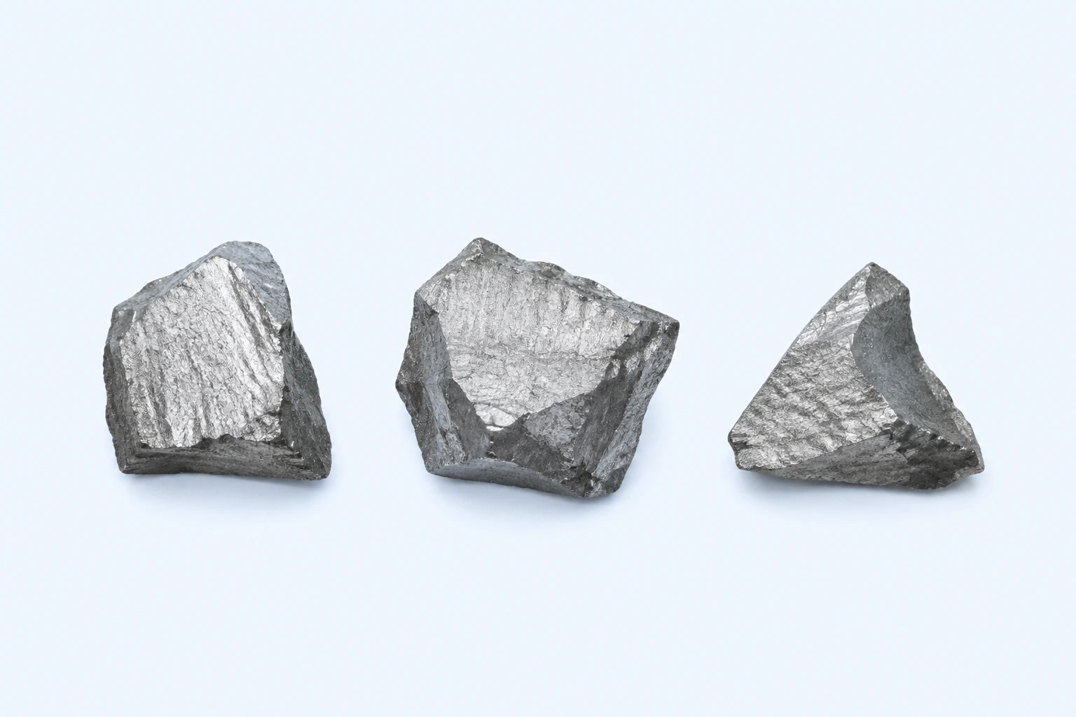
Elemental manganese (Mn), shown as three representative hard brittle gray metallic fragments. Form and surface vary with purity, processing, and oxidation; this is not ore, analytical reference material, or a photograph.
Scientific media record2 verified identifiers
- Subject
- Manganeseelement
- Identifiers
- Atomic number 25PubChem CID:23930
- Review
- Editorial review completeIdentifiers authority-verified · Accessibility validated · · manganese|manganese-technical-specimen-v1.webp
- Digital source
- Trained-algorithmic mediaCreated with a trained generative algorithm and reviewed by WikiBiome for subject identity, scientific framing, identifiers, provenance, and accessibility.
- Scientific basis
- Manganese — PubChem ElementManganese, PubChem CID 23930
- License
- CC BY-SA 4.0Created
Manganese is an essential trace element with a narrow therapeutic window: required as a cofactor for critical enzymes including manganese (Mn)-SOD (SOD2), pyruvate carboxylase, arginase, and ribonucleotide reductase, yet profoundly neurotoxic at elevated levels.[1]Martin & Waters 2022 — Manganese Homeostasis, Stress, and Pathogenesis in BacteriaJulia E. Martin, Lauren S. Waters · 2022Open reference 1 ↓
It occupies a unique position in metal biology for three reasons that go beyond what standard references cover.
First, manganese sits at the bottom of the Irving-Williams series (manganese(II) < iron(II) (Fe2+) < cobalt(II) (Co2+) < nickel(II) (Ni2+) < copper(II) (Cu2+) < zinc(II) (Zn2+)), meaning it forms the weakest complexes with biological ligands among the transition metals.[2]Robinson & Glasfeld 2020 — Metalation and Mis-metalation: Nature's Challenge in Metal CoordinationNigel J. Robinson, Andrea Glasfeld · 2020Open reference 2 ↓
Cells compensate by maintaining manganese at the highest free cytosolic concentration of any transition metal—approximately 10^-6 M—so that correct metalation occurs by mass action before stronger-binding metals can compete.[3]Helmann 2025 — Metals in Motion: Understanding Labile Metal Pools in BacteriaJohn D. Helmann · 2025Open reference 3 ↓[4]Lenner 2025 — Compatibility of Intracellular Binding: Evolutionary Design Principles for Metal SensorsNicolas Lenner, Logan Chariker, Stanislas Leibler · 2025Open reference 4 ↓
This makes manganese homeostasis uniquely sensitive to perturbation: even small shifts in competing metal concentrations (zinc, iron) can redirect manganese from its intended enzyme targets.
Second, manganese is the only essential transition metal that can functionally substitute for iron in a wide range of mononuclear enzymes under Oxidative Stress—a survival strategy exploited by diverse bacteria from E. coli to Salmonella to Lactococcus lactis.[5]Rohaun 2024 — Microbes Vary Strategically in Their Metalation of Mononuclear EnzymesSanjay Kumar Rohaun, Ramakrishnan Sethu, James A Imlay · 2024Open reference 5 ↓[6]McEwan 2024 — Metalloproteome Plasticity: A Factor in Bacterial Pathogen Adaptive Responses?Alastair G. McEwan · 2024Open reference 6 ↓
This cambialistic interchangeability means manganese biology cannot be understood in isolation from iron biology.
Third, manganese-dependent enzymes—particularly superoxide dismutase—are the primary targets of host nutritional immunity via calprotectin-mediated manganese sequestration, making manganese the metallic battleground where host-pathogen metal warfare is most directly observed.[7]Bushman 2025 — The Exploitation of Nutrient Metals by Bacteria for Survival and Infection in the GutSummer D Bushman, Eric P Skaar, N Luisa Hiller · 2025Open reference 7 ↓[8]Metal Ion Acquisition in Staphylococcus aureus: Overcoming Nutritional ImmunityJames E. Cassat, Eric P. Skaar · 2012Open reference 8 ↓
Evidence map82 cited passagesInspect provenance +
Manganese is an essential trace element with a narrow therapeutic window: required as a cofactor for critical enzymes including Mn-SOD (SOD2), pyruvate carboxylase, arginase, and ribonucleotide reductase, yet profoundly neurotoxic at elevated levels. It occupies a unique position in metal biology for three reasons that go beyond what standard references cove
First, Mn sits at the bottom of the Irving-Williams series (Mn2+ < Fe2+ < Co2+ < Ni2+ < Cu2+ < Zn2+), meaning it forms the weakest complexes with biological ligands among the transition metals. Cells compensate by maintaining Mn at the highest free cytosolic concentration of any transition metal—approximately 10^-6 M—so that correct metalation occurs b
Second, Mn is the only essential transition metal that can functionally substitute for iron in a wide range of mononuclear enzymes under oxidative stress—a survival strategy exploited by diverse bacteria from E. coli to Salmonella to Lactococcus lactis,. This cambialistic interchangeability means Mn biology cannot be understood in isolation from iron biol
Third, Mn-dependent enzymes—particularly superoxide dismutase—are the primary targets of host nutritional immunity via calprotectin-mediated Mn sequestration, making Mn the metallic battleground where host-pathogen metal warfare is most directly observed,.
Mn-SOD (SOD2)—the primary mitochondrial antioxidant enzyme, dismutating superoxide to hydrogen peroxide. SODs bind their metal cofactor irreversibly, making them a permanent Mn sink during Mn limitation. When Mn-SOD is mis-metalated with iron, it loses superoxide dismutation activity entirely because the redox potential is wrong for the Mn active site geo
Ribonucleotide reductase (RNR)—essential for DNA synthesis and therefore cell division; Mn-dependent in many pathogenic species.
Pyruvate carboxylase—a key enzyme in gluconeogenesis; Mn deficiency impairs glucose metabolism.
Arginase—Mn-dependent enzyme in the urea cycle.
Phosphatases—including PhpP in Streptococcus pneumoniae, which requires Mn for catalytic activity but is rendered completely inactive when mis-metalated with Zn.
Mn is found in approximately 8% of known metalloproteins, compared to iron's ~25% and magnesium's ~40%.
One of the most remarkable aspects of Mn biology—and one largely absent from standard references—is the phenomenon of cambialistic enzymes that function with either Mn or Fe at the active site. The Imlay lab demonstrated that ribulose-5-phosphate 3-epimerase (Rpe), a universally conserved enzyme, is metalated with iron in E. coli and Bacteroides, with
Iron-metalated enzymes have the highest catalytic turnover but are instantly inactivated by 0.1 mM H2O2 through Fenton chemistry at the active site.
Mn-metalated enzymes are fully resistant to peroxide but have intermediate catalytic rates.
Organisms in oxidizing environments (aerobes like B. subtilis, L. lactis) constitutively use Mn to avoid iron-mediated oxidative damage—sacrificing catalytic efficiency for survival.
E. coli conditionally switches Rpe metalation from Fe to Mn under H2O2 stress via the OxyR-MntH regulatory circuit: OxyR senses peroxide, induces MntH (Mn importer), and the rising Mn pool displaces iron from newly synthesized enzymes,. Salmonella Typhimurium performs a similar switch inside macrophages, increasing Mn uptake via MntH and SitABC while reducin
Staphylococcus aureus possesses both the Mn-specific SodA and the cambialistic SodM, which functions with either Fe or Mn. Under Mn limitation, the small RNA RsaC suppresses SodA translation, freeing Mn for other essential processes, while SodM is upregulated to maintain some SOD activity using available iron.
Beyond enzyme cofactors, Mn2+ forms low-molecular-weight complexes with metabolites (phosphate, carboxylates, amino acids, peptides) that scavenge superoxide non-enzymatically. In Borrelia burgdorferi—a pathogen that has eliminated iron from its biology entirely—MnSOD at the cell surface handles extracellular superoxide while cytoplasmic Mn-metabolite
Baby food jars from Spain contained Mn at 40 times recommended values, posing neurotoxic risk to infants.
German total diet study: infant formula Mn concentration of 840 ug/kg; 95th percentile exposure approached the safe level of intake.
Italian baby food cereal creams contained highest Mn among food categories (8.40 mg/kg).
Welding is the primary occupational exposure route, particularly flux core arc welding (FCAW) in confined spaces.
Mean cumulative Mn exposure of 1.0 mg Mn/m3-year in the Racette welding cohort.
The Crohn's disease-associated SNP rs13107325 in SLC39A8 (ZIP8) alters Mn handling at the colonic mucosal-luminal interface. Homozygous A391T mice exhibit higher Mn in bulk colon tissue with reduced luminal Mn availability, altering the metal environment accessible to gut microbes. This demonstrates that host genetics can reshape microbial metal access indep
Mn exposure reshapes gut microbial communities with sex-specific effects. Fecal microbiota transplant (FMT) can alleviate Mn-induced neurotoxicity in rats, demonstrating that Mn's neurological effects are partly mediated through the gut-brain axis,.
Showing 24 of 82 evidence-bearing passages. Every remaining citation is still indexed in the reference record below.
Contents
1. Biological Roles2. Dietary and Environmental Sources3. Microbiome Interactions4. Nutritional Immunity5. Mis-Metallation: When Manganese is the Wrong Metal6. Conditions Associated7. Interactions with Other Metals8. Biomarkers9. Key Studies10. Open Questions11. Cross-ReferencesBiological Roles#
Enzyme Cofactor#
manganese (Mn) serves as the essential cofactor for several classes of metalloenzymes. manganese-SOD (SOD2)—the primary mitochondrial antioxidant enzyme, dismutating superoxide to hydrogen peroxide. SODs bind their metal cofactor irreversibly, making them a permanent manganese sink during manganese limitation.[9]McFarlane 2025 — A Manganese-Sparing Response Balances Competing Cellular Demands to Enable Staphylococcus aureus InfectionRiley A McFarlane, Jana N Radin, Rafat Mazgaj et al. · 2025Open reference 9 ↓
When manganese-SOD is mis-metalated with iron, it loses superoxide dismutation activity entirely because the redox potential is wrong for the manganese active site geometry.[2]Robinson & Glasfeld 2020 — Metalation and Mis-metalation: Nature's Challenge in Metal CoordinationNigel J. Robinson, Andrea Glasfeld · 2020Open reference 2 ↓
Ribonucleotide reductase (RNR)—essential for DNA synthesis and therefore cell division; manganese-dependent in many pathogenic species.[1]Martin & Waters 2022 — Manganese Homeostasis, Stress, and Pathogenesis in BacteriaJulia E. Martin, Lauren S. Waters · 2022Open reference 1 ↓ Pyruvate carboxylase—a key enzyme in gluconeogenesis; manganese deficiency impairs glucose metabolism.[10]Metals in the pathogenesis of type 2 diabetesAbdul Rehman Khan, Fazli Rabbi Awan · 2014Open reference 10 ↓
Arginase—manganese-dependent enzyme in the urea cycle.[11]Capdevila 2024 — Bacterial Metallostasis: Metal Sensing, Metalloproteome Remodeling, and Metal TraffickingDaiana A. Capdevila, Johnma J. Rondon, Katherine A. Edmonds et al. · 2024Open reference 11 ↓
Phosphatases—including PhpP in Streptococcus pneumoniae, which requires manganese for catalytic activity but is rendered completely inactive when mis-metalated with zinc (Zn).[1]Martin & Waters 2022 — Manganese Homeostasis, Stress, and Pathogenesis in BacteriaJulia E. Martin, Lauren S. Waters · 2022Open reference 1 ↓
manganese is found in approximately 8% of known metalloproteins, compared to iron's ~25% and magnesium's ~40%.[6]McEwan 2024 — Metalloproteome Plasticity: A Factor in Bacterial Pathogen Adaptive Responses?Alastair G. McEwan · 2024Open reference 6 ↓
Cambialistic Enzymes: The Fe-to-Mn Switch#
One of the most remarkable aspects of manganese (Mn) biology—and one largely absent from standard references—is the phenomenon of cambialistic enzymes that function with either manganese or iron (Fe) at the active site.
The Imlay lab demonstrated that ribulose-5-phosphate 3-epimerase (Rpe), a universally conserved enzyme, is metalated with iron in E. coli and Bacteroides, with manganese in B. subtilis and L. lactis, and with zinc in S. cerevisiae—despite having identical metal-coordinating residues across all organisms.[5]Rohaun 2024 — Microbes Vary Strategically in Their Metalation of Mononuclear EnzymesSanjay Kumar Rohaun, Ramakrishnan Sethu, James A Imlay · 2024Open reference 5 ↓
When Rpe genes from manganese-using organisms are expressed in E. coli, they uniformly load with iron, proving that the intracellular metal pool—not the protein sequence—determines cofactor identity.
This has profound ecological consequences. Iron-metalated enzymes have the highest catalytic turnover but are instantly inactivated by 0.1 mM H2O2 through Fenton chemistry at the active site.[5]Rohaun 2024 — Microbes Vary Strategically in Their Metalation of Mononuclear EnzymesSanjay Kumar Rohaun, Ramakrishnan Sethu, James A Imlay · 2024Open reference 5 ↓
manganese-metalated enzymes are fully resistant to peroxide but have intermediate catalytic rates.[5]Rohaun 2024 — Microbes Vary Strategically in Their Metalation of Mononuclear EnzymesSanjay Kumar Rohaun, Ramakrishnan Sethu, James A Imlay · 2024Open reference 5 ↓
Organisms in oxidizing environments (aerobes like B. subtilis, L. lactis) constitutively use manganese to avoid iron-mediated oxidative damage—sacrificing catalytic efficiency for survival.[5]Rohaun 2024 — Microbes Vary Strategically in Their Metalation of Mononuclear EnzymesSanjay Kumar Rohaun, Ramakrishnan Sethu, James A Imlay · 2024Open reference 5 ↓
E. coli conditionally switches Rpe metalation from iron to manganese under H2O2 stress via the OxyR-MntH regulatory circuit: OxyR senses peroxide, induces MntH (manganese importer), and the rising manganese pool displaces iron from newly synthesized enzymes.[5]Rohaun 2024 — Microbes Vary Strategically in Their Metalation of Mononuclear EnzymesSanjay Kumar Rohaun, Ramakrishnan Sethu, James A Imlay · 2024Open reference 5 ↓[6]McEwan 2024 — Metalloproteome Plasticity: A Factor in Bacterial Pathogen Adaptive Responses?Alastair G. McEwan · 2024Open reference 6 ↓
Salmonella Typhimurium performs a similar switch inside macrophages, increasing manganese uptake via MntH and SitABC while reducing iron use—a deliberate metalloproteome remodeling under host immune pressure.[6]McEwan 2024 — Metalloproteome Plasticity: A Factor in Bacterial Pathogen Adaptive Responses?Alastair G. McEwan · 2024Open reference 6 ↓
Staphylococcus aureus possesses both the manganese-specific SodA and the cambialistic SodM, which functions with either iron or manganese.
Under manganese limitation, the small RNA RsaC suppresses SodA translation, freeing manganese for other essential processes, while SodM is upregulated to maintain some SOD activity using available iron.[9]McFarlane 2025 — A Manganese-Sparing Response Balances Competing Cellular Demands to Enable Staphylococcus aureus InfectionRiley A McFarlane, Jana N Radin, Rafat Mazgaj et al. · 2025Open reference 9 ↓
Non-Enzymatic Antioxidant Role#
Beyond enzyme cofactors, manganese(II) (Mn2+) forms low-molecular-weight complexes with metabolites (phosphate, carboxylates, amino acids, peptides) that scavenge superoxide non-enzymatically.
In Borrelia burgdorferi—a pathogen that has eliminated iron from its biology entirely—MnSOD at the cell surface handles extracellular superoxide while cytoplasmic manganese-metabolite complexes (H-manganese) handle intracellular superoxide.[12]Londono 2025 — EPR Spectroscopy Reveals Antioxidant Manganese Defenses in the Lyme Disease Pathogen Borrelia burgdorferiAndres F Londono, Ajay Sharma, Venkatesan Kathiresan et al. · 2025Open reference 12 ↓
The ratio of enzyme-bound manganese (L-manganese) to metabolite-bound manganese (H-manganese) predicts radiation resistance across all domains of life: Deinococcus radiodurans has >90% H-manganese while E. coli has only ~10%, explaining their vastly different radiation tolerance.[12]Londono 2025 — EPR Spectroscopy Reveals Antioxidant Manganese Defenses in the Lyme Disease Pathogen Borrelia burgdorferiAndres F Londono, Ajay Sharma, Venkatesan Kathiresan et al. · 2025Open reference 12 ↓
Dietary and Environmental Sources#
Dietary#
Found in grains, legumes, nuts, tea, and leafy vegetables.
Baby food jars from Spain contained manganese (Mn) at 40 times recommended values, posing neurotoxic risk to infants.[13]Baby Food Jars as a Dietary Source of Essential (K, Na, Ca, Mg, Fe, Zn, Cu, Co, Mo, Mn) and Toxic Elements (Al, Cd, Pb, B, Ba, V, Sr, Li, Ni)Gonzalez-Suarez S, Paz-Montelongo S, Niebla-Canelo D et al. · 2022Open reference 13 ↓
German total diet study: infant formula manganese concentration of 840 ug/kg; 95th percentile exposure approached the safe level of intake.[14]The contribution of infant formula to the food survey-based dietary exposure of nine selected elementsHopfner T, Wollenberg M, Jager A et al. · 2025Open reference 14 ↓
Italian baby food cereal creams contained highest manganese among food categories (8.40 mg/kg).[15]Meli 2024 — Chemical characterization of baby food consumed in ItalyMaria Assunta Meli, Donatella Desideri, Davide Sisti et al. · 2024Open reference 15 ↓
Occupational#
Welding is the primary occupational exposure route, particularly flux core arc welding (FCAW) in confined spaces.[16]Dose-Dependent Progression of Parkinsonism in Manganese-Exposed WeldersBrad A. Racette, Susan Searles Nielsen, Susan R. Criswell et al. · 2017Open reference 16 ↓ Mining, battery manufacturing, and steel production are additional high-exposure occupations. Mean cumulative manganese (Mn) exposure of 1.0 mg manganese/m3-year in the Racette welding cohort.[16]Dose-Dependent Progression of Parkinsonism in Manganese-Exposed WeldersBrad A. Racette, Susan Searles Nielsen, Susan R. Criswell et al. · 2017Open reference 16 ↓
Environmental#
Drinking water contamination from natural geological sources and industrial discharge. manganese (Mn) is a component of the gasoline additive MMT (methylcyclopentadienyl manganese tricarbonyl). Lithium-ion battery recycling represents an emerging exposure source.
Genetic Determinants of Exposure#
The Crohn's disease-associated SNP rs13107325 in SLC39A8 (ZIP8) alters manganese (Mn) handling at the colonic mucosal-luminal interface. Homozygous A391T mice exhibit higher manganese in bulk colon tissue with reduced luminal manganese availability, altering the metal environment accessible to gut microbes.[17]ZIP8 A391T Crohn's Disease-Linked Risk Variant Induces Colonic Metal Ion Dyshomeostasis, Microbiome Compositional Shifts, and InflammationYang JC, Zhao M, Chernikova D et al. · 2024Open reference 17 ↓
This demonstrates that host genetics can reshape microbial metal access independently of dietary exposure.
Microbiome Interactions#
Metals as Selective Pressures on Gut Microbiota#
manganese (Mn) exposure reshapes gut microbial communities with sex-specific effects.[18]Influence of Toxic Metal Exposure on the Gut Microbiota (Review)Federica Giambo, Sebastiano Italia, Michele Teodoro et al. · 2021Open reference 18 ↓
Fecal microbiota transplant (FMT) can alleviate manganese-induced neurotoxicity in rats, demonstrating that manganese's neurological effects are partly mediated through the gut-brain axis.[19]Toxic and Essential Metals: Metabolic Interactions with the Gut Microbiota and Health ImplicationsQinheng Zhu, Boyan Chen, Fu Zhang et al. · 2024Open reference 19 ↓[18]Influence of Toxic Metal Exposure on the Gut Microbiota (Review)Federica Giambo, Sebastiano Italia, Michele Teodoro et al. · 2021Open reference 18 ↓
In the infant gut, manganese drives specific taxonomic selection: serum manganese was identified as a key contributor to Burkholderia-Caballeronia-Paraburkholderia abundance (posterior inclusion probability = 0.535), and synergistic manganese-copper (Cu) interactions shaped Clostridium sensu stricto abundance.[20]Yan 2025 — Association Between Infants' Serum Levels of 26 Metals and Gut Microbiota: A Hospital-Based Cross-Sectional Study in ChinaXing Yan, Jun Qiu, Ruiwen Huang et al. · 2025Open reference 20 ↓
Prenatal manganese exposure (measured in maternal hair) was associated with increased Bifidobacterium relative abundance in 3-month-old infants.[21]Xiong 2025 — Prenatal Exposure to Trace Elements Impacts Mother-Infant Gut Microbiome, Metabolome and Resistome During the First Year of LifeShimao Xiong, Bing Xie, Naiyi Yin et al. · 2025Open reference 21 ↓
The ZIP8 A391T Crohn's risk variant, which reduces luminal manganese availability, resulted in decreased Lactobacillus abundance in mice—consistent with Lactobacillus species being manganese-dependent organisms that rely on manganese rather than iron for their antioxidant strategy.[17]ZIP8 A391T Crohn's Disease-Linked Risk Variant Induces Colonic Metal Ion Dyshomeostasis, Microbiome Compositional Shifts, and InflammationYang JC, Zhao M, Chernikova D et al. · 2024Open reference 17 ↓
Bacterial Mn Acquisition Systems#
Pathogenic bacteria have evolved multiple manganese (Mn) import systems to ensure supply of this critical cofactor:
| System | Type | Organisms | Notes |
|---|---|---|---|
| MntABC | ABC transporter | S. aureus, S. pneumoniae | High-affinity; primary route[8]Metal Ion Acquisition in Staphylococcus aureus: Overcoming Nutritional ImmunityJames E. Cassat, Eric P. Skaar · 2012Open reference 8 ↓ |
| MntH (NRAMP) | Proton-coupled | E. coli, Salmonella, Streptococcus | Induced by OxyR under oxidative stress[6]McEwan 2024 — Metalloproteome Plasticity: A Factor in Bacterial Pathogen Adaptive Responses?Alastair G. McEwan · 2024Open reference 6 ↓ |
| SitABC | ABC transporter | Salmonella | Upregulated during macrophage residence[6]McEwan 2024 — Metalloproteome Plasticity: A Factor in Bacterial Pathogen Adaptive Responses?Alastair G. McEwan · 2024Open reference 6 ↓ |
| PsaA/PsaBCA | ABC transporter | S. pneumoniae, GBS | Sole high-affinity manganese importer—single point of failure[22]Eijkelkamp et al. 2014 — Extracellular Zinc Competitively Inhibits Manganese Uptake in Streptococcus pneumoniaeBart A. Eijkelkamp, Jacqueline R. Morey, Stephanie L. Neville et al. · 2014Open reference 22 ↓ |
| TmpA | ZIP family | S. sanguinis | Alternative manganese import[23]Metal Homeostasis in Pathogenic StreptococciAkbari MS, Doran KS, Burcham LR · 2022Open reference 23 ↓ |
Bacteria also require manganese export to avoid toxicity. MntE (CDF pump) and MgtA (P-type ATPase) export excess manganese in streptococci; loss of MntE renders S. pneumoniae hypersensitive to manganese (growth abolished at 700 uM).[24]Opoku 2024 — Calcium Rescues Streptococcus pneumoniae D39 delta-mntE Manganese-Sensitive Growth PhenotypeReuben Opoku, Edgar Carrasco, Nicholas R De Lay et al. · 2024Open reference 24 ↓
In E. coli, MntP is the primary manganese efflux pump, regulated by both the MntR metalloregulator and a yybP-ykoY manganese-sensing riboswitch in its 5'-UTR.[25]Prakash 2024 — Rho and Riboswitch-Dependent Regulations of mntP Gene Expression Evade Manganese and Membrane ToxicitiesAnand Prakash, Arunima Kalita, Kanika Bhardwaj et al. · 2024Open reference 25 ↓
Mn-Sensing Riboswitches#
Bacteria sense manganese (Mn) through the yybP-ykoY riboswitch family—the largest metal-sensing riboswitch family known, with over 1,000 unique representatives across bacterial phyla.[26]Stephen 2025 — Structurally Distinct Manganese-Sensing Riboswitch Aptamers Regulate Different Expression Platform ArchitecturesChristine Stephen, Danea E Palmer, Clarisa Bautista et al. · 2025Open reference 26 ↓
These RNA-based sensors detect the labile manganese(II) pool co-transcriptionally—metal ion sampling begins before the riboswitch RNA is fully synthesized.[26]Stephen 2025 — Structurally Distinct Manganese-Sensing Riboswitch Aptamers Regulate Different Expression Platform ArchitecturesChristine Stephen, Danea E Palmer, Clarisa Bautista et al. · 2025Open reference 26 ↓
The E. coli alx riboswitch uniquely integrates two orthogonal signals—manganese(II) concentration and pH—through a single RNA element.
At alkaline pH (where manganese toxicity is greatest because manganese(II) oxidizes to DNA-cleaving manganese(IV)), the riboswitch becomes 1,000-fold more sensitive to manganese(II), enabling response to physiologically relevant fluctuations specifically when export is most needed.[27]Palmer 2026 — pH-Dependent Allosteric Remodeling of a Bacterial Riboswitch Couples Alkaline Activation to Metal SensingDanea Palmer, Adrien Chauvier, Tomas F D Silva et al. · 2026Open reference 27 ↓
Mn-Dependent Virulence in Key Pathogens#
Staphylococcus aureus requires manganese (Mn) for SodA/SodM superoxide dismutase activity and oxidative stress defense against host immune attack.[8]Metal Ion Acquisition in Staphylococcus aureus: Overcoming Nutritional ImmunityJames E. Cassat, Eric P. Skaar · 2012Open reference 8 ↓ Under host calprotectin-mediated manganese starvation, the small RNA RsaC activates a "manganese-sparing response"—suppressing the manganese-hungry SodA to redistribute scarce manganese to other essential enzymes.
RsaC is required for virulence in both subcutaneous and systemic mouse infection models.[9]McFarlane 2025 — A Manganese-Sparing Response Balances Competing Cellular Demands to Enable Staphylococcus aureus InfectionRiley A McFarlane, Jana N Radin, Rafat Mazgaj et al. · 2025Open reference 9 ↓
RsaC joins a growing family of metal-responsive sRNAs: RyhB (iron (Fe)-sparing in E. coli), s-SodF (nickel (Ni)-SOD suppression in Streptomyces), and NikS (nickel-responsive in H. pylori).[9]McFarlane 2025 — A Manganese-Sparing Response Balances Competing Cellular Demands to Enable Staphylococcus aureus InfectionRiley A McFarlane, Jana N Radin, Rafat Mazgaj et al. · 2025Open reference 9 ↓
Streptococcus pneumoniae depends on PsaA as its sole high-affinity manganese importer. Zinc competitively inhibits manganese uptake through PsaA with an EC50 of 30.2 uM zinc (Zn) at 1 uM manganese.
When manganese import is blocked, pneumococcal SOD loads with iron instead—a classic mis-metallation event that renders the enzyme inactive against superoxide, leaving the bacterium vulnerable to oxidative killing by host phagocytes.[22]Eijkelkamp et al. 2014 — Extracellular Zinc Competitively Inhibits Manganese Uptake in Streptococcus pneumoniaeBart A. Eijkelkamp, Jacqueline R. Morey, Stephanie L. Neville et al. · 2014Open reference 22 ↓
The zinc:manganese ratio, not the absolute concentration of either metal, determines bacterial vulnerability.[22]Eijkelkamp et al. 2014 — Extracellular Zinc Competitively Inhibits Manganese Uptake in Streptococcus pneumoniaeBart A. Eijkelkamp, Jacqueline R. Morey, Stephanie L. Neville et al. · 2014Open reference 22 ↓
Group B Streptococcus (S. agalactiae) is a manganese-centric organism that relies on manganese for SOD activity and oxidative stress defense; host manganese restriction via calprotectin is a potent anti-GBS strategy.[28]Goh 2024 — An Opportunistic Pathogen Under Stress: How Group B Streptococcus Responds to Cytotoxic Reactive Species and Conditions of Metal Ion Imbalance to SurviveKelvin G K Goh, Devika Desai, Ruby Thapa et al. · 2024Open reference 28 ↓
Excess zinc(II) in phagosomes displaces manganese(II) from SodA and other manganese-dependent enzymes, compounding the damage.[28]Goh 2024 — An Opportunistic Pathogen Under Stress: How Group B Streptococcus Responds to Cytotoxic Reactive Species and Conditions of Metal Ion Imbalance to SurviveKelvin G K Goh, Devika Desai, Ruby Thapa et al. · 2024Open reference 28 ↓
Borrelia burgdorferi has taken the most extreme approach: eliminating iron entirely from its biology.
With no iron-dependent enzymes, B. burgdorferi avoids Fenton chemistry and circumvents host nutritional immunity targeting iron—but at the cost of absolute dependence on manganese for its antioxidant defense.[12]Londono 2025 — EPR Spectroscopy Reveals Antioxidant Manganese Defenses in the Lyme Disease Pathogen Borrelia burgdorferiAndres F Londono, Ajay Sharma, Venkatesan Kathiresan et al. · 2025Open reference 12 ↓
Nutritional Immunity#
The host's primary weapon against manganese (Mn)-dependent pathogens is calprotectin (S100A8/S100A9), which comprises 40-50% of neutrophil cytoplasmic protein and can reach >1 mg/mL at infection sites.[7]Bushman 2025 — The Exploitation of Nutrient Metals by Bacteria for Survival and Infection in the GutSummer D Bushman, Eric P Skaar, N Luisa Hiller · 2025Open reference 7 ↓
Calprotectin binds both manganese and zinc (Zn) to create metal-free zones at abscesses, rendering S. aureus virtually devoid of manganese.[7]Bushman 2025 — The Exploitation of Nutrient Metals by Bacteria for Survival and Infection in the GutSummer D Bushman, Eric P Skaar, N Luisa Hiller · 2025Open reference 7 ↓[8]Metal Ion Acquisition in Staphylococcus aureus: Overcoming Nutritional ImmunityJames E. Cassat, Eric P. Skaar · 2012Open reference 8 ↓
The effectiveness of calprotectin against manganese-dependent pathogens depends on the pathogen's metal flexibility.
Strictly manganese-dependent SODs (as in most Streptococcus species) are catastrophically disabled by manganese restriction.[1]Martin & Waters 2022 — Manganese Homeostasis, Stress, and Pathogenesis in BacteriaJulia E. Martin, Lauren S. Waters · 2022Open reference 1 ↓ Cambialistic SODs (as in S. aureus SodM) retain some activity using iron, providing partial resistance.[9]McFarlane 2025 — A Manganese-Sparing Response Balances Competing Cellular Demands to Enable Staphylococcus aureus InfectionRiley A McFarlane, Jana N Radin, Rafat Mazgaj et al. · 2025Open reference 9 ↓
Organisms with both manganese-SOD and iron (Fe)-SOD (as in E. coli) are least affected by manganese restriction alone.[1]Martin & Waters 2022 — Manganese Homeostasis, Stress, and Pathogenesis in BacteriaJulia E. Martin, Lauren S. Waters · 2022Open reference 1 ↓
The bacterial cell wall also serves as a manganese reservoir: peptidoglycan and teichoic acids bind divalent cations including manganese, buffering against host-imposed restriction.
S. aureus can evolve resistance to metal chelators by reconfiguring its cell wall to increase surface calcium, effectively substituting calcium(II) (Ca2+) for manganese(II) in maintaining structural integrity.[29]Paterson 2025 — Enhanced Resistance of Metal Sequestering Agents by Reconfiguration of the Staphylococcus aureus Cell WallJoy R Paterson, Joshua M Wadsworth, Rebecca J Lee et al. · 2025Open reference 29 ↓
Exogenous calcium(II) can also rescue S. pneumoniae from manganese toxicity without reducing intracellular manganese levels, suggesting functional compensation at calcium-dependent enzyme active sites.[24]Opoku 2024 — Calcium Rescues Streptococcus pneumoniae D39 delta-mntE Manganese-Sensitive Growth PhenotypeReuben Opoku, Edgar Carrasco, Nicholas R De Lay et al. · 2024Open reference 24 ↓
Mis-Metallation: When Manganese is the Wrong Metal#
Mn Excess Mis-Metalates Iron Sensors#
Elevated manganese (Mn) occupies the iron (Fe)-sensing Fur (ferric uptake regulator) protein, causing iron import genes to remain repressed even when iron is needed.
Unincorporated intracellular iron then generates toxic hydroxyl radicals via Fenton chemistry—a non-obvious cascade linking manganese excess to oxidative stress through iron dysregulation rather than direct manganese toxicity.[1]Martin & Waters 2022 — Manganese Homeostasis, Stress, and Pathogenesis in BacteriaJulia E. Martin, Lauren S. Waters · 2022Open reference 1 ↓
This Fur mis-metallation has been demonstrated in both pathogenic bacteria and cyanobacteria: in Synechocystis, excess manganese produces a transcriptome that partly resembles iron limitation even though iron levels are unchanged, because manganese-bound Fur erroneously signals iron sufficiency.[30]Reis 2024 — Study of Excess Manganese Stress Response Highlights the Central Role of Manganese Exporter Mnx for Holding Manganese Homeostasis in Synechocystis sp. PCC 6803Mara Reis, Sanja Zenker, Prisca Viehoever et al. · 2024Open reference 30 ↓
Cells respond to manganese-driven mis-metallation by upregulating ribosomes, proteases, and chaperones—investing in protein quality control to replace mis-metalated proteins with correctly metalated copies.[30]Reis 2024 — Study of Excess Manganese Stress Response Highlights the Central Role of Manganese Exporter Mnx for Holding Manganese Homeostasis in Synechocystis sp. PCC 6803Mara Reis, Sanja Zenker, Prisca Viehoever et al. · 2024Open reference 30 ↓
The Irving-Williams Vulnerability#
According to the Irving-Williams series, manganese(II) (Mn2+) binds proteins more weakly than any other transition metal, meaning it is the most easily displaced from binding sites when competing metals are present.[2]Robinson & Glasfeld 2020 — Metalation and Mis-metalation: Nature's Challenge in Metal CoordinationNigel J. Robinson, Andrea Glasfeld · 2020Open reference 2 ↓
Cells maintain manganese at the highest free cytosolic concentration to compensate, but when this balance is disrupted—by host zinc (Zn) flooding, for example—manganese-dependent enzymes become mis-metalated.
PerR regulator in Bacillus: zinc displaces the correct manganese/iron (Fe) cofactor, constitutively repressing catalase while derepressing heme biosynthesis, flooding the cell with pro-oxidant heme and no defense.[31]Chandrangsu & Helmann 2016 — Intracellular Zn Intoxication Mis-metalates PerR, Causing Heme Toxicity and Oxidative DeathPete Chandrangsu, John D. Helmann · 2016Open reference 31 ↓
PsaA-imported manganese in pneumococcus: zinc blocks the importer, forcing SOD to load with iron (inactive for superoxide dismutation).[22]Eijkelkamp et al. 2014 — Extracellular Zinc Competitively Inhibits Manganese Uptake in Streptococcus pneumoniaeBart A. Eijkelkamp, Jacqueline R. Morey, Stephanie L. Neville et al. · 2014Open reference 22 ↓
Cross-metal displacement in C. elegans: chronic zinc exposure significantly decreases manganese levels (p=0.001), while manganese and iron exposure decreases zinc levels—demonstrating that excess of any single transition metal cascades into disruption of the entire metallome.[32]Blume 2026 — Combined Metallomics and Metabolomics Reveal Impact of Metal Homeostasis on Biological Pathways in C. elegansBastian Blume, Philippe Schmitt-Kopplin, Bernhard Michalke · 2026Open reference 32 ↓
Bacterial manganese-sensing metalloregulators (MntR) use O-rich coordination sites to achieve specificity for Mn2+—the weakest binder—without being overwhelmed by stronger-binding metals.[4]Lenner 2025 — Compatibility of Intracellular Binding: Evolutionary Design Principles for Metal SensorsNicolas Lenner, Logan Chariker, Stanislas Leibler · 2025Open reference 4 ↓[33]Nies 2025 — A Flow Equilibrium Model Controlling Cytoplasmic Transition Metal Cation Pools and Preventing Mis-MetalationDietrich H Nies, Julie A Maupin-Furlow · 2025Open reference 33 ↓
Mn Toxicity from Excess#
Uncontrolled manganese (Mn) accumulation is itself toxic. In S. pneumoniae lacking the MntE efflux transporter, excess manganese (300-700 uM) impairs growth, increases capsule production, and reduces biofilm formation.[24]Opoku 2024 — Calcium Rescues Streptococcus pneumoniae D39 delta-mntE Manganese-Sensitive Growth PhenotypeReuben Opoku, Edgar Carrasco, Nicholas R De Lay et al. · 2024Open reference 24 ↓
In E. coli, the MntP efflux pump is essential; its regulation by a manganese-sensing riboswitch and Rho-dependent transcription termination prevents both manganese accumulation (too little MntP) and membrane protein toxicity (too much MntP).[25]Prakash 2024 — Rho and Riboswitch-Dependent Regulations of mntP Gene Expression Evade Manganese and Membrane ToxicitiesAnand Prakash, Arunima Kalita, Kanika Bhardwaj et al. · 2024Open reference 25 ↓
At elevated pH, manganese(II) oxidizes to manganese(IV), which cleaves RNA and DNA, and manganese(II) hydroxide increases reactive oxygen species generation.[27]Palmer 2026 — pH-Dependent Allosteric Remodeling of a Bacterial Riboswitch Couples Alkaline Activation to Metal SensingDanea Palmer, Adrien Chauvier, Tomas F D Silva et al. · 2026Open reference 27 ↓
In metabolite-depleted cells, excess free manganese(II) that cannot form protective metabolite complexes becomes directly cytotoxic through off-target binding—a form of self-poisoning observed in stationary-phase B. burgdorferi.[12]Londono 2025 — EPR Spectroscopy Reveals Antioxidant Manganese Defenses in the Lyme Disease Pathogen Borrelia burgdorferiAndres F Londono, Ajay Sharma, Venkatesan Kathiresan et al. · 2025Open reference 12 ↓
Conditions Associated#
Parkinsonism and Neurotoxicity#
manganese (Mn) neurotoxicity is the most thoroughly documented health effect. Dose-dependent parkinsonism in welders shows an annual UPDRS3 increase of 0.24 points per mg manganese/m3-year of cumulative exposure (p<0.001); a worker with 20 years of welding would show nearly a 7-point increase.[16]Dose-Dependent Progression of Parkinsonism in Manganese-Exposed WeldersBrad A. Racette, Susan Searles Nielsen, Susan R. Criswell et al. · 2017Open reference 16 ↓
The phenotype predominantly affects upper limb bradykinesia, rigidity, and impaired speech/facial expression.[16]Dose-Dependent Progression of Parkinsonism in Manganese-Exposed WeldersBrad A. Racette, Susan Searles Nielsen, Susan R. Criswell et al. · 2017Open reference 16 ↓ Workers whose baseline examination was within 5 years of first manganese exposure showed dramatically higher progression (4.45 vs 0.23 UPDRS3/year), suggesting an early vulnerability window.[16]Dose-Dependent Progression of Parkinsonism in Manganese-Exposed WeldersBrad A. Racette, Susan Searles Nielsen, Susan R. Criswell et al. · 2017Open reference 16 ↓
FCAW in confined spaces showed a 6.7-fold higher progression rate than non-confined FCAW.[16]Dose-Dependent Progression of Parkinsonism in Manganese-Exposed WeldersBrad A. Racette, Susan Searles Nielsen, Susan R. Criswell et al. · 2017Open reference 16 ↓
manganese accumulates preferentially in the globus pallidus and striatum of the basal ganglia, unlike most toxic metals that target cortical regions.[34]Common and Trace Metals in Alzheimer's and Parkinson's DiseasesDoroszkiewicz J, Farhan JA, Mroczko J et al. · 2023Open reference 34 ↓
Mechanism involves oxidative stress in dopaminergic neurons through mitochondrial accumulation, electron transport chain disruption, and ROS generation via Fenton-like chemistry.[34]Common and Trace Metals in Alzheimer's and Parkinson's DiseasesDoroszkiewicz J, Farhan JA, Mroczko J et al. · 2023Open reference 34 ↓
In the metal-driven PD framework, manganese acts alongside iron and nickel to reshape gut microbial communities, with downstream Neuroinflammation converging on dopaminergic neuron vulnerability.[35]Microbial Metallomics and Parkinson's Disease: A Unified Metal-Driven Framework Linking Ferroptosis, Dysbiosis, and alpha-Synuclein PathologyKaren Pendergrass · 2025Open reference 35 ↓
Alzheimer's Disease#
manganese (Mn) impairs autophagy at low concentrations; Drp1 inhibition is protective against manganese-induced autophagic impairment.[36]Associations of Environmental Exposure to Arsenic, Manganese, Lead, and Cadmium with Alzheimer's Disease: A Review of Recent Evidence from Mechanistic StudiesGiasuddin Ahmed, Md. Shiblur Rahaman, Enrique Perez et al. · 2025Open reference 36 ↓ Acute manganese exposure increases cortical GLAST expression and seizure susceptibility in APP/PSEN1 mice.[36]Associations of Environmental Exposure to Arsenic, Manganese, Lead, and Cadmium with Alzheimer's Disease: A Review of Recent Evidence from Mechanistic StudiesGiasuddin Ahmed, Md. Shiblur Rahaman, Enrique Perez et al. · 2025Open reference 36 ↓
manganese primarily affects the basal ganglia (parkinsonism) rather than cortical regions (AD), distinguishing its neurodegeneration pattern from lead or mercury.[37]Heavy Metals Exposure and Alzheimer's Disease and Related DementiasBakulski KM, Seo YA, Hickman RC et al. · 2020Open reference 37 ↓ manganese alterations in brain tissue of dementia patients connect to SOD2 cofactor function and mitochondrial antioxidant defense.[38]Scholefield et al. 2024 — Brain Metallomic Signatures Distinguish DLB from AD and PDDMelissa Scholefield, Stephanie J. Church, Jingshu Xu et al. · 2024Open reference 38 ↓
Inflammatory Bowel Disease#
Plasma manganese (Mn) was significantly lower in ulcerative colitis patients (1.4 ug/L) compared to healthy controls (2.4 ug/L, p=0.041).[39]Clinical and inflammatory biomarkers of inflammatory bowel diseases are linked to plasma trace elements and toxic metals; new insights into an old conceptAmerikanou C, Karavoltsos S, Gioxari A et al. · 2022Open reference 39 ↓
The ZIP8 A391T Crohn's disease risk variant reduces luminal manganese availability in the colon, reshaping the microbiome—with age-dependent microbiome shifts (R2 increasing from 3% at 2 months to 9% at 12 months) and spontaneous Metal-Driven Inflammation developing by 10 months.[17]ZIP8 A391T Crohn's Disease-Linked Risk Variant Induces Colonic Metal Ion Dyshomeostasis, Microbiome Compositional Shifts, and InflammationYang JC, Zhao M, Chernikova D et al. · 2024Open reference 17 ↓
Lactobacillus depletion in ZIP8 mutant mice is consistent with the manganese-dependent antioxidant biology of lactic acid bacteria.[17]ZIP8 A391T Crohn's Disease-Linked Risk Variant Induces Colonic Metal Ion Dyshomeostasis, Microbiome Compositional Shifts, and InflammationYang JC, Zhao M, Chernikova D et al. · 2024Open reference 17 ↓
Breast Cancer#
A meta-analysis of 36 case-control studies (n=4,151) found serum manganese (Mn) significantly lower in breast cancer patients (SMD: -2.95, 95% CI: -4.26 to -1.64, Asian studies).[40]Relationships Between Biological Heavy Metals and Breast Cancer: A Systematic Review and Meta-AnalysisLiu L, Chen J, Liu C et al. · 2022Open reference 40 ↓
manganese depletion disrupts MnSOD antioxidant function, converging with the copper (Cu) elevation and zinc (Zn) depletion also observed in the same meta-analysis.[40]Relationships Between Biological Heavy Metals and Breast Cancer: A Systematic Review and Meta-AnalysisLiu L, Chen J, Liu C et al. · 2022Open reference 40 ↓
PCOS and Reproductive Health#
Serum manganese (Mn) was significantly lower in both obese and non-obese PCOS patients compared to controls (0.086 vs 0.225 ug/dl), possibly reflecting impaired MnSOD antioxidant capacity.[41]A Comparative Study of Blood Levels of Manganese, Some Macroelements and Heavy Metals in Obese and Non-Obese Polycystic Ovary Syndrome PatientsSarah H. Mhaibes, Mohammed A. Taher, Ala H. Badr · 2017Open reference 41 ↓
A more recent Slovenian case-control study (n=70) found no significant manganese differences, though this study prioritized copper (Cu) and molybdenum (Mo) findings.[42]Smovršnik 2025 — Association of Trace Elements with Polycystic Ovary Syndrome in Women — A Case-Control StudyTinkara Smovršnik, Bojana Pinter, Milena Horvat et al. · 2025Open reference 42 ↓
Thyroid Function#
Elevated manganese (Mn) levels found in autoimmune hypothyroidism; manganese affects deiodinase (DIO) activity and T4 to T3 conversion.[43]Effects of Trace Elements on Endocrine Function and Pathogenesis of Thyroid Diseases — A Literature ReviewBrylinski L, Kostelecka K, Wolinski F et al. · 2025Open reference 43 ↓ Higher blood manganese observed in thyroid cancer patients.[43]Effects of Trace Elements on Endocrine Function and Pathogenesis of Thyroid Diseases — A Literature ReviewBrylinski L, Kostelecka K, Wolinski F et al. · 2025Open reference 43 ↓
Cancer#
Serum manganese (Mn) decreased in prostate cancer patients (0.001 vs 0.0024 ug/ml, p<0.005), potentially impairing MnSOD-mediated mitochondrial antioxidant defense.[44]Serum Levels of Selenium, Zinc, Copper, Manganese, and Iron in Prostate Cancer PatientsSaleh A. K. Saleh, Heba M. Adly, Altaf A. Abdelkhaliq et al. · 2020Open reference 44 ↓
manganese elevated 1.26-fold in lung cancer serum; aluminum (Al)/manganese ratios serve as potential LC biomarkers (AUC close to 1).[45]Metallomic Signatures of Lung Cancer and Chronic Obstructive Pulmonary DiseaseBelen Callejon-Leblic, Saida Sanchez Espirilla, Carolina Gotera-Rivera et al. · 2023Open reference 45 ↓ In COPD-to-LC transition, manganese showed dramatically altered profiles (0.41-fold decrease), suggesting progressive manganese dyshomeostasis.[45]Metallomic Signatures of Lung Cancer and Chronic Obstructive Pulmonary DiseaseBelen Callejon-Leblic, Saida Sanchez Espirilla, Carolina Gotera-Rivera et al. · 2023Open reference 45 ↓
Renal Injury#
In a longitudinal study (n=384, 4 repeated measurements), urinary manganese (Mn) was associated with renal biomarkers including UACR (beta=2.60), though with complex dose-response relationships at high levels.[46]Complex interplay of heavy metals and renal injury: New perspectives from longitudinal epidemiological evidenceYin G, Zhao S, Zhao M et al. · 2024Open reference 46 ↓
Perinatal Exposure#
One longitudinal cohort found a positive association between 3rd trimester blood manganese (Mn) and continuous EPDS (depression) scores (beta=0.13, 95% CI: 0.04-0.21).[47]Exposure to environmental chemicals and perinatal psychopathologyJacobson MH, Ghassabian A, Gore AC et al. · 2022Open reference 47 ↓ manganese treatment (50-300 uM) induces cytochrome C release, caspase activation, and protein aggregation in dopaminergic neurons.[48]Environmental pollutants as risk factors for neurodegenerative disorders: Alzheimer and Parkinson diseasesChin-Chan M, Navarro-Yepes J, Quintanilla-Vega B · 2015Open reference 48 ↓
A meta-analysis of prenatal metal exposure and ASD found no consistent association for manganese specifically, though other metals (cadmium) showed significant effects.[49]Dou 2024 — Exposure to Heavy Metals in Utero and Autism Spectrum Disorder at Age 3: A Meta-Analysis of Two Longitudinal CohortsJohn F. Dou, Rebecca J. Schmidt, Heather E. Volk et al. · 2024Open reference 49 ↓
Cross-Condition Pattern: Mn Depletion#
A notable cross-condition pattern emerges from the evidence: manganese (Mn) depletion appears in breast cancer,[40]Relationships Between Biological Heavy Metals and Breast Cancer: A Systematic Review and Meta-AnalysisLiu L, Chen J, Liu C et al. · 2022Open reference 40 ↓ ulcerative colitis,[39]Clinical and inflammatory biomarkers of inflammatory bowel diseases are linked to plasma trace elements and toxic metals; new insights into an old conceptAmerikanou C, Karavoltsos S, Gioxari A et al. · 2022Open reference 39 ↓ PCOS,[41]A Comparative Study of Blood Levels of Manganese, Some Macroelements and Heavy Metals in Obese and Non-Obese Polycystic Ovary Syndrome PatientsSarah H. Mhaibes, Mohammed A. Taher, Ala H. Badr · 2017Open reference 41 ↓ and prostate cancer.[44]Serum Levels of Selenium, Zinc, Copper, Manganese, and Iron in Prostate Cancer PatientsSaleh A. K. Saleh, Heba M. Adly, Altaf A. Abdelkhaliq et al. · 2020Open reference 44 ↓
In each case, manganese depletion is accompanied by impaired MnSOD function and oxidative stress—suggesting a shared vulnerability axis where reduced manganese-dependent antioxidant capacity contributes to disease progression.
Interactions with Other Metals#
Iron#
manganese (Mn) competes with iron for DMT1 transport; iron status affects manganese absorption and vice versa.[50]Mechanism of Iron Ion Homeostasis in Intestinal Immunity and Gut Microbiota RemodelingHonghong Bao, Yi Wang, Hanlin Xiong et al. · 2024Open reference 50 ↓
manganese and iron (Fe) are functionally interchangeable in many mononuclear enzymes, with organisms switching between them depending on oxidative stress conditions.[5]Rohaun 2024 — Microbes Vary Strategically in Their Metalation of Mononuclear EnzymesSanjay Kumar Rohaun, Ramakrishnan Sethu, James A Imlay · 2024Open reference 5 ↓
Iron supplementation rescues manganese-limitation growth defects in S. aureus because many enzymes can use iron or manganese interchangeably—a beneficial mis-metallation.[9]McFarlane 2025 — A Manganese-Sparing Response Balances Competing Cellular Demands to Enable Staphylococcus aureus InfectionRiley A McFarlane, Jana N Radin, Rafat Mazgaj et al. · 2025Open reference 9 ↓
Iron treatment shifts manganese speciation from low-molecular-weight to high-molecular-weight fractions in C. elegans, indicating competition for binding sites.[32]Blume 2026 — Combined Metallomics and Metabolomics Reveal Impact of Metal Homeostasis on Biological Pathways in C. elegansBastian Blume, Philippe Schmitt-Kopplin, Bernhard Michalke · 2026Open reference 32 ↓
Loss of both SODs in E. coli (manganese-SodA and iron-SodB) causes superoxide to attack iron-sulfur clusters, releasing free iron that fuels Fenton chemistry—and triggers enterobactin (siderophore) upregulation to recapture iron.[51]Nong 2026 — Despite Inducing Antioxidant Regulation, Superoxide Dismutase Deficiency Makes E. coli More Sensitive to Hydrogen PeroxideYuejuan Nong, Jiaxin Qiao, Yixuan Zhao et al. · 2026Open reference 51 ↓
Zinc#
zinc (Zn) competitively inhibits manganese (Mn) uptake through PsaA in pneumococcus (EC50 = 30.2 uM at 1 uM manganese).[22]Eijkelkamp et al. 2014 — Extracellular Zinc Competitively Inhibits Manganese Uptake in Streptococcus pneumoniaeBart A. Eijkelkamp, Jacqueline R. Morey, Stephanie L. Neville et al. · 2014Open reference 22 ↓
Host macrophages flood phagosomes with zinc, displacing manganese from enzymes following the Irving-Williams series.[28]Goh 2024 — An Opportunistic Pathogen Under Stress: How Group B Streptococcus Responds to Cytotoxic Reactive Species and Conditions of Metal Ion Imbalance to SurviveKelvin G K Goh, Devika Desai, Ruby Thapa et al. · 2024Open reference 28 ↓[52]Sullivan 2024 — Resisting Death by Metal: Metabolism and Cu/Zn Homeostasis in BacteriaMatthew J. Sullivan, Ignacio Teran, Kelvin GK Goh et al. · 2024Open reference 52 ↓
Chronic zinc exposure decreases manganese levels significantly (p=0.001) in C. elegans.[32]Blume 2026 — Combined Metallomics and Metabolomics Reveal Impact of Metal Homeostasis on Biological Pathways in C. elegansBastian Blume, Philippe Schmitt-Kopplin, Bernhard Michalke · 2026Open reference 32 ↓ The zinc:manganese ratio—not the absolute concentration of either metal—determines bacterial vulnerability.[22]Eijkelkamp et al. 2014 — Extracellular Zinc Competitively Inhibits Manganese Uptake in Streptococcus pneumoniaeBart A. Eijkelkamp, Jacqueline R. Morey, Stephanie L. Neville et al. · 2014Open reference 22 ↓
Calcium#
calcium(II) (Ca2+) completely rescues the manganese (Mn)-sensitive growth defect of S. pneumoniae MntE mutants without reducing intracellular manganese, suggesting functional compensation at enzyme active sites.[24]Opoku 2024 — Calcium Rescues Streptococcus pneumoniae D39 delta-mntE Manganese-Sensitive Growth PhenotypeReuben Opoku, Edgar Carrasco, Nicholas R De Lay et al. · 2024Open reference 24 ↓
S. pneumoniae requires calcium(II) as an obligatory micronutrient (minimum 150 uM for viability); a yybP-ykoY riboswitch senses both manganese and calcium(II), suggesting shared sensing mechanisms.[24]Opoku 2024 — Calcium Rescues Streptococcus pneumoniae D39 delta-mntE Manganese-Sensitive Growth PhenotypeReuben Opoku, Edgar Carrasco, Nicholas R De Lay et al. · 2024Open reference 24 ↓
Chelator-resistant S. aureus compensates for manganese depletion by increasing surface-associated calcium.[29]Paterson 2025 — Enhanced Resistance of Metal Sequestering Agents by Reconfiguration of the Staphylococcus aureus Cell WallJoy R Paterson, Joshua M Wadsworth, Rebecca J Lee et al. · 2025Open reference 29 ↓
Copper#
copper (Cu)-manganese (Mn) correlation (r=0.61) in baby food matrices suggests shared contamination sources.[15]Meli 2024 — Chemical characterization of baby food consumed in ItalyMaria Assunta Meli, Donatella Desideri, Davide Sisti et al. · 2024Open reference 15 ↓ Copper-BMDC antimicrobial treatment significantly decreases manganese levels in S. aureus, compromising SOD-dependent antioxidant defense.[53]Sanchez-Rosario 2026 — N-benzyl-N-methyldithiocarbamate (BMDC) Combines with Metals to Produce Antimicrobial and Anti-Biofilm Activity Against MRSA and S. epidermidisYamil Sanchez-Rosario, Natasha R Cornejo, Isaiah S Gonzalez et al. · 2026Open reference 53 ↓
cobalt (Co)-exposure with iron and nickel in welding fumes creates complex mixture effects.[16]Dose-Dependent Progression of Parkinsonism in Manganese-Exposed WeldersBrad A. Racette, Susan Searles Nielsen, Susan R. Criswell et al. · 2017Open reference 16 ↓
Biomarkers#
Blood manganese (Mn) reflects recent exposure but has limited utility for cumulative assessment.[37]Heavy Metals Exposure and Alzheimer's Disease and Related DementiasBakulski KM, Seo YA, Hickman RC et al. · 2020Open reference 37 ↓ Cumulative manganese exposure (mg manganese/m3-year) calculated from work histories is the gold standard in occupational studies.[16]Dose-Dependent Progression of Parkinsonism in Manganese-Exposed WeldersBrad A. Racette, Susan Searles Nielsen, Susan R. Criswell et al. · 2017Open reference 16 ↓
UPDRS3 motor assessment by movement disorders specialists serves as the clinical outcome measure.[16]Dose-Dependent Progression of Parkinsonism in Manganese-Exposed WeldersBrad A. Racette, Susan Searles Nielsen, Susan R. Criswell et al. · 2017Open reference 16 ↓
Serum manganese levels significantly altered in prostate cancer and lung cancer, suggesting diagnostic potential in metallomic panels.[44]Serum Levels of Selenium, Zinc, Copper, Manganese, and Iron in Prostate Cancer PatientsSaleh A. K. Saleh, Heba M. Adly, Altaf A. Abdelkhaliq et al. · 2020Open reference 44 ↓[45]Metallomic Signatures of Lung Cancer and Chronic Obstructive Pulmonary DiseaseBelen Callejon-Leblic, Saida Sanchez Espirilla, Carolina Gotera-Rivera et al. · 2023Open reference 45 ↓
manganese quotas vary across host body sites: ~650 nM in nasopharynx, ~1100 nM in lung, ~400-700 nM in blood.[24]Opoku 2024 — Calcium Rescues Streptococcus pneumoniae D39 delta-mntE Manganese-Sensitive Growth PhenotypeReuben Opoku, Edgar Carrasco, Nicholas R De Lay et al. · 2024Open reference 24 ↓
Key Studies#
| Study | Design | Key Finding | Evidence Level |
|---|---|---|---|
| [5]Rohaun 2024 — Microbes Vary Strategically in Their Metalation of Mononuclear EnzymesSanjay Kumar Rohaun, Ramakrishnan Sethu, James A Imlay · 2024Open reference 5 ↓ | In-vitro | Metal pool, not protein, determines enzyme cofactor identity | In-vitro |
| [2]Robinson & Glasfeld 2020 — Metalation and Mis-metalation: Nature's Challenge in Metal CoordinationNigel J. Robinson, Andrea Glasfeld · 2020Open reference 2 ↓ | Review | Irving-Williams series governs mis-metallation; MnSOD routinely mis-metalated with iron (Fe) in E. coli | Expert-opinion |
| [16]Dose-Dependent Progression of Parkinsonism in Manganese-Exposed WeldersBrad A. Racette, Susan Searles Nielsen, Susan R. Criswell et al. · 2017Open reference 16 ↓ | Prospective cohort | Dose-dependent UPDRS3 progression in welders (0.24 points/mg manganese (Mn)/m3-year) | Prospective-cohort |
| [9]McFarlane 2025 — A Manganese-Sparing Response Balances Competing Cellular Demands to Enable Staphylococcus aureus InfectionRiley A McFarlane, Jana N Radin, Rafat Mazgaj et al. · 2025Open reference 9 ↓ | Animal model | RsaC sRNA enables manganese-sparing response required for S. aureus virulence | Animal-model |
| [22]Eijkelkamp et al. 2014 — Extracellular Zinc Competitively Inhibits Manganese Uptake in Streptococcus pneumoniaeBart A. Eijkelkamp, Jacqueline R. Morey, Stephanie L. Neville et al. · 2014Open reference 22 ↓ | In-vitro | zinc (Zn):manganese ratio determines pneumococcal survival via PsaA competition | In-vitro |
| [12]Londono 2025 — EPR Spectroscopy Reveals Antioxidant Manganese Defenses in the Lyme Disease Pathogen Borrelia burgdorferiAndres F Londono, Ajay Sharma, Venkatesan Kathiresan et al. · 2025Open reference 12 ↓ | Quasi-experimental | Dual enzymatic and non-enzymatic manganese antioxidant systems in iron-free B. burgdorferi | Quasi-experimental |
| [40]Relationships Between Biological Heavy Metals and Breast Cancer: A Systematic Review and Meta-AnalysisLiu L, Chen J, Liu C et al. · 2022Open reference 40 ↓ | Meta-analysis | manganese significantly depleted in breast cancer (SMD: -2.95) across 36 studies | Systematic-review |
| [17]ZIP8 A391T Crohn's Disease-Linked Risk Variant Induces Colonic Metal Ion Dyshomeostasis, Microbiome Compositional Shifts, and InflammationYang JC, Zhao M, Chernikova D et al. · 2024Open reference 17 ↓ | Animal model | ZIP8 Crohn's risk variant alters colonic manganese, reshapes microbiome, causes age-dependent inflammation | Cross-sectional |
| [26]Stephen 2025 — Structurally Distinct Manganese-Sensing Riboswitch Aptamers Regulate Different Expression Platform ArchitecturesChristine Stephen, Danea E Palmer, Clarisa Bautista et al. · 2025Open reference 26 ↓ | In-vitro | yybP-ykoY riboswitches sense manganese co-transcriptionally; >1,000 members across bacteria | In-vitro |
| [7]Bushman 2025 — The Exploitation of Nutrient Metals by Bacteria for Survival and Infection in the GutSummer D Bushman, Eric P Skaar, N Luisa Hiller · 2025Open reference 7 ↓ | Animal model | Calprotectin at >1 mg/mL sequesters manganese/zinc; weaponized at infection sites | Animal-model |
Open Questions#
Unresolved questions identified by the current evidence record.
01Whether manganese (Mn)-induced parkinsonism is truly distinct from idiopathic Parkinson's disease or represents an accelerated/modified form of the same pathology.+
The current WikiBiome record identifies this as an unresolved evidence gap.
02The role of Gut Microbiome-mediated manganese (Mn) metabolism in modulating neurotoxicity risk, and whether probiotic interventions could be protective.+
The current WikiBiome record identifies this as an unresolved evidence gap.
03Whether the dramatically elevated manganese (Mn) in baby food (40x recommended) translates to neurodevelopmental risk at population level.+
The current WikiBiome record identifies this as an unresolved evidence gap.
04The significance of low manganese (Mn) in PCOS—is it a cause (reduced MnSOD capacity) or consequence of the disease?+
The current WikiBiome record identifies this as an unresolved evidence gap.
05Whether dietary metal ratios (e.g., high zinc (Zn) relative to manganese (Mn) in processed food) create chronic low-grade mis-metallation in commensal organisms.[2]Robinson & Glasfeld 2020 — Metalation and Mis-metalation: Nature's Challenge in Metal CoordinationNigel J. Robinson, Andrea Glasfeld · 2020Open reference 2 ↓+
The current WikiBiome record identifies this as an unresolved evidence gap.
06How gut lumen manganese (Mn):zinc (Zn):iron (Fe) ratios differ in dysbiotic vs. healthy microbiomes and whether these ratios predict community structure.[1]Martin & Waters 2022 — Manganese Homeostasis, Stress, and Pathogenesis in BacteriaJulia E. Martin, Lauren S. Waters · 2022Open reference 1 ↓+
The current WikiBiome record identifies this as an unresolved evidence gap.
07Whether the cross-condition manganese (Mn) depletion pattern (breast cancer, UC, PCOS, prostate cancer) reflects a shared MnSOD vulnerability or independent mechanisms.+
The current WikiBiome record identifies this as an unresolved evidence gap.
Cross-References#
Ferroptosis—manganese (Mn)-driven oxidative stress converges with iron-dependent ferroptotic pathways in neurodegeneration. Parkinson's Disease—manganese-induced parkinsonism is the prototype occupational neurodegenerative syndrome. Alzheimer's Disease—manganese impairs autophagy and glutamate homeostasis in AD models.
Gut-Brain Axis—manganese reshapes gut microbiota with downstream neurological consequences. Nutritional Immunity (Metal Sequestration)—host calprotectin sequesters manganese from pathogens; primary immune metal-restriction strategy. Iron—shared DMT1 transport; cambialistic enzyme interchangeability; Fur mis-metallation cascade.
Zinc—zinc (Zn):manganese ratio as determinant of bacterial survival; PsaA competitive inhibition. Nickel—co-exposure in welding fumes; shared gut microbiome disruption. Mis-Metallation—manganese at bottom of Irving-Williams series makes it most vulnerable to displacement; Fur mis-metallation by manganese excess.
Polycystic Ovary Syndrome—manganese depletion may impair antioxidant defense. oxidative stress—central mechanism of both manganese toxicity (via mitochondrial disruption) and manganese benefit (SOD cofactor). Crohn's Disease—ZIP8 A391T genetic variant alters colonic manganese availability and microbiome.
Breast Cancer—manganese depletion pattern across meta-analytic evidence. Staphylococcus aureus—manganese-sparing response (RsaC), calprotectin resistance, cell wall manganese reservoir. Streptococcus pneumoniae—PsaA as sole manganese importer; zinc competitive inhibition; MntE/MgtA export.
Calcium—functional compensation for manganese; shared riboswitch sensing.
References 53
Numbered by first appearance in the article, then reconciled with its declared source list.
- 1
Julia E. Martin, Lauren S. Waters (2022). Martin & Waters 2022 — Manganese Homeostasis, Stress, and Pathogenesis in Bacteria. Frontiers in Molecular Biosciences.
- 2
Nigel J. Robinson, Andrea Glasfeld (2020). Robinson & Glasfeld 2020 — Metalation and Mis-metalation: Nature's Challenge in Metal Coordination. Journal of Biological Inorganic Chemistry.
- 3
John D. Helmann (2025). Helmann 2025 — Metals in Motion: Understanding Labile Metal Pools in Bacteria. Biochemistry.
- 4
Nicolas Lenner, Logan Chariker, Stanislas Leibler (2025). Lenner 2025 — Compatibility of Intracellular Binding: Evolutionary Design Principles for Metal Sensors. Proceedings of the National Academy of Sciences.
- 5
Sanjay Kumar Rohaun, Ramakrishnan Sethu, James A Imlay (2024). Rohaun 2024 — Microbes Vary Strategically in Their Metalation of Mononuclear Enzymes. Proceedings of the National Academy of Sciences.
- 6
Alastair G. McEwan (2024). McEwan 2024 — Metalloproteome Plasticity: A Factor in Bacterial Pathogen Adaptive Responses?. Emerging Topics in Life Sciences.
- 7
Summer D Bushman, Eric P Skaar, N Luisa Hiller (2025). Bushman 2025 — The Exploitation of Nutrient Metals by Bacteria for Survival and Infection in the Gut. PLOS Pathogens.
- 8
★James E. Cassat, Eric P. Skaar (2012). Metal Ion Acquisition in Staphylococcus aureus: Overcoming Nutritional Immunity. Seminars in Immunopathology.
- 9
Riley A McFarlane, Jana N Radin, Rafat Mazgaj et al. (2025). McFarlane 2025 — A Manganese-Sparing Response Balances Competing Cellular Demands to Enable Staphylococcus aureus Infection. mBio.
- 10
★Abdul Rehman Khan, Fazli Rabbi Awan (2014). Metals in the pathogenesis of type 2 diabetes. Journal of Diabetes and Metabolic Disorders.
- 11
Daiana A. Capdevila, Johnma J. Rondon, Katherine A. Edmonds et al. (2024). Capdevila 2024 — Bacterial Metallostasis: Metal Sensing, Metalloproteome Remodeling, and Metal Trafficking. Chemical Reviews.
- 12
Andres F Londono, Ajay Sharma, Venkatesan Kathiresan et al. (2025). Londono 2025 — EPR Spectroscopy Reveals Antioxidant Manganese Defenses in the Lyme Disease Pathogen Borrelia burgdorferi. mBio.
- 13
Gonzalez-Suarez S, Paz-Montelongo S, Niebla-Canelo D et al. (2022). Baby Food Jars as a Dietary Source of Essential (K, Na, Ca, Mg, Fe, Zn, Cu, Co, Mo, Mn) and Toxic Elements (Al, Cd, Pb, B, Ba, V, Sr, Li, Ni). Applied Sciences.
- 14
Hopfner T, Wollenberg M, Jager A et al. (2025). The contribution of infant formula to the food survey-based dietary exposure of nine selected elements. Journal of Environmental Exposure Assessment.
- 15
Maria Assunta Meli, Donatella Desideri, Davide Sisti et al. (2024). Meli 2024 — Chemical characterization of baby food consumed in Italy. PLOS ONE.
- 16
Brad A. Racette, Susan Searles Nielsen, Susan R. Criswell et al. (2017). Dose-Dependent Progression of Parkinsonism in Manganese-Exposed Welders. Neurology.
- 17
★Yang JC, Zhao M, Chernikova D et al. (2024). ZIP8 A391T Crohn's Disease-Linked Risk Variant Induces Colonic Metal Ion Dyshomeostasis, Microbiome Compositional Shifts, and Inflammation. Digestive Diseases and Sciences.
- 18
★Federica Giambo, Sebastiano Italia, Michele Teodoro et al. (2021). Influence of Toxic Metal Exposure on the Gut Microbiota (Review). World Academy of Sciences Journal.
- 19
★Qinheng Zhu, Boyan Chen, Fu Zhang et al. (2024). Toxic and Essential Metals: Metabolic Interactions with the Gut Microbiota and Health Implications. Frontiers in Nutrition.
- 20
Xing Yan, Jun Qiu, Ruiwen Huang et al. (2025). Yan 2025 — Association Between Infants' Serum Levels of 26 Metals and Gut Microbiota: A Hospital-Based Cross-Sectional Study in China. Frontiers in Microbiology.
- 21
Shimao Xiong, Bing Xie, Naiyi Yin et al. (2025). Xiong 2025 — Prenatal Exposure to Trace Elements Impacts Mother-Infant Gut Microbiome, Metabolome and Resistome During the First Year of Life. Nature Communications.
- 22
Bart A. Eijkelkamp, Jacqueline R. Morey, Stephanie L. Neville et al. (2014). Eijkelkamp et al. 2014 — Extracellular Zinc Competitively Inhibits Manganese Uptake in Streptococcus pneumoniae. PLoS ONE.
- 23
Akbari MS, Doran KS, Burcham LR (2022). Metal Homeostasis in Pathogenic Streptococci. Microorganisms.
- 24
Reuben Opoku, Edgar Carrasco, Nicholas R De Lay et al. (2024). Opoku 2024 — Calcium Rescues Streptococcus pneumoniae D39 delta-mntE Manganese-Sensitive Growth Phenotype. Microorganisms.
- 25
Anand Prakash, Arunima Kalita, Kanika Bhardwaj et al. (2024). Prakash 2024 — Rho and Riboswitch-Dependent Regulations of mntP Gene Expression Evade Manganese and Membrane Toxicities. Journal of Biological Chemistry.
- 26
Christine Stephen, Danea E Palmer, Clarisa Bautista et al. (2025). Stephen 2025 — Structurally Distinct Manganese-Sensing Riboswitch Aptamers Regulate Different Expression Platform Architectures. Nucleic Acids Research.
- 27
Danea Palmer, Adrien Chauvier, Tomas F D Silva et al. (2026). Palmer 2026 — pH-Dependent Allosteric Remodeling of a Bacterial Riboswitch Couples Alkaline Activation to Metal Sensing. bioRxiv.
- 28
Kelvin G K Goh, Devika Desai, Ruby Thapa et al. (2024). Goh 2024 — An Opportunistic Pathogen Under Stress: How Group B Streptococcus Responds to Cytotoxic Reactive Species and Conditions of Metal Ion Imbalance to Survive. FEMS Microbiology Reviews.
- 29
Joy R Paterson, Joshua M Wadsworth, Rebecca J Lee et al. (2025). Paterson 2025 — Enhanced Resistance of Metal Sequestering Agents by Reconfiguration of the Staphylococcus aureus Cell Wall. npj Antimicrobials and Resistance.
- 30
Mara Reis, Sanja Zenker, Prisca Viehoever et al. (2024). Reis 2024 — Study of Excess Manganese Stress Response Highlights the Central Role of Manganese Exporter Mnx for Holding Manganese Homeostasis in Synechocystis sp. PCC 6803. Microbiology.
- 31
Pete Chandrangsu, John D. Helmann (2016). Chandrangsu & Helmann 2016 — Intracellular Zn Intoxication Mis-metalates PerR, Causing Heme Toxicity and Oxidative Death. PLoS Genetics.
- 32
Bastian Blume, Philippe Schmitt-Kopplin, Bernhard Michalke (2026). Blume 2026 — Combined Metallomics and Metabolomics Reveal Impact of Metal Homeostasis on Biological Pathways in C. elegans. Analytical and Bioanalytical Chemistry.
- 33
Dietrich H Nies, Julie A Maupin-Furlow (2025). Nies 2025 — A Flow Equilibrium Model Controlling Cytoplasmic Transition Metal Cation Pools and Preventing Mis-Metalation. Journal of Bacteriology.
- 34
Doroszkiewicz J, Farhan JA, Mroczko J et al. (2023). Common and Trace Metals in Alzheimer's and Parkinson's Diseases. International Journal of Molecular Sciences.
- 35
★Karen Pendergrass (2025). Microbial Metallomics and Parkinson's Disease: A Unified Metal-Driven Framework Linking Ferroptosis, Dysbiosis, and alpha-Synuclein Pathology. Conference Presentation.
- 36
★Giasuddin Ahmed, Md. Shiblur Rahaman, Enrique Perez et al. (2025). Associations of Environmental Exposure to Arsenic, Manganese, Lead, and Cadmium with Alzheimer's Disease: A Review of Recent Evidence from Mechanistic Studies. Journal of Xenobiotics.
- 37
★Bakulski KM, Seo YA, Hickman RC et al. (2020). Heavy Metals Exposure and Alzheimer's Disease and Related Dementias. Journal of Alzheimer's Disease.
- 38
★Melissa Scholefield, Stephanie J. Church, Jingshu Xu et al. (2024). Scholefield et al. 2024 — Brain Metallomic Signatures Distinguish DLB from AD and PDD. Frontiers in Neuroscience.
- 39
Amerikanou C, Karavoltsos S, Gioxari A et al. (2022). Clinical and inflammatory biomarkers of inflammatory bowel diseases are linked to plasma trace elements and toxic metals; new insights into an old concept. Frontiers in Nutrition.
- 40
★Liu L, Chen J, Liu C et al. (2022). Relationships Between Biological Heavy Metals and Breast Cancer: A Systematic Review and Meta-Analysis. Frontiers in Nutrition.
- 41
Sarah H. Mhaibes, Mohammed A. Taher, Ala H. Badr (2017). A Comparative Study of Blood Levels of Manganese, Some Macroelements and Heavy Metals in Obese and Non-Obese Polycystic Ovary Syndrome Patients. Iraqi Journal of Pharmaceutical Sciences.
- 42
Tinkara Smovršnik, Bojana Pinter, Milena Horvat et al. (2025). Smovršnik 2025 — Association of Trace Elements with Polycystic Ovary Syndrome in Women — A Case-Control Study. Metabolites.
- 43
★Brylinski L, Kostelecka K, Wolinski F et al. (2025). Effects of Trace Elements on Endocrine Function and Pathogenesis of Thyroid Diseases — A Literature Review. Nutrients.
- 44
★Saleh A. K. Saleh, Heba M. Adly, Altaf A. Abdelkhaliq et al. (2020). Serum Levels of Selenium, Zinc, Copper, Manganese, and Iron in Prostate Cancer Patients. Current Urology.
- 45
Belen Callejon-Leblic, Saida Sanchez Espirilla, Carolina Gotera-Rivera et al. (2023). Metallomic Signatures of Lung Cancer and Chronic Obstructive Pulmonary Disease. International Journal of Molecular Sciences.
- 46
Yin G, Zhao S, Zhao M et al. (2024). Complex interplay of heavy metals and renal injury: New perspectives from longitudinal epidemiological evidence. Ecotoxicology and Environmental Safety.
- 47
Jacobson MH, Ghassabian A, Gore AC et al. (2022). Exposure to environmental chemicals and perinatal psychopathology. Biochemical Pharmacology.
- 48
★Chin-Chan M, Navarro-Yepes J, Quintanilla-Vega B (2015). Environmental pollutants as risk factors for neurodegenerative disorders: Alzheimer and Parkinson diseases. Frontiers in Cellular Neuroscience.
- 49
John F. Dou, Rebecca J. Schmidt, Heather E. Volk et al. (2024). Dou 2024 — Exposure to Heavy Metals in Utero and Autism Spectrum Disorder at Age 3: A Meta-Analysis of Two Longitudinal Cohorts. Environmental Health.
- 50
★Honghong Bao, Yi Wang, Hanlin Xiong et al. (2024). Mechanism of Iron Ion Homeostasis in Intestinal Immunity and Gut Microbiota Remodeling. International Journal of Molecular Sciences.
- 51
Yuejuan Nong, Jiaxin Qiao, Yixuan Zhao et al. (2026). Nong 2026 — Despite Inducing Antioxidant Regulation, Superoxide Dismutase Deficiency Makes E. coli More Sensitive to Hydrogen Peroxide. Frontiers in Microbiology.
- 52
Matthew J. Sullivan, Ignacio Teran, Kelvin GK Goh et al. (2024). Sullivan 2024 — Resisting Death by Metal: Metabolism and Cu/Zn Homeostasis in Bacteria. Emerging Topics in Life Sciences.
- 53
Yamil Sanchez-Rosario, Natasha R Cornejo, Isaiah S Gonzalez et al. (2026). Sanchez-Rosario 2026 — N-benzyl-N-methyldithiocarbamate (BMDC) Combines with Metals to Produce Antimicrobial and Anti-Biofilm Activity Against MRSA and S. epidermidis. mSphere.
Article network
Mentioned here 18
Pages linking here 50
Connect the evidence
Publicly readable discussion by ORCID-authenticated researchers. Questions, interpretation, methods, corrections, and new evidence stay attached to this record.
No discussion yet. Start with a precise question or a source-backed observation.
Activity and accepted changes
Accepted researcher context, editorial status, public discussion, and upstream Git revisions are shown together. Pending, declined, and withdrawn proposals remain private.
- published revision
Add reviewed neuroinflammation coverage batch
Karen Pendergrass · +1 −1
Inspect exact Git diff ↗ - published revision
Backfill oxidative stress concept links
Karen Pendergrass · +2 −2
Inspect exact Git diff ↗ - published revision
Backfill gut microbiome concept links
Karen Pendergrass · +1 −1
Inspect exact Git diff ↗ - published revision
Backfill inflammation concept links
Karen Pendergrass · +1 −1
Inspect exact Git diff ↗ - published revision
massive wiki expansion: 149 stubs fixed, 100+ new pages, Rule 15 scan, keystone papers
WikiBiome Deploy Bot · +47 −47
Inspect exact Git diff ↗ - published revision
ops: token-saving levers (batch 5, audit 3, alt-night maintenance, sonnet) + accumulated ingest work
WikiBiome Deploy Bot · +220 −62
Inspect exact Git diff ↗ - published revision
Deepen metal/concept entities + 8 new sources for T1D/schizophrenia
WikiBiome Deploy Bot · +4 −1
Inspect exact Git diff ↗ - published revision
WikiBiome update — 2026-04-15 17:23
WikiBiome Deploy Bot · +1 −1
Inspect exact Git diff ↗ - published revision
wiki: bulk entity upgrades, new article pages, and site regeneration
WikiBiome Deploy Bot · +6 −0
Inspect exact Git diff ↗ - published revision
WikiBiome v2 migration: signature pages + safety fixes + gap analysis
WikiBiome Deploy Bot · +1 −0
Inspect exact Git diff ↗ - published revision
WikiBiome update — integrity fixes, metallomic diet pages, cross-condition analyses
WikiBiome Deploy Bot · +43 −41
Inspect exact Git diff ↗ - published revision
WikiBiome v7 — interactive microbiome metallomics encyclopedia
Karen Pendergrass · +120 −0
Inspect exact Git diff ↗