
 metals · microbes · host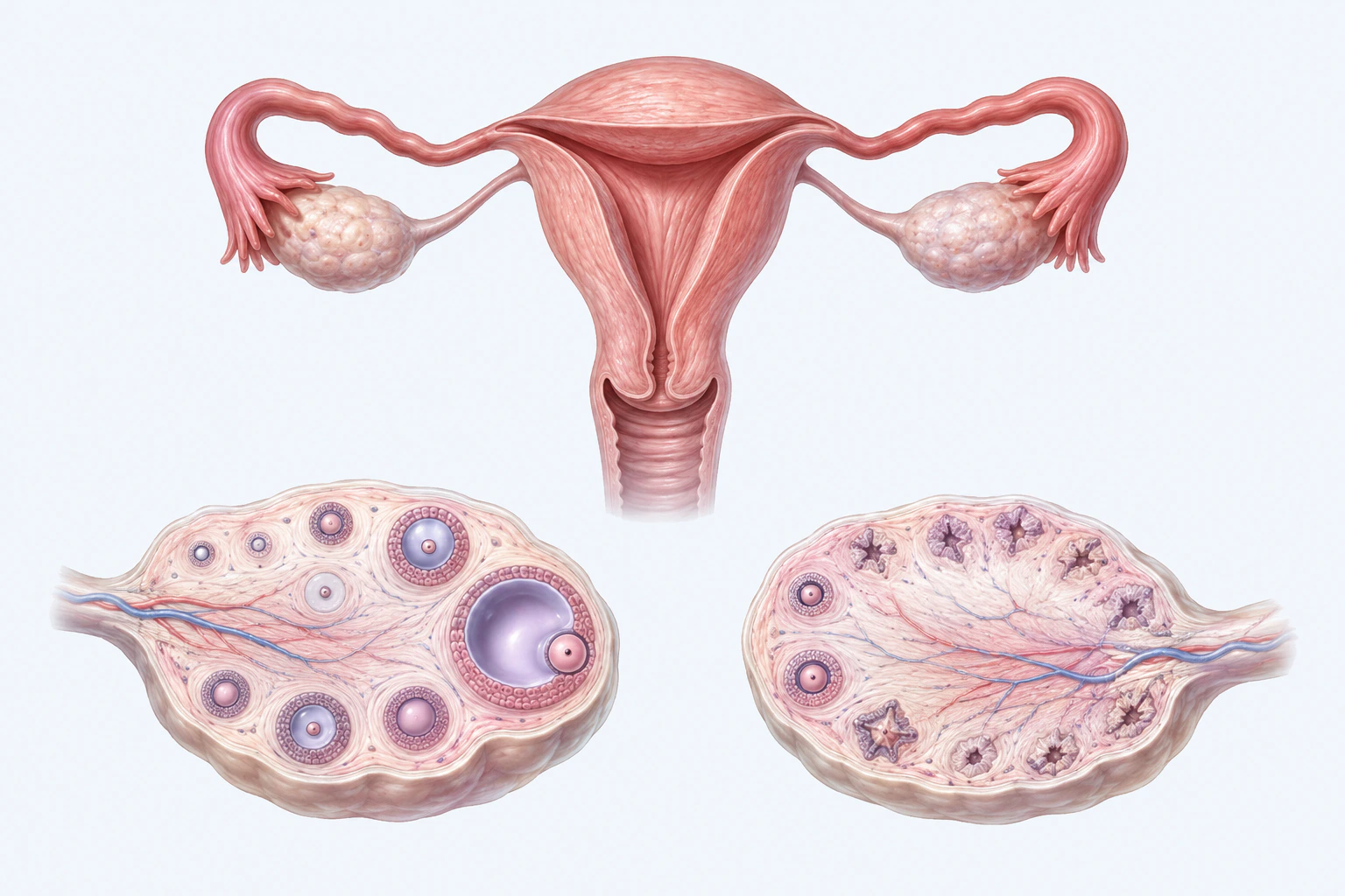
PCOS orientation using follicles rather than cysts, without follicle-count, ovarian-reserve, phenotype, endocrine-state, fertility, or diagnostic claims.
Scientific media record1 verified identifier
- Subject
- Polycystic Ovary Syndromecondition
- Identifiers
- MeSH:D011085
- Review
- Editorial review completeIdentifiers authority-verified · Accessibility validated · · pcos|pcos-pathology-v1.webp
- Digital source
- Trained-algorithmic mediaCreated with a trained generative algorithm and reviewed by WikiBiome for subject identity, scientific framing, identifiers, provenance, and accessibility.
- Scientific basis
- Polycystic Ovary Syndrome — MeSHPolycystic Ovary Syndrome
- License
- CC BY-SA 4.0Created
A metabolic-endocrine disorder affecting 6-20% of reproductive-age women worldwide, characterized by hyperandrogenism, oligo/anovulation, and polycystic ovary morphology (Rotterdam criteria).[1]Adherence to the Mediterranean Diet, Dietary Patterns and Body Composition in Women with Polycystic Ovary Syndrome (PCOS)Barrea L, Arnone A, Annunziata G et al. · 2019Open reference 1 ↓ PCOS is the leading cause of anovulatory infertility.[2]Fecal microbiota transplantation from patients with polycystic ovary syndrome induces metabolic disorders and ovarian dysfunction in germ-free miceHuang F, Deng Y, Zhou M et al. · 2024Open reference 2 ↓
From a Metallomics perspective, PCOS is among the most thoroughly studied diseases, with ~30 source pages documenting a distinctive metallomic signature of Copper elevation, toxic metal burden, antioxidant depletion, and metalloestrogen activity.
Evidence map98 cited passagesInspect provenance +
A metabolic-endocrine disorder affecting 6-20% of reproductive-age women worldwide, characterized by hyperandrogenism, oligo/anovulation, and polycystic ovary morphology (Rotterdam criteria). PCOS is the leading cause of anovulatory infertility. From a metallomics perspective, PCOS is among the most thoroughly studied diseases, with ~30 source pages document
Meta-analysis of 9 studies (n=2,274) confirmed elevated Cu with SMD = 0.51 (95% CI 0.30-0.72, p < 0.0001). When the sole contradictory study was removed, SMD increased to 0.59 with reduced heterogeneity (I2 from 78% to 43%).
Replicated in a large n=766 IVF cohort.
Confirmed in Slovenian case-control: Cu serum 1028 vs 940 mcg/L (p = 0.026).
Cu correlates with beef consumption in PCOS women (r = 0.36).
Cu's estrogen-like activity may contribute to LH and ACTH release via pituitary effects.
All four major toxic metals (As, Cd, Pb, Hg) significantly elevated in PCOS patients (p < 0.001 for all).
Strong positive intercorrelations among all four metals suggest co-exposure patterns.
Antimony (Sb) also elevated, with positive correlation to HOMA-IR and fasting glucose, suggesting a role as endocrine-disruptor.
Cd correlates positively with fasting glucose, insulin, and HOMA-IR.
nickel notably elevated: PCOS median 0.046 g/L vs controls 0.014 g/L (p < 0.05).
Ni correlates with estradiol and LH in erythrocytes of PCOS women.
Zn elevated in PCOS in (0.659 vs 0.442 g/L, p < 0.05).
Zn depleted in PCOS in (1350 vs 1598 ppb, p = 0.010).
Mn significantly elevated in PCOS: 0.0098 vs 0.0056 g/L.
First reported association: lower whole blood and serum Mo in PCOS (p = 0.024, 0.011).
SOD significantly decreased: 9.30 vs 17.39 IU/mL (p < 0.001).
GSH significantly decreased: 6.24 vs 8.09 mg/mL (p < 0.001).
TAS and TOS/OSI abnormal.
Toxic metals (As, Pb, Hg) negatively correlate with GSH levels.
Cd binds ERa with Kd nearly equivalent to estradiol (4.5 x 10^-10 M), activating ER target genes at concentrations as low as 1 uM.
Ni binds ERa noncompetitively, increasing MCF-7 cell growth 2-5 fold at 10^-9 to 10^-6 M.
Cu may also have estrogen-like activity, contributing to endocrine disruption in PCOS.
High-fiber diets and acarbose remodel gut microbiota and improve PCOS outcomes.
Showing 24 of 98 evidence-bearing passages. Every remaining citation is still indexed in the reference record below.
One disease. Five evidence layers.
A generated systems view of the metals, organisms, host sequestration signals, ecological conditions, and microbial functions indexed for Polycystic Ovary Syndrome.
Evidence layer
Taxonomic signature
Organisms reported as enriched or depleted, with their indexed functional context kept beside the name.Gram-negative pathogen — metal-dependent enzymes, estrogen deconjugation via beta-glucuronidase, siderophore production, contributes to lipopolysaccharide (LPS) burden
Gram-negative — enriched in PCOS dysbiosis, associated with elevated androgens and reduced Lactobacillus dominance
Gram-negative strict anaerobe — induced/enriched by PCOS dysbiosis, responds negatively to high-fiber + acarbose intervention
Enriched in PCOS fecal microbiota; transfers PCOS phenotype via FMT
Enriched in PCOS; associated with metabolic dysfunction in FMT transfer
Enriched in PCOS fecal microbiota
Fungal genus uniquely enriched in PCOS; distinguishes PCOS from healthy controls in multi-omics analysis
Enriched in PCOS; correlated with insulin resistance markers; inhibited by fiber intervention
Vaginal/gut SCFA producer — depleted in PCOS, associated with dysbiosis and elevated androgens; Lactobacillus-dominated microbiota protective
SCFA producer, butyrate manufacturer — depleted in PCOS dysbiosis, enriched by probiotic/prebiotic intervention; restores gut barrier function
Major butyrate producer depleted in PCOS; loss reduces SCFA-mediated insulin sensitization and anti-inflammatory signaling
Gram-negative — inhibited by high-fiber + acarbose intervention; associated with PCOS dysbiosis
Depleted in estrogen-deficient states (ovariectomy models); loss disrupts mucosal barrier and glucose metabolism
Evidence layer
Nutritional immunity
Host metal-withholding, inflammatory, antioxidant, and microbial-metabolite signals indexed in the signature.Elevated host signals
5Depleted protective signals
6Evidence layer
Ecological state
The environmental conditions that connect the organism-level observations into a system.Evidence layer
Virulence functions
Microbial structures, enzymes, and acquisition systems implicated by the linked evidence.The disease record, in full.
The original WikiBiome disease narrative remains intact beneath the generated signature atlas.
Metallomic Signature#
The Metal-Disease Matrix: A Cross-Source Synthesis identifies PCOS's metallomic profile as: nickel (Ni) ↑, copper (Cu) ↑, zinc (Zn) ↑↓, iron (Fe) ↑, lead (Pb) ↑, cadmium (Cd) ↑, mercury (Hg) ↑, As ↑.
Copper: The Most Robust Finding#
Serum copper is significantly elevated in PCOS—the single most replicated metallomic finding in this disease.
Meta-analysis of 9 studies (n=2,274) confirmed elevated copper (Cu) with SMD = 0.51 (95% CI 0.30-0.72, p < 0.0001).[3]Serum Copper Level and Polycystic Ovarian Syndrome: A Meta-AnalysisJiang Q, Zhang F, Han L et al. · 2021Open reference 3 ↓ When the sole contradictory study[4]Are Heavy Metal Exposure and Trace Element Levels Related to Metabolic and Endocrine Problems in Polycystic Ovary Syndrome?Kirmizi DA, Baser E, Turksoy VA et al. · 2020Open reference 4 ↓ was removed, SMD increased to 0.59 with reduced heterogeneity (I2 from 78% to 43%).
Replicated in a large n=766 IVF cohort.[5]Serum Copper Assessment in Patients with Polycystic Ovary Syndrome and Tubal Infertility: A Retrospective 5-Year StudyLiu Y, Zhang W, Liu Z et al. · 2024Open reference 5 ↓ Confirmed in Slovenian case-control: copper serum 1028 vs 940 mcg/L (p = 0.026).[6]Association of Trace Elements with Polycystic Ovary Syndrome in Women -- A Case-Control StudySmovrsnik T, Pinter B, Horvat M et al. · 2025Open reference 6 ↓ copper correlates with beef consumption in PCOS women (r = 0.36).[6]Association of Trace Elements with Polycystic Ovary Syndrome in Women -- A Case-Control StudySmovrsnik T, Pinter B, Horvat M et al. · 2025Open reference 6 ↓
copper's estrogen-like activity may contribute to LH and ACTH release via pituitary effects.[3]Serum Copper Level and Polycystic Ovarian Syndrome: A Meta-AnalysisJiang Q, Zhang F, Han L et al. · 2021Open reference 3 ↓
Toxic Metals: As, Cd, Pb, Hg All Elevated#
All four major toxic metals (arsenic (As), cadmium (Cd), lead (Pb), mercury (Hg)) significantly elevated in PCOS patients (p < 0.001 for all).[7]Antioxidant Status in Relation to Heavy Metals Induced Oxidative Stress in Patients with Polycystic Ovarian Syndrome (PCOS)Manal Abudawood, Hajera Tabassum, Atheer H. Alanazi et al. · 2021Open reference 7 ↓
Strong positive intercorrelations among all four metals suggest co-exposure patterns.[7]Antioxidant Status in Relation to Heavy Metals Induced Oxidative Stress in Patients with Polycystic Ovarian Syndrome (PCOS)Manal Abudawood, Hajera Tabassum, Atheer H. Alanazi et al. · 2021Open reference 7 ↓ Antimony (antimony (Sb)) also elevated, with positive correlation to HOMA-IR and fasting glucose, suggesting a role as endocrine-disruptor.[4]Are Heavy Metal Exposure and Trace Element Levels Related to Metabolic and Endocrine Problems in Polycystic Ovary Syndrome?Kirmizi DA, Baser E, Turksoy VA et al. · 2020Open reference 4 ↓
cadmium correlates positively with fasting glucose, insulin, and HOMA-IR.[4]Are Heavy Metal Exposure and Trace Element Levels Related to Metabolic and Endocrine Problems in Polycystic Ovary Syndrome?Kirmizi DA, Baser E, Turksoy VA et al. · 2020Open reference 4 ↓
Nickel Elevation#
Nickel notably elevated: PCOS median 0.046 g/L vs controls 0.014 g/L (p < 0.05).[8]Serum Micro- and Macroelements Levels in Women with Polycystic Ovary Syndrome Associated with Pelvic Inflammatory DiseaseTatarchuk TF, Kosei NV, Vetokh HV et al. · 2016Open reference 8 ↓ nickel (Ni) correlates with estradiol and LH in erythrocytes of PCOS women.[9]Levels of Trace Elements in Erythrocytes as Endocrine Disruptors in Obese and Nonobese Women with Polycystic Ovary SyndromeKamila Pokorska-Niewiada, Agnieszka Brodowska, Jacek Brodowski et al. · 2022Open reference 9 ↓
This is one of the few diseases where nickel has been directly measured and found elevated, making it critical for this wiki's nickel toxicology focus.
Zinc: Conflicting Evidence#
zinc (Zn) elevated in PCOS in[8]Serum Micro- and Macroelements Levels in Women with Polycystic Ovary Syndrome Associated with Pelvic Inflammatory DiseaseTatarchuk TF, Kosei NV, Vetokh HV et al. · 2016Open reference 8 ↓ (0.659 vs 0.442 g/L, p < 0.05). zinc depleted in PCOS in[4]Are Heavy Metal Exposure and Trace Element Levels Related to Metabolic and Endocrine Problems in Polycystic Ovary Syndrome?Kirmizi DA, Baser E, Turksoy VA et al. · 2020Open reference 4 ↓ (1350 vs 1598 ppb, p = 0.010).
The conflicting zinc findings may reflect geographic, dietary, or methodological differences; copper (Cu)/zinc ratio may be a more reliable marker than either element alone.
Manganese Elevation#
manganese (Mn) significantly elevated in PCOS: 0.0098 vs 0.0056 g/L.[8]Serum Micro- and Macroelements Levels in Women with Polycystic Ovary Syndrome Associated with Pelvic Inflammatory DiseaseTatarchuk TF, Kosei NV, Vetokh HV et al. · 2016Open reference 8 ↓
Molybdenum Depletion (Novel Finding)#
First reported association: lower whole blood and serum molybdenum (Mo) in PCOS (p = 0.024, 0.011).[6]Association of Trace Elements with Polycystic Ovary Syndrome in Women -- A Case-Control StudySmovrsnik T, Pinter B, Horvat M et al. · 2025Open reference 6 ↓ molybdenum serves as cofactor for xanthine oxidase; copper (Cu)-molybdenum antagonism (excess copper decreases molybdenum absorption) may explain this finding.
Antioxidant Collapse#
SOD significantly decreased: 9.30 vs 17.39 IU/mL (p < 0.001).[7]Antioxidant Status in Relation to Heavy Metals Induced Oxidative Stress in Patients with Polycystic Ovarian Syndrome (PCOS)Manal Abudawood, Hajera Tabassum, Atheer H. Alanazi et al. · 2021Open reference 7 ↓ GSH significantly decreased: 6.24 vs 8.09 mg/mL (p < 0.001).[7]Antioxidant Status in Relation to Heavy Metals Induced Oxidative Stress in Patients with Polycystic Ovarian Syndrome (PCOS)Manal Abudawood, Hajera Tabassum, Atheer H. Alanazi et al. · 2021Open reference 7 ↓ TAS and TOS/OSI abnormal.[4]Are Heavy Metal Exposure and Trace Element Levels Related to Metabolic and Endocrine Problems in Polycystic Ovary Syndrome?Kirmizi DA, Baser E, Turksoy VA et al. · 2020Open reference 4 ↓
Toxic metals (arsenic (As), lead (Pb), mercury (Hg)) negatively correlate with GSH levels.[7]Antioxidant Status in Relation to Heavy Metals Induced Oxidative Stress in Patients with Polycystic Ovarian Syndrome (PCOS)Manal Abudawood, Hajera Tabassum, Atheer H. Alanazi et al. · 2021Open reference 7 ↓
Metalloestrogen Connection#
Cadmium and Nickel act as Metalloestrogens, binding estrogen receptor alpha (ERa) and activating estrogen-responsive gene expression. cadmium (Cd) binds ERa with Kd nearly equivalent to estradiol (4.5 x 10^-10 M), activating ER target genes at concentrations as low as 1 uM.[10]Role of Cadmium and Nickel in Estrogen Receptor Signaling and Breast Cancer: Metalloestrogens or Not?Aquino NB, Sevigny MB, Sabangan J et al. · 2012Open reference 10 ↓
nickel (Ni) binds ERa noncompetitively, increasing MCF-7 cell growth 2-5 fold at 10^-9 to 10^-6 M.[10]Role of Cadmium and Nickel in Estrogen Receptor Signaling and Breast Cancer: Metalloestrogens or Not?Aquino NB, Sevigny MB, Sabangan J et al. · 2012Open reference 10 ↓ copper (Cu) may also have estrogen-like activity, contributing to endocrine disruption in PCOS.[3]Serum Copper Level and Polycystic Ovarian Syndrome: A Meta-AnalysisJiang Q, Zhang F, Han L et al. · 2021Open reference 3 ↓
The metalloestrogen hypothesis provides a mechanistic bridge between environmental metal exposure and PCOS's core feature of hormonal dysregulation. See Dietary Metal Paradoxes: When Healthy Foods and Good Intentions Backfire for how "healthy" plant-based foods simultaneously increase metalloestrogen exposure.
Gut Microbiome Connection#
PCOS is increasingly recognized as a disease with a strong Gut Microbiome component. High-fiber diets and acarbose remodel gut microbiota and improve PCOS outcomes.[11]High-Fiber Diet or Combined With Acarbose Alleviates Heterogeneous Phenotypes of Polycystic Ovary Syndrome by Regulating Gut MicrobiotaWang X, Xu T, Liu R et al. · 2022Open reference 11 ↓ Probiotics significantly decrease FPG, FBI, TG, and inflammatory markers (CRP, MDA) across multiple meta-analyses.[12]The effects of probiotics, prebiotics, and synbiotics on polycystic ovarian syndrome: an overview of systematic reviewsAngoorani P, Ejtahed H-S, Ettehad Marvasti F et al. · 2023Open reference 12 ↓
Prebiotics decrease BMI, waist circumference, and FPG more effectively than probiotics alone.[12]The effects of probiotics, prebiotics, and synbiotics on polycystic ovarian syndrome: an overview of systematic reviewsAngoorani P, Ejtahed H-S, Ettehad Marvasti F et al. · 2023Open reference 12 ↓
Vitamin D + probiotic co-supplementation (L. acidophilus, B. bifidum, L. reuteri, L. fermentum) for 12 weeks significantly reduced testosterone, hirsutism, hs-CRP, and MDA while increasing TAC and GSH.[13]Vitamin D and probiotic co-supplementation affects mental health, hormonal, inflammatory and oxidative stress parameters in women with polycystic ovary syndromeOstadmohammadi V, Jamilian M, Bahmani F et al. · 2019Open reference 13 ↓
FMT transfer of the PCOS microbiome to germ-free mice induced insulin resistance, ovarian dysfunction, and obese-like phenotype—direct causal evidence that gut Dysbiosis drives PCOS pathology.[2]Fecal microbiota transplantation from patients with polycystic ovary syndrome induces metabolic disorders and ovarian dysfunction in germ-free miceHuang F, Deng Y, Zhou M et al. · 2024Open reference 2 ↓
Combined bacteriome-mycobiome-metabolome analysis reveals hyperandrogenemia as a central driver of PCOS dysbiosis, with Mortierella enrichment distinguishing PCOS from healthy controls.[14]Alterations of bacteriome, mycobiome and metabolome characteristics in PCOS patients with normal/overweight individualsYin G, Chen F, Chen G et al. · 2022Open reference 14 ↓
Lactobacillus depletion and altered Bifidobacterium abundance are replicated findings; restoration with specific probiotic strains improves androgen and metabolic markers.[13]Vitamin D and probiotic co-supplementation affects mental health, hormonal, inflammatory and oxidative stress parameters in women with polycystic ovary syndromeOstadmohammadi V, Jamilian M, Bahmani F et al. · 2019Open reference 13 ↓[15]Probiotics and Polycystic Ovary Syndrome: A Perspective for Management in Adolescents with ObesityCalcaterra V, Rossi V, Massini G et al. · 2023Open reference 15 ↓
The metal-microbiome interaction is bidirectional: toxic metals reshape gut flora (favoring metal-tolerant species), and dysbiotic gut flora impairs metal detoxification and antioxidant capacity.
Environmental Metal Exposure Links#
PCOS women show elevated serum levels of multiple environmental contaminants simultaneously, with strong positive intercorrelations suggesting common exposure sources.[7]Antioxidant Status in Relation to Heavy Metals Induced Oxidative Stress in Patients with Polycystic Ovarian Syndrome (PCOS)Manal Abudawood, Hajera Tabassum, Atheer H. Alanazi et al. · 2021Open reference 7 ↓
Dietary sources dominate: beef consumption predicts copper (Cu) levels; cereals and boiled vegetables predict molybdenum (Mo) levels.[6]Association of Trace Elements with Polycystic Ovary Syndrome in Women -- A Case-Control StudySmovrsnik T, Pinter B, Horvat M et al. · 2025Open reference 6 ↓ Nickel-containing foods (legumes, whole grains, cocoa, nuts) are among the most common exposure routes.[8]Serum Micro- and Macroelements Levels in Women with Polycystic Ovary Syndrome Associated with Pelvic Inflammatory DiseaseTatarchuk TF, Kosei NV, Vetokh HV et al. · 2016Open reference 8 ↓
mercury (Hg) positively correlates with fasting blood sugar and HbA1c; cadmium (Cd) correlates with total cholesterol—linking specific metals to specific metabolic derangements.[7]Antioxidant Status in Relation to Heavy Metals Induced Oxidative Stress in Patients with Polycystic Ovarian Syndrome (PCOS)Manal Abudawood, Hajera Tabassum, Atheer H. Alanazi et al. · 2021Open reference 7 ↓
Dietary Metal Paradoxes#
Full evidence is maintained on the canonical paradox article.
Insulin Resistance as Central Mediator#
Insulin resistance (IR) is the metabolic hub connecting metal exposure to PCOS pathology.
antimony (Sb) and cadmium (Cd) both correlate positively with HOMA-IR.[4]Are Heavy Metal Exposure and Trace Element Levels Related to Metabolic and Endocrine Problems in Polycystic Ovary Syndrome?Kirmizi DA, Baser E, Turksoy VA et al. · 2020Open reference 4 ↓ mercury (Hg) correlates with fasting blood sugar and HbA1c.[7]Antioxidant Status in Relation to Heavy Metals Induced Oxidative Stress in Patients with Polycystic Ovarian Syndrome (PCOS)Manal Abudawood, Hajera Tabassum, Atheer H. Alanazi et al. · 2021Open reference 7 ↓ Probiotics and dietary interventions improve PCOS outcomes primarily through IR reduction.[12]The effects of probiotics, prebiotics, and synbiotics on polycystic ovarian syndrome: an overview of systematic reviewsAngoorani P, Ejtahed H-S, Ettehad Marvasti F et al. · 2023Open reference 12 ↓
Gut microbiome transfer recapitulates the insulin-resistant, obese, anovulatory phenotype in germ-free mice, implicating microbiome-insulin signaling as causal.[2]Fecal microbiota transplantation from patients with polycystic ovary syndrome induces metabolic disorders and ovarian dysfunction in germ-free miceHuang F, Deng Y, Zhou M et al. · 2024Open reference 2 ↓
Metal-induced Oxidative Stress impairs insulin signaling, creating a vicious cycle: oxidative stress -> IR -> hyperandrogenism -> anovulation.[7]Antioxidant Status in Relation to Heavy Metals Induced Oxidative Stress in Patients with Polycystic Ovarian Syndrome (PCOS)Manal Abudawood, Hajera Tabassum, Atheer H. Alanazi et al. · 2021Open reference 7 ↓
Current Interventions with Metal Relevance#
| Intervention | Evidence | Metal Mechanism |
|---|---|---|
| Mediterranean diet | Strong (RCT + observational)[1]Adherence to the Mediterranean Diet, Dietary Patterns and Body Composition in Women with Polycystic Ovary Syndrome (PCOS)Barrea L, Arnone A, Annunziata G et al. · 2019Open reference 1 ↓ | Anti-inflammatory; improves copper (Cu)/zinc (Zn) ratio; antioxidant-rich |
| Ketogenic/low-carb diet | Moderate (pilot RCTs)[17]Ketogenic diet improves fertility in patients with polycystic ovary syndrome: a brief reportTsushima Y, Nachawi N, Pantalone KM et al. · 2024Open reference 17 ↓ | Reduces insulin resistance; may lower metal absorption |
| Probiotics | Strong (8 meta-analyses)[12]The effects of probiotics, prebiotics, and synbiotics on polycystic ovarian syndrome: an overview of systematic reviewsAngoorani P, Ejtahed H-S, Ettehad Marvasti F et al. · 2023Open reference 12 ↓ | Metal sequestration; gut barrier repair; antioxidant restoration |
| Vitamin D + probiotics | Strong (RCT)[13]Vitamin D and probiotic co-supplementation affects mental health, hormonal, inflammatory and oxidative stress parameters in women with polycystic ovary syndromeOstadmohammadi V, Jamilian M, Bahmani F et al. · 2019Open reference 13 ↓ | Restores GSH/TAC; reduces MDA; lowers CRP |
| Zinc supplementation | Moderate (extrapolated from related conditions) | Counters copper excess; restores copper/zinc superoxide dismutase (Cu/Zn-SOD); competes with cadmium (Cd) for binding sites |
| High-fiber diet | Moderate[11]High-Fiber Diet or Combined With Acarbose Alleviates Heterogeneous Phenotypes of Polycystic Ovary Syndrome by Regulating Gut MicrobiotaWang X, Xu T, Liu R et al. · 2022Open reference 11 ↓ | Microbiota remodeling; SCFA production; but increases nickel (Ni)/cadmium exposure |
| Low-nickel diet | Theoretical (no PCOS-specific trials) | Would reduce metalloestrogen burden and nickel-driven Metal-Driven Inflammation |
Open Questions#
Unresolved questions identified by the current evidence record.
01Does nickel sensitization modify PCOS severity?+
Nickel allergy affects up to 17.6% of women; no study has examined whether nickel-allergic PCOS patients have worse outcomes or respond differently to dietary intervention.
02Is the copper (Cu)/zinc (Zn) ratio a better biomarker than either element alone?+
Conflicting zinc (Zn) results suggest the ratio captures the relevant biology more reliably.
03Can low-nickel diets improve PCOS outcomes?+
Given nickel (Ni) elevation[8]Serum Micro- and Macroelements Levels in Women with Polycystic Ovary Syndrome Associated with Pelvic Inflammatory DiseaseTatarchuk TF, Kosei NV, Vetokh HV et al. · 2016Open reference 8 ↓ and metalloestrogen activity,[10]Role of Cadmium and Nickel in Estrogen Receptor Signaling and Breast Cancer: Metalloestrogens or Not?Aquino NB, Sevigny MB, Sabangan J et al. · 2012Open reference 10 ↓ this is a testable hypothesis with no current trial data.
04Does antimony (antimony (Sb)) deserve more attention?+
Only one study measured it,[4]Are Heavy Metal Exposure and Trace Element Levels Related to Metabolic and Endocrine Problems in Polycystic Ovary Syndrome?Kirmizi DA, Baser E, Turksoy VA et al. · 2020Open reference 4 ↓ but its correlation with HOMA-IR is provocative.
05What is the optimal probiotic strain for metal-burdened PCOS patients?+
Metal-sequestering Lactobacillus strains exist but have not been tested specifically in PCOS.
06Causal direction: Are elevated metals a cause of PCOS, a consequence of metabolic dysfunction, or both?+
Longitudinal and Mendelian randomization studies are needed.
Comorbidities#
Type 2 Diabetes—PCOS women have 4-8x increased T2D risk; shared insulin resistance as the central metabolic defect; shared cadmium (Cd) and mercury (Hg) correlations with HOMA-IR and fasting glucose; shared gut dysbiosis with SCFA producer depletion; metformin treats both.
Cardiovascular Disease—PCOS is an independent CVD risk factor; shared metabolic inflammation, dyslipidemia, and TMAO pathway activation; copper (Cu) elevation in both conditions; shared endothelial dysfunction from oxidative stress.
Obesity—40-80% of PCOS patients are overweight/obese; adipose tissue aromatase amplifies hyperandrogenism; shared insulin resistance and gut dysbiosis; weight loss improves ovulation, androgen levels, and metabolic parameters.
Depression—depression prevalence is 2-3x higher in PCOS; shared tryptophan pathway disruption and gut-brain axis dysfunction; hyperandrogenism-driven hirsutism and acne contribute to body image distress; shared inflammatory markers (elevated CRP, IL-6).
Endometriosis—20% comorbidity rate; both are estrogen-related conditions with shared metalloestrogen exposure (cadmium, nickel (Ni)); opposing estrogen dynamics (excess in endometriosis vs dysregulated in PCOS) may reflect different manifestations of the same metalloestrogen disruption.
Connections#
- Metals: Copper, Nickel, Zinc, Cadmium, Lead, Mercury, Arsenic, Manganese
- Concepts: Metalloestrogens, oxidative stress, Insulin Resistance, Mis-Metallation
- Analyses: Metal-Disease Matrix: A Cross-Source Synthesis, Dietary Metal Paradoxes: When Healthy Foods and Good Intentions Backfire
- Related diseases: Breast Cancer (shared metalloestrogen pathway), Type 2 Diabetes (shared insulin resistance and metal signature), Autism Spectrum Disorder (shared zinc (Zn) depletion and toxic metal burden)
- Pathogens: Indirect—gut dysbiosis patterns overlap with metal-driven dysbiosis described in
- Interventions: Mediterranean Diet, Probiotics, Ketogenic Diet, Zinc Supplementation
- Prebiotics—prebiotic fiber supports gut microbiome restoration and insulin sensitization in PCOS
- Environmental Metal Exposure—dietary and cosmetic metal exposures contribute to PCOS metal burden
- Biomarkers—copper (Cu)/zinc ratio, toxic metal panels, and hormonal markers for PCOS diagnosis and monitoring
- The Exposome—PCOS as a disease shaped by the full exposome: dietary metals, endocrine disruptors, and microbiome
References 25
Numbered by first appearance in the article, then reconciled with its declared source list.
- 1
Barrea L, Arnone A, Annunziata G et al. (2019). Adherence to the Mediterranean Diet, Dietary Patterns and Body Composition in Women with Polycystic Ovary Syndrome (PCOS). Nutrients.
- 2
Huang F, Deng Y, Zhou M et al. (2024). Fecal microbiota transplantation from patients with polycystic ovary syndrome induces metabolic disorders and ovarian dysfunction in germ-free mice. BMC Microbiology.
- 3
Jiang Q, Zhang F, Han L et al. (2021). Serum Copper Level and Polycystic Ovarian Syndrome: A Meta-Analysis. Gynecologic and Obstetric Investigation.
- 4
★Kirmizi DA, Baser E, Turksoy VA et al. (2020). Are Heavy Metal Exposure and Trace Element Levels Related to Metabolic and Endocrine Problems in Polycystic Ovary Syndrome?. Biological Trace Element Research.
- 5
Liu Y, Zhang W, Liu Z et al. (2024). Serum Copper Assessment in Patients with Polycystic Ovary Syndrome and Tubal Infertility: A Retrospective 5-Year Study. Food Science & Nutrition.
- 6
Smovrsnik T, Pinter B, Horvat M et al. (2025). Association of Trace Elements with Polycystic Ovary Syndrome in Women -- A Case-Control Study. Metabolites.
- 7
Manal Abudawood, Hajera Tabassum, Atheer H. Alanazi et al. (2021). Antioxidant Status in Relation to Heavy Metals Induced Oxidative Stress in Patients with Polycystic Ovarian Syndrome (PCOS). Scientific Reports.
- 8
Tatarchuk TF, Kosei NV, Vetokh HV et al. (2016). Serum Micro- and Macroelements Levels in Women with Polycystic Ovary Syndrome Associated with Pelvic Inflammatory Disease. Reproductive Endocrinology.
- 9
Kamila Pokorska-Niewiada, Agnieszka Brodowska, Jacek Brodowski et al. (2022). Levels of Trace Elements in Erythrocytes as Endocrine Disruptors in Obese and Nonobese Women with Polycystic Ovary Syndrome. International Journal of Environmental Research and Public Health.
- 10
★Aquino NB, Sevigny MB, Sabangan J et al. (2012). Role of Cadmium and Nickel in Estrogen Receptor Signaling and Breast Cancer: Metalloestrogens or Not?. Journal of Environmental Science and Health Part C - Environmental Carcinogenesis and Ecotoxicology Reviews.
- 11
Wang X, Xu T, Liu R et al. (2022). High-Fiber Diet or Combined With Acarbose Alleviates Heterogeneous Phenotypes of Polycystic Ovary Syndrome by Regulating Gut Microbiota. Frontiers in Endocrinology.
- 12
Angoorani P, Ejtahed H-S, Ettehad Marvasti F et al. (2023). The effects of probiotics, prebiotics, and synbiotics on polycystic ovarian syndrome: an overview of systematic reviews. Frontiers in Medicine.
- 13
Ostadmohammadi V, Jamilian M, Bahmani F et al. (2019). Vitamin D and probiotic co-supplementation affects mental health, hormonal, inflammatory and oxidative stress parameters in women with polycystic ovary syndrome. Journal of Ovarian Research.
- 14
Yin G, Chen F, Chen G et al. (2022). Alterations of bacteriome, mycobiome and metabolome characteristics in PCOS patients with normal/overweight individuals. Journal of Ovarian Research.
- 15
Calcaterra V, Rossi V, Massini G et al. (2023). Probiotics and Polycystic Ovary Syndrome: A Perspective for Management in Adolescents with Obesity. Nutrients.
- 16
Mavropoulos JC, Yancy WS, Hepburn J et al. (2005). The effects of a low-carbohydrate, ketogenic diet on the polycystic ovary syndrome: A pilot study. Nutrition & Metabolism.
- 17
Tsushima Y, Nachawi N, Pantalone KM et al. (2024). Ketogenic diet improves fertility in patients with polycystic ovary syndrome: a brief report. Frontiers in Nutrition.
- 18
Leung WT, Tang Z, Feng Y et al. (2022). Lower Fiber Consumption in Women with Polycystic Ovary Syndrome: A Meta-Analysis of Observational Studies. Nutrients.
- 19
Kurdoglu Z, Kurdoglu M, Demir H et al. (2012). Serum Trace Elements and Heavy Metals in Polycystic Ovary Syndrome. Human & Experimental Toxicology.
- 20
Sarah H. Mhaibes, Mohammed A. Taher, Ala H. Badr (2017). A Comparative Study of Blood Levels of Manganese, Some Macroelements and Heavy Metals in Obese and Non-Obese Polycystic Ovary Syndrome Patients. Iraqi Journal of Pharmaceutical Sciences.
- 21
Mei S, Ding J, Wang K et al. (2022). Mediterranean Diet Combined With a Low-Carbohydrate Dietary Pattern in the Treatment of Overweight Polycystic Ovary Syndrome Patients. Frontiers in Nutrition.
- 22
Cutler DA, Pride SM, Cheung AP (2019). Low intakes of dietary fiber and magnesium are associated with insulin resistance and hyperandrogenism in polycystic ovary syndrome: A cohort study. Food Science & Nutrition.
- 23
Smovrsnik T, Virant-Klun I, Pinter B (2023). Heavy Metals and Essential Elements in Association with Oxidative Stress in Women with Polycystic Ovary Syndrome -- A Systematic Review. Antioxidants.
- 24
Song He, Hao Li, Zehui Yu et al. (2021). The Gut Microbiome and Sex Hormone-Related Diseases. Frontiers in Microbiology.
- 25
Georgina Quaranta, Mauro Pittiruti, Brunella Posteraro et al. (2019). Quaranta 2019 — FMT as a Potential Tool for Female Reproductive Tract Diseases (Review). Frontiers in Immunology.
Article network
Mentioned here 18
Pages linking here 56
Connect the evidence
Publicly readable discussion by ORCID-authenticated researchers. Questions, interpretation, methods, corrections, and new evidence stay attached to this record.
No discussion yet. Start with a precise question or a source-backed observation.
Activity and accepted changes
Accepted researcher context, editorial status, public discussion, and upstream Git revisions are shown together. Pending, declined, and withdrawn proposals remain private.
- published revision
Strengthen Metallomics and link high-leverage contexts
Karen Pendergrass · +1 −1
Inspect exact Git diff ↗ - published revision
Backfill oxidative stress concept links
Karen Pendergrass · +2 −2
Inspect exact Git diff ↗ - published revision
Backfill gut microbiome concept links
Karen Pendergrass · +1 −1
Inspect exact Git diff ↗ - published revision
Backfill inflammation concept links
Karen Pendergrass · +1 −1
Inspect exact Git diff ↗ - published revision
Complete corpus-wide Dysbiosis linking
Karen Pendergrass · +1 −1
Inspect exact Git diff ↗ - published revision
massive wiki expansion: 149 stubs fixed, 100+ new pages, Rule 15 scan, keystone papers
WikiBiome Deploy Bot · +14 −14
Inspect exact Git diff ↗ - published revision
ingest: quaranta-2019-fmt-female-reproductive-tract-diseases
WikiBiome Deploy Bot · +2 −2
Inspect exact Git diff ↗ - published revision
ingest: 87 papers (17 endometriosis, 67 erectile dysfunction, 3 cross-condition)
WikiBiome Deploy Bot · +1 −1
Inspect exact Git diff ↗ - published revision
nightly maintenance: 94 stub demotions, 181 source_count fixes, 22 auto-discovered stubs, 5 adversarial audits, 3 boundary fixes, 3 evidence-level corrections
WikiBiome Deploy Bot · +1 −0
Inspect exact Git diff ↗ - published revision
semantic integrity pass: boundary fixes, 10 interventions, 31 STOPs, 2 supersessions, keystone revalidation
WikiBiome Deploy Bot · +1 −1
Inspect exact Git diff ↗ - published revision
cycle 1: health check + lint fixes + 8 ingests + 2 stubs + gestational-diabetes signature
WikiBiome Deploy Bot · +1 −0
Inspect exact Git diff ↗ - published revision
Batch: fix 1025 broken wikilinks, wire 13 STOP pages, deepen PPD/GERD/T1D/8 microbes
WikiBiome Deploy Bot · +3 −0
Inspect exact Git diff ↗ - published revision
Deep citation pass on 10 disease entities + expand 3 thin entities
WikiBiome Deploy Bot · +6 −2
Inspect exact Git diff ↗ - published revision
Deepen metal/concept entities + 8 new sources for T1D/schizophrenia
WikiBiome Deploy Bot · +296 −0
Inspect exact Git diff ↗ - published revision
WikiBiome update — 2026-04-15 17:23
WikiBiome Deploy Bot · +2 −3
Inspect exact Git diff ↗ - published revision
v2 migration Priority 2: All 29 disease entity pages upgraded with associated_conditions, seo_target, wikipedia_differentiation
WikiBiome Deploy Bot · +6 −0
Inspect exact Git diff ↗