

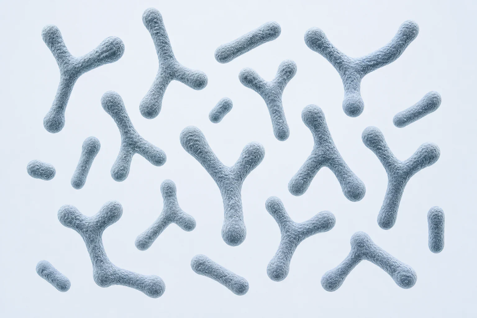
Representative Bifidobacterium morphology, including irregular bifid and club-shaped rods. Cell shape varies by species and culture conditions; this is an educational reconstruction, not a micrograph.
Scientific media record1 verified identifier
- Subject
- Bifidobacteriumtaxon · genus
- Identifiers
- NCBITaxon:1678
- Review
- Editorial review completeIdentifiers authority-verified · Accessibility validated · · bifidobacterium|bifidobacterium-morphology-v1.webp
- Digital source
- Trained-algorithmic mediaCreated with a trained generative algorithm and reviewed by WikiBiome for subject identity, scientific framing, identifiers, provenance, and accessibility.
- Scientific basis
- Bifidobacterium — NCBI TaxonomyBifid Shape Is Intrinsic to Bifidobacterium adolescentis
- License
- CC BY-SA 4.0Created
A genus of Gram-positive, obligate anaerobic bacteria that dominate the infant Gut Microbiome and remain important commensals throughout life.
Bifidobacterium species are distinctive in the Metallomics context because some species possess nickel (Ni)-dependent Urease—an unusual feature for a commensal genus—while the genus as a whole functions as a key probiotic with metal-binding and detoxification properties.
Evidence map18 cited passagesInspect provenance +
Some Bifidobacterium species carry urease genes and produce active Ni-dependent urease.
This commensal urease activity has implications for nickel restriction strategies: dietary nickel limitation aimed at pathogen urease could also affect beneficial Bifidobacterium urease, potentially causing unintended dysbiosis.
Bifidobacterium species demonstrate metal-binding capacity at the cell surface, contributing to heavy metal sequestration in the gut lumen.
B. longum, B. breve, and B. lactis have been studied for cadmium, lead, and mercury binding capacity.
Bifidobacterium (including B. longum subsp. infantis, B. breve, and B. longum) can dominate breastfed infant gut communities; the cited review used the historical shorthand “B. infantis”.
Human milk oligosaccharides (HMOs) selectively feed Bifidobacterium, establishing early colonization dominance.
This dominance creates an acid-producing, nickel-independent microbial environment that naturally suppresses Ni-enzyme-dependent pathogens.
Formula-fed infants have lower Bifidobacterium and higher Proteobacteriaceae—a shift compounded by formula's higher nickel content.
The convergence of reduced Bifidobacterium, increased dietary nickel, and enrichment of Ni-urease pathogens in formula-fed preterm infants may explain NEC susceptibility.
Probiotic supplementation with Bifidobacterium (often combined with Lactobacillus) reduces NEC incidence in very low birth weight preterm infants.
Obesity and metabolic syndrome: inversely correlated with BMI; depleted in metabolic dysfunction.
Type 1 diabetes: depleted in children progressing to T1D.
Autism spectrum disorder: multiple studies report altered Bifidobacterium in ASD.
Long COVID: depleted in the gut microbiome of Long COVID patients; Mendelian randomization supports a causal relationship.
Necrotizing enterocolitis: depleted in preterm infants who develop NEC, alongside reduced SCFA producers.
Iron supplementation effects: excess iron in infant formula may suppress Bifidobacterium while promoting Enterobacteriaceae.
Acetate production strengthens gut barrier integrity and provides substrate for butyrate production by cross-feeding partners like faecalibacterium prausnitzii.
Competes with pathogens for ecological niches without requiring nickel-dependent virulence factors.
Contents
1. Nickel-Dependent Urease in Select Species2. Metal Binding and Detoxification3. Infant Gut Colonization4. Depletion Across Disease States5. SCFA Production and Immune Modulation6. ConnectionsNickel-Dependent Urease in Select Species#
Some Bifidobacterium species carry urease genes and produce active nickel (Ni)-dependent urease.[1]Role of Nickel in Microbial PathogenesisRobert J. Maier, Stéphane L. Benoit · 2019Open reference 1 ↓
In the commensal context, urease likely serves for nitrogen acquisition (urea is abundant in the gut lumen at ~2-6 mM) rather than as a virulence factor.
This commensal urease activity has implications for nickel restriction strategies: dietary nickel limitation aimed at pathogen urease could also affect beneficial Bifidobacterium urease, potentially causing unintended Dysbiosis.[1]Role of Nickel in Microbial PathogenesisRobert J. Maier, Stéphane L. Benoit · 2019Open reference 1 ↓
This dual-use problem—pathogen vs. commensal urease—is a key challenge for anti-nickel therapeutic approaches.
Metal Binding and Detoxification#
Bifidobacterium species demonstrate metal-binding capacity at the cell surface, contributing to heavy metal sequestration in the gut lumen.[2]Gut Microbiota: A Target for Heavy Metal Toxicity and a Probiotic Protective StrategyHui Duan, Leilei Yu, Fengwei Tian et al. · 2020Open reference 2 ↓
Cell wall peptidoglycan and exopolysaccharides provide carboxyl and phosphoryl groups that chelate divalent metal cations.
B. longum, B. breve, and B. lactis have been studied for cadmium, lead, and mercury binding capacity.[3]Heavy Metal-Gut Microbiota Interactions: Probiotics Modulation and Biosensors DetectionLiliana Anchidin-Norocel, Oana C. Iatcu, Andrei Lobiuc et al. · 2025Open reference 3 ↓
Combined with Lactobacillus, Bifidobacterium forms the core of traditional probiotic metal detoxification strategies.
Infant Gut Colonization#
Bifidobacterium (including B. longum subsp. infantis, B. breve, and B. longum) can dominate breastfed infant gut communities; the cited review used the historical shorthand “B. infantis”.[4]Torrazza 2013 — Intestinal Microbial Ecology and Environmental Factors Affecting NECTorrazza RM, Ukhanova M, Wang X et al. · 2013Open reference 4 ↓
Human milk oligosaccharides (HMOs) selectively feed Bifidobacterium, establishing early colonization dominance.[5]Sami 2023 — Human Milk Nutrients Preventing NECSami et al. · 2023Open reference 5 ↓[6]Chapman 2026 — Non-toxigenic Clostridia Metabolize HMOs and Suppress Pathobionts in NECChapman et al. · 2026Open reference 6 ↓
This dominance creates an acid-producing, nickel-independent microbial environment that naturally suppresses nickel (Ni)-enzyme-dependent pathogens.[7]Nickel as a Catalytic Driver of Necrotizing Enterocolitis: Dietary Nickel, Microbial Metallomics, and the Activation of Nickel-Dependent Virulence Pathways in the Preterm GutKaren Pendergrass · 2026Open reference 7 ↓ Formula-fed infants have lower Bifidobacterium and higher Proteobacteriaceae—a shift compounded by formula's higher nickel content.[4]Torrazza 2013 — Intestinal Microbial Ecology and Environmental Factors Affecting NECTorrazza RM, Ukhanova M, Wang X et al. · 2013Open reference 4 ↓
The convergence of reduced Bifidobacterium, increased dietary nickel, and enrichment of nickel-urease pathogens in formula-fed preterm infants may explain NEC susceptibility.[7]Nickel as a Catalytic Driver of Necrotizing Enterocolitis: Dietary Nickel, Microbial Metallomics, and the Activation of Nickel-Dependent Virulence Pathways in the Preterm GutKaren Pendergrass · 2026Open reference 7 ↓
Probiotic supplementation with Bifidobacterium (often combined with Lactobacillus) reduces NEC incidence in very low birth weight preterm infants.[8]Zhou 2023 — Probiotics Prevent NEC in VLBW (Network Meta-Analysis)Zhou et al. · 2023Open reference 8 ↓
Depletion Across Disease States#
IBD: reduced in both Crohn's disease and ulcerative colitis.
Obesity and metabolic syndrome: inversely correlated with BMI; depleted in metabolic dysfunction.[9]Heavy Metals, Microbial Metallomics, and the US Obesity Epidemic: A Mechanistic Examination of a Population-Level Metabolic DisruptionKaren Pendergrass · 2026Open reference 9 ↓[10]Ismail 2022 — Does the Gut Microbiome Play a Role in Obesity in Type 1 Diabetes? Unanswered Questions and ReviewHeba M. Ismail, Carmella Evans-Molina · 2022Open reference 10 ↓
Type 1 diabetes: depleted in children progressing to T1D.[11]Belteky 2023 — Infant Gut Microbiome Composition Correlated with Type 1 Diabetes Acquisition: The ABIS StudyMalin Belteky, Patricia L. Milletich, Angelica P. Ahrens et al. · 2023Open reference 11 ↓[12]de Goffau 2014 — Aberrant Gut Microbiota Composition at the Onset of Type 1 Diabetes in Young ChildrenMarcus C. de Goffau, Susana Fuentes, Bartholomeus van den Bogert et al. · 2014Open reference 12 ↓
Autism spectrum disorder: multiple studies report altered Bifidobacterium in ASD.[13]Zhang 2022 — Probiotics in Children with ASD: RCT Study ProtocolLingling Zhang, Yiran Xu, Hongwei Li et al. · 2022Open reference 13 ↓
Long COVID: depleted in the gut microbiome of Long COVID patients; Mendelian randomization supports a causal relationship.[14]Li et al 2024 — The Causal Role of Gut Microbiota in Susceptibility of Long COVID: A Mendelian Randomization StudyZuming Li, Qinghua Xia, Jieni Feng et al. · 2024Open reference 14 ↓[15]Rego & Araújo-Filho 2024 — The Impact of Gut Microbiota on Long COVID: Insights and ChallengesAmália Cinthia Meneses do Rêgo, Irami Araújo-Filho · 2024Open reference 15 ↓
Necrotizing enterocolitis: depleted in preterm infants who develop NEC, alongside reduced SCFA producers.[16]Liu 2022 — Gut microbiota and SCFAs as early predictive biomarkers for neonatal NEC (pilot)Xiao-Chen Liu, Ting-Ting Du, Xiong Gao et al. · 2022Open reference 16 ↓[17]Lin 2025 — Integrated serum metabolomics and fecal microbiome in NEC infantsZhi-ying Lin, Shan-shan He, Zi-tong Mo et al. · 2025Open reference 17 ↓
Allergic disease: early-life Bifidobacterium depletion associated with increased allergy risk. Iron supplementation effects: excess iron in infant formula may suppress Bifidobacterium while promoting Enterobacteriaceae.[18]Mechanism of Iron Ion Homeostasis in Intestinal Immunity and Gut Microbiota RemodelingHonghong Bao, Yi Wang, Hanlin Xiong et al. · 2024Open reference 18 ↓
SCFA Production and Immune Modulation#
Produces acetate and lactate via the "bifid shunt" (fructose-6-phosphate phosphoketolase pathway).
Acetate production strengthens gut barrier integrity and provides substrate for Butyrate production by cross-feeding partners like Faecalibacterium prausnitzii.[3]Heavy Metal-Gut Microbiota Interactions: Probiotics Modulation and Biosensors DetectionLiliana Anchidin-Norocel, Oana C. Iatcu, Andrei Lobiuc et al. · 2025Open reference 3 ↓
Promotes regulatory T cell development and anti-inflammatory IL-10 production. Competes with pathogens for ecological niches without requiring nickel-dependent virulence factors.[2]Gut Microbiota: A Target for Heavy Metal Toxicity and a Probiotic Protective StrategyHui Duan, Leilei Yu, Fengwei Tian et al. · 2020Open reference 2 ↓
Connections#
- Urease—nickel (Ni)-urease in some species; complicates anti-nickel strategies
- Nickel—some species nickel-dependent via urease; the commensal collateral damage concern
- Gut-Metal-Microbiome Interactions—metal-binding probiotic and metal-sensitive commensal
- Iron—suppressed by excess iron supplementation
- Lactobacillus—complementary probiotic genus; co-depleted in disease
- Faecalibacterium prausnitzii—metabolic cross-feeding via acetate-butyrate axis
- Akkermansia muciniphila—co-protective commensal; both depleted in disease
- Nutritional Immunity (Metal Sequestration)—the commensal-pathogen urease dilemma
- dysbiosis—depletion is a consistent disease-associated signature
- Metal-Driven Inflammation—anti-inflammatory via SCFA production and Treg promotion
References 18
Numbered by first appearance in the article, then reconciled with its declared source list.
- 1
★Robert J. Maier, Stéphane L. Benoit (2019). Role of Nickel in Microbial Pathogenesis. Inorganics.
- 2
★Hui Duan, Leilei Yu, Fengwei Tian et al. (2020). Gut Microbiota: A Target for Heavy Metal Toxicity and a Probiotic Protective Strategy. Science of the Total Environment.
- 3
★Liliana Anchidin-Norocel, Oana C. Iatcu, Andrei Lobiuc et al. (2025). Heavy Metal-Gut Microbiota Interactions: Probiotics Modulation and Biosensors Detection. Biosensors.
- 4
Torrazza RM, Ukhanova M, Wang X et al. (2013). Torrazza 2013 — Intestinal Microbial Ecology and Environmental Factors Affecting NEC. PLoS ONE.
- 5
Sami et al. (2023). Sami 2023 — Human Milk Nutrients Preventing NEC. Frontiers in Pediatrics.
- 6
Chapman et al. (2026). Chapman 2026 — Non-toxigenic Clostridia Metabolize HMOs and Suppress Pathobionts in NEC. Nature Microbiology.
- 7
Karen Pendergrass (2026). Nickel as a Catalytic Driver of Necrotizing Enterocolitis: Dietary Nickel, Microbial Metallomics, and the Activation of Nickel-Dependent Virulence Pathways in the Preterm Gut. Zenodo Preprint.
- 8
Zhou et al. (2023). Zhou 2023 — Probiotics Prevent NEC in VLBW (Network Meta-Analysis). Frontiers in Pediatrics.
- 9
★Karen Pendergrass (2026). Heavy Metals, Microbial Metallomics, and the US Obesity Epidemic: A Mechanistic Examination of a Population-Level Metabolic Disruption. Zenodo Preprint.
- 10
Heba M. Ismail, Carmella Evans-Molina (2022). Ismail 2022 — Does the Gut Microbiome Play a Role in Obesity in Type 1 Diabetes? Unanswered Questions and Review. Frontiers in Cellular and Infection Microbiology.
- 11
Malin Belteky, Patricia L. Milletich, Angelica P. Ahrens et al. (2023). Belteky 2023 — Infant Gut Microbiome Composition Correlated with Type 1 Diabetes Acquisition: The ABIS Study. Diabetologia.
- 12
Marcus C. de Goffau, Susana Fuentes, Bartholomeus van den Bogert et al. (2014). de Goffau 2014 — Aberrant Gut Microbiota Composition at the Onset of Type 1 Diabetes in Young Children. Diabetologia.
- 13
Lingling Zhang, Yiran Xu, Hongwei Li et al. (2022). Zhang 2022 — Probiotics in Children with ASD: RCT Study Protocol. PLOS ONE.
- 14
Zuming Li, Qinghua Xia, Jieni Feng et al. (2024). Li et al 2024 — The Causal Role of Gut Microbiota in Susceptibility of Long COVID: A Mendelian Randomization Study. Frontiers in Microbiology.
- 15
Amália Cinthia Meneses do Rêgo, Irami Araújo-Filho (2024). Rego & Araújo-Filho 2024 — The Impact of Gut Microbiota on Long COVID: Insights and Challenges. Journal of Scientific Case Reports.
- 16
Xiao-Chen Liu, Ting-Ting Du, Xiong Gao et al. (2022). Liu 2022 — Gut microbiota and SCFAs as early predictive biomarkers for neonatal NEC (pilot). Frontiers in Microbiology.
- 17
Zhi-ying Lin, Shan-shan He, Zi-tong Mo et al. (2025). Lin 2025 — Integrated serum metabolomics and fecal microbiome in NEC infants. Frontiers in Microbiology.
- 18
★Honghong Bao, Yi Wang, Hanlin Xiong et al. (2024). Mechanism of Iron Ion Homeostasis in Intestinal Immunity and Gut Microbiota Remodeling. International Journal of Molecular Sciences.
Article network
Mentioned here 13
Pages linking here 109
Connect the evidence
Publicly readable discussion by ORCID-authenticated researchers. Questions, interpretation, methods, corrections, and new evidence stay attached to this record.
No discussion yet. Start with a precise question or a source-backed observation.
Activity and accepted changes
Accepted researcher context, editorial status, public discussion, and upstream Git revisions are shown together. Pending, declined, and withdrawn proposals remain private.
- published revision
Strengthen Metallomics and link high-leverage contexts
Karen Pendergrass · +1 −1
Inspect exact Git diff ↗ - published revision
Backfill gut microbiome concept links
Karen Pendergrass · +1 −1
Inspect exact Git diff ↗ - published revision
Complete corpus-wide Dysbiosis linking
Karen Pendergrass · +2 −2
Inspect exact Git diff ↗ - published revision
Canonicalize Bifidobacterium infantis
Karen Pendergrass · +1 −1
Inspect exact Git diff ↗ - published revision
massive wiki expansion: 149 stubs fixed, 100+ new pages, Rule 15 scan, keystone papers
WikiBiome Deploy Bot · +13 −13
Inspect exact Git diff ↗ - published revision
nightly maintenance: 94 stub demotions, 181 source_count fixes, 22 auto-discovered stubs, 5 adversarial audits, 3 boundary fixes, 3 evidence-level corrections
WikiBiome Deploy Bot · +1 −0
Inspect exact Git diff ↗ - published revision
11 new sources for Hashimotos/MS/CVD + 10 microbe entity citation pass
WikiBiome Deploy Bot · +12 −8
Inspect exact Git diff ↗ - published revision
wiki: bulk entity upgrades, new article pages, and site regeneration
WikiBiome Deploy Bot · +6 −0
Inspect exact Git diff ↗ - published revision
WikiBiome update — integrity fixes, metallomic diet pages, cross-condition analyses
WikiBiome Deploy Bot · +9 −7
Inspect exact Git diff ↗ - published revision
WikiBiome v7 — interactive microbiome metallomics encyclopedia
Karen Pendergrass · +65 −0
Inspect exact Git diff ↗