

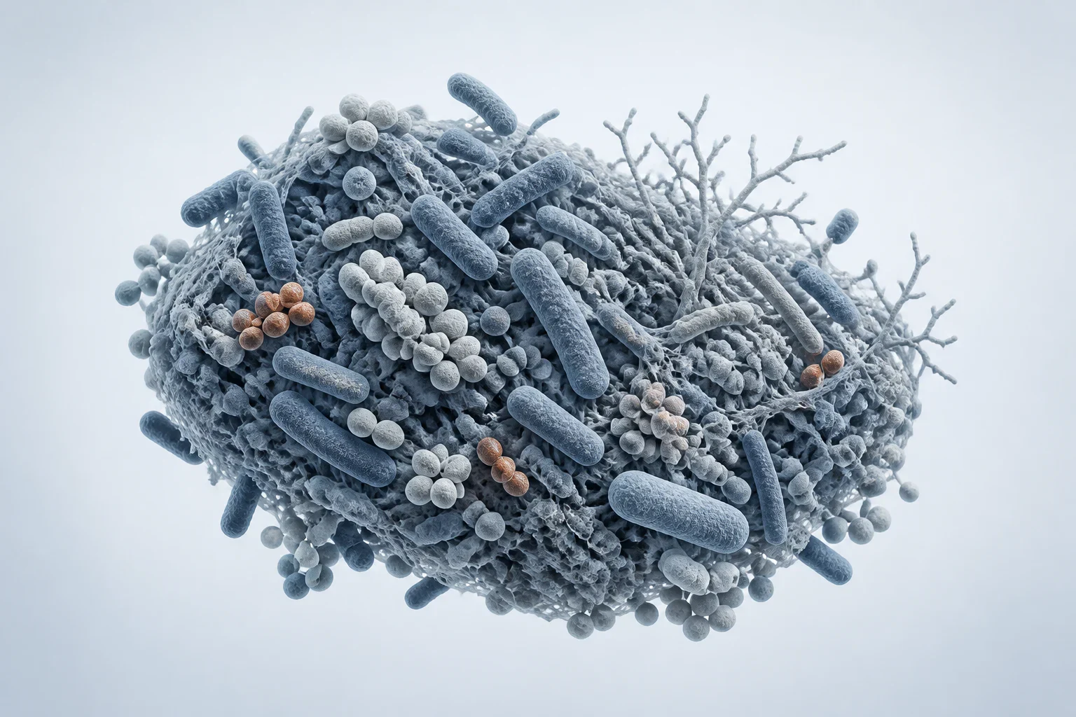
Representative mixed microbial morphologies arranged as an uneven community. Dysbiosis is an ecological state rather than a single visible structure; this reconstruction is not a diagnostic micrograph.
Scientific media record1 verified identifier
- Subject
- Dysbiosiscondition
- Identifiers
- MeSH:D064806
- Review
- Editorial review completeIdentifiers authority-verified · Accessibility validated · · dysbiosis|dysbiosis-microbial-community-v2.webp
- Digital source
- Trained-algorithmic mediaCreated with a trained generative algorithm and reviewed by WikiBiome for subject identity, scientific framing, identifiers, provenance, and accessibility.
- Scientific basis
- Dysbiosis — Medical Subject HeadingsGut dysbiosis: Ecological causes and causative effects on human disease
- License
- CC BY-SA 4.0Created
Disruption of the normal composition and metabolic function of microbial communities, particularly the Gut Microbiome.
In the Metallomics context, dysbiosis is both a consequence of metal toxicity and an amplifier of further disease—creating vicious cycles where metal exposure, microbial imbalance, barrier breakdown, and systemic Metal-Driven Inflammation feed forward into progressive pathology.
With inbound links from virtually every page in this wiki, dysbiosis is the single most cross-referenced concept in WikiBiome, reflecting its role as the central mediating process between environmental metal exposure and human disease.
Evidence map12 cited passagesInspect provenance +
The primary focus of this wiki. Across toxic metals (As, Cd, Pb, Hg, Ni), exposure consistently produces a recognizable dysbiotic signature,. Even essential metals (Fe, Zn, Cu) cause dysbiosis when supplemented in excess or when their homeostasis is disrupted. See the Metal-Specific Patterns section below for details.
Broad-spectrum antibiotics are the most acute cause of dysbiosis. A single course of ciprofloxacin or amoxicillin can reduce alpha diversity by 25-50%, with recovery taking weeks to months and sometimes remaining incomplete. Repeated antibiotic courses produce cumulative damage. Early-life antibiotic exposure is associated with increased risk of IBD, asthma,
Across toxic metals (As, Cd, Pb, Hg, Ni), exposure consistently produces a recognizable dysbiotic signature,:
Cadmium: dose-dependent and sex-dependent effects; 42 genera altered; enhances mammary tumorigenesis through microbiome-mediated pathways. Akkermansia muciniphila is particularly sensitive to low-dose Cd.
Lead: time-dependent changes; reduces Ruminococcus, Coprococcus, Oscillospira, Blautia; prenatal exposure alters childhood gut microbiome.
Barrier breakdown increases metal absorption (germ-free mice absorb significantly more heavy metals than conventional mice).
This cycle explains why even low-level chronic metal exposure can produce progressively worsening health effects over time. The ZIP8 A391T variant in Crohn's disease provides direct genetic evidence: metal transport dysfunction causes microbiome shifts at 2 months but inflammation only at 10 months—dysbiosis is the cause, not the consequence.
Shannon index, Chao1 richness, and observed OTUs measure within-sample diversity. Reduced alpha diversity is the most consistent marker of dysbiosis across diseases. The most severe reductions are seen in Crohn's disease, followed by ulcerative colitis, CRC, and neurodegenerative diseases.
Neurodegenerative disease: PD and AD patients show dysbiotic patterns consistent with metal-driven shifts; the gut-first hypothesis for PD places dysbiosis as the initiating event.
Autoimmune disease: dysbiosis precedes or accompanies IBD, RA, thyroid autoimmunity, and MS. In Crohn's, dysbiosis persists even during endoscopic remission.
Nickel allergy (SNAS): intestinal dysbiosis documented in patients with systemic nickel allergy syndrome.
Metal binding: Lactobacillus and Bifidobacterium species can biosorb heavy metals, reducing absorption.
Contents
1. Definition and Scope2. Causes of Dysbiosis3. Consequences of Dysbiosis4. Metal-Induced Dysbiosis: The Common Pattern5. Metal-Specific Dysbiotic Patterns6. The Dysbiosis-Disease Vicious Cycle7. Diagnostic Markers of Dysbiosis8. Dysbiosis Across Disease Domains9. Restoration Strategies10. ConnectionsDefinition and Scope#
Dysbiosis encompasses three overlapping disturbances:
- Loss of beneficial organisms—reduced abundance and diversity of commensals, particularly SCFA-producing Firmicutes (Faecalibacterium, Roseburia, Lachnospiraceae, Ruminococcus).
- Expansion of pathobionts—overgrowth of potentially harmful organisms that are normally kept in check by commensal competition and host immunity (Enterobacteriaceae, Escherichia-Shigella, Fusobacterium, Candida).
- Loss of microbial metabolic function—reduced production of beneficial metabolites (Short-Chain Fatty Acids (SCFAs), indoles, secondary bile acids) and increased production of harmful metabolites (LPS, TMAO, Ammonia, hydrogen sulfide).
These three dimensions do not always co-occur. A community can lose diversity without gaining specific pathobionts, or harbor pathobionts without a dramatic drop in alpha diversity. The most severe clinical dysbiosis involves all three.
Causes of Dysbiosis#
Metals and Environmental Toxicants#
The primary focus of this wiki. Across toxic metals (arsenic (As), cadmium (Cd), lead (Pb), mercury (Hg), nickel (Ni)), exposure consistently produces a recognizable dysbiotic signature.[1]Toxic and Essential Metals: Metabolic Interactions with the Gut Microbiota and Health ImplicationsQinheng Zhu, Boyan Chen, Fu Zhang et al. · 2024Open reference 1 ↓[2]Influence of Toxic Metal Exposure on the Gut Microbiota (Review)Federica Giambo, Sebastiano Italia, Michele Teodoro et al. · 2021Open reference 2 ↓
Even essential metals (iron (Fe), zinc (Zn), copper (Cu)) cause dysbiosis when supplemented in excess or when their homeostasis is disrupted. See the Metal-Specific Patterns section below for details.
Antibiotics#
Broad-spectrum antibiotics are the most acute cause of dysbiosis. A single course of ciprofloxacin or amoxicillin can reduce alpha diversity by 25-50%, with recovery taking weeks to months and sometimes remaining incomplete.[3]Role of Microbiome and Antibiotics in Autoimmune DiseasesVangoitsenhoven R, Cresci GAM · 2020Open reference 3 ↓ Repeated antibiotic courses produce cumulative damage.
Early-life antibiotic exposure is associated with increased risk of IBD, asthma, obesity, and ASD—all diseases with strong dysbiosis signatures in this wiki.
Diet#
Low-fiber, high-fat, high-sugar Western diets starve SCFA-producing bacteria of fermentable substrate, reducing Butyrate output and shifting communities toward Bacteroides-dominant profiles. Conversely, high-fiber and Mediterranean-pattern diets support microbial diversity and SCFA production.
Specific dietary metals (nickel in legumes and nuts, iron in red meat, cadmium in leafy greens) introduce metal-driven selection pressures superimposed on macronutrient effects.
Stress and the HPA Axis#
Psychological and physiological stress activate the hypothalamic-pituitary-adrenal axis, releasing cortisol and catecholamines that alter gut motility, reduce mucosal blood flow, increase intestinal permeability, and directly modulate microbial gene expression. Stress-induced dysbiosis is particularly relevant to the Gut-Brain Axis and to stress-associated diseases like Depression, anxiety, and IBS.
Consequences of Dysbiosis#
Gut Barrier Failure#
Loss of SCFA-producing bacteria—particularly butyrate producers like Faecalibacterium prausnitzii and Roseburia—starves colonocytes of their preferred energy source. Butyrate fuels 60-70% of colonocyte metabolism and is required for tight junction protein expression (claudins, occludin, ZO-1) and mucin production.
Without adequate butyrate, the epithelial barrier becomes permeable ("leaky gut"), measurable by the lactulose/mannitol ratio test. Barrier failure permits translocation of bacteria, LPS, food antigens, and metals into the lamina propria and systemic circulation.
Systemic Inflammation#
Dysbiosis drives chronic low-grade inflammation through multiple routes. LPS translocation: Gram-negative pathobiont enrichment increases endotoxin load; LPS crossing the compromised barrier activates TLR4/NF-kB Signaling Pathway on macrophages, hepatocytes, and microglia. Cytokine cascades: NF-kB activation drives transcription of IL-6, TNF-alpha, IL-1beta, and COX-2.
NLRP3 inflammasome: Bacterial products and damage-associated molecular patterns activate the NLRP3 inflammasome, driving IL-1beta and IL-18 maturation. Molecular mimicry: Bacterial antigens can cross-react with host tissues, triggering autoimmune responses (relevant to RA, MS, Hashimoto's, Graves').
Immune Dysregulation#
A healthy microbiome educates and calibrates the immune system. Dysbiosis disrupts this calibration. Treg/Th17 imbalance: SCFA-producing Clostridia clusters IV and XIVa induce regulatory T cells via butyrate-mediated HDAC inhibition.
Their loss shifts the balance toward pro-inflammatory Th17 responses.
IgA dysregulation: Secretory IgA normally coats and contains commensal bacteria; dysbiosis alters IgA targeting, permitting pathobiont escape. Impaired oral tolerance: Disrupted microbial education of the immune system increases susceptibility to food allergies, autoimmunity, and chronic inflammation.
Metabolite Shifts#
Dysbiosis alters the entire gut metabolome. Reduced SCFAs: Butyrate, propionate, and acetate production declines as their producers are lost. Increased TMAO precursors: Expansion of TMA lyase-positive organisms (Enterobacteriaceae, Desulfovibrio) increases trimethylamine production, contributing to cardiovascular risk via hepatic TMAO conversion.
Altered bile acid metabolism: Loss of 7-alpha-dehydroxylating bacteria changes the primary-to-secondary bile acid ratio, affecting FXR/TGR5 signaling. Tryptophan pathway shifts: Reduced indole-3-propionic acid (IPA) and indole-3-aldehyde production diminishes AhR-mediated anti-inflammatory signaling. Increased uremic toxins: Indoxyl sulfate and p-cresol sulfate accumulate, contributing to kidney and cardiovascular damage.
Metal-Induced Dysbiosis: The Common Pattern#
Across toxic metals (arsenic (As), cadmium (Cd), lead (Pb), mercury (Hg), nickel (Ni)), exposure consistently produces a recognizable dysbiotic signature:[1]Toxic and Essential Metals: Metabolic Interactions with the Gut Microbiota and Health ImplicationsQinheng Zhu, Boyan Chen, Fu Zhang et al. · 2024Open reference 1 ↓[2]Influence of Toxic Metal Exposure on the Gut Microbiota (Review)Federica Giambo, Sebastiano Italia, Michele Teodoro et al. · 2021Open reference 2 ↓
What Decreases#
SCFA-producing commensals: Faecalibacterium, Lachnospiraceae, Blautia, Ruminococcus, Lactobacillus, Bifidobacterium. Microbial diversity: alpha diversity consistently reduced, especially with cadmium (Cd) exposure. Butyrate production: loss of butyrate-producing bacteria impairs gut barrier integrity and anti-inflammatory signaling.
Akkermansia muciniphila: particularly sensitive to low-dose cadmium; loss compromises mucus layer maintenance.
What Increases#
Enterobacteriaceae: gram-negative, LPS-producing, siderophore-equipped pathobionts that thrive in metal-rich, inflammation-rich environments. Escherichia-Shigella: consistently enriched across multiple metals. Metal-tolerant species: organisms with efflux pumps, metal-binding proteins, and biofilm capacity outcompete sensitive commensals.
LPS burden: gram-negative enrichment increases endotoxin load, driving systemic inflammation via TLR4/NF-kB Signaling Pathway.
Metal-Specific Dysbiotic Patterns#
Arsenic: increases Bacteroidetes and Bilophila; perturbs bile acid homeostasis; Faecalibacterium is essential for arsenic biotransformation via arsM methyltransferase.
Cadmium: dose-dependent and sex-dependent effects; 42 genera altered; enhances mammary tumorigenesis through microbiome-mediated pathways.[4]Cadmium Exposure Induces Changes in Gut Microbial Composition and Metabolic Function in Long-Tailed Dwarf Hamsters, Cricetulus longicaudatusMengfan Tao, Kanglin Cao, Xinsheng Pu et al. · 2024Open reference 4 ↓ Akkermansia muciniphila is particularly sensitive to low-dose cadmium (Cd).
Lead: time-dependent changes; reduces Ruminococcus, Coprococcus, Oscillospira, Blautia; prenatal exposure alters childhood gut microbiome.[5]Interaction of Heavy Metal Lead with Gut Microbiota: Implications for Autism Spectrum DisorderTizabi Y, Bennani S, El Kouhen N et al. · 2023Open reference 5 ↓ Mercury: methylmercury concentrates in the gut lumen; reduces Lactobacillus and Bifidobacterium; increases Desulfovibrio and Clostridium.
Nickel: occupational exposure increases Parabacteroides, Escherichia-Shigella; decreases Lactobacillus; nickel (Ni)-dependent virulence enzymes (Urease, Hydrogenase) in gut pathogens contribute to ammonia-mediated epithelial damage.
Iron: both deficiency and excess are dysbiotic; supplementation increases Enterobacteriaceae, decreases Lactobacillus. The iron paradox is especially acute in IBD—see Crohn's Disease.
The Dysbiosis-Disease Vicious Cycle#
Metal-induced dysbiosis is self-amplifying:
- Metal exposure kills sensitive commensals, favoring metal-tolerant pathobionts.
- Loss of SCFA producers weakens the gut epithelial barrier (butyrate fuels colonocytes and maintains tight junctions).
- Barrier breakdown increases metal absorption (germ-free mice absorb significantly more Heavy Metals than conventional mice).[6]Gut Microbiota: A Target for Heavy Metal Toxicity and a Probiotic Protective StrategyHui Duan, Leilei Yu, Fengwei Tian et al. · 2020Open reference 6 ↓
- Increased metal absorption further disrupts the microbiome systemically.
- LPS translocation through the leaky barrier activates systemic inflammation.
- Inflammation reinforces dysbiosis: hepcidin elevation, calprotectin release, and Oxidative Stress further alter the luminal metal environment, selecting against commensals.
- Disease progression generates additional stressors (medications, surgical interventions, malnutrition) that deepen the dysbiotic state.
This cycle explains why even low-level chronic metal exposure can produce progressively worsening health effects over time.
The ZIP8 A391T variant in Crohn's disease provides direct genetic evidence: metal transport dysfunction causes microbiome shifts at 2 months but inflammation only at 10 months—dysbiosis is the cause, not the consequence.[7]ZIP8 A391T Crohn's Disease-Linked Risk Variant Induces Colonic Metal Ion Dyshomeostasis, Microbiome Compositional Shifts, and InflammationYang JC, Zhao M, Chernikova D et al. · 2024Open reference 7 ↓
Diagnostic Markers of Dysbiosis#
Alpha Diversity#
Shannon index, Chao1 richness, and observed OTUs measure within-sample diversity. Reduced alpha diversity is the most consistent marker of dysbiosis across diseases. The most severe reductions are seen in Crohn's disease, followed by ulcerative colitis, CRC, and neurodegenerative diseases.[8]Diagnosis of Crohn's Disease and Ulcerative Colitis Using the MicrobiomeKang DY, Park JL, Yeo MK et al. · 2023Open reference 8 ↓
Beta Diversity#
Bray-Curtis dissimilarity and UniFrac distances measure between-sample community differences. Disease-associated communities cluster separately from healthy controls in ordination plots (PCoA, NMDS), confirming compositional shifts beyond simple diversity loss.
Firmicutes/Bacteroidetes (F/B) Ratio#
Historically used as a simple dysbiosis metric—decreased F/B ratio in IBD and obesity, increased in some metabolic diseases. However, the F/B ratio is now recognized as overly simplistic; phylum-level changes obscure functionally important genus-level shifts.
Specific Taxonomic Markers#
Faecalibacterium prausnitzii depletion: the single most consistent marker across IBD, CRC, metabolic disease, and neurodegeneration. Enterobacteriaceae enrichment: marks metal-driven and inflammation-driven dysbiosis. Akkermansia muciniphila: depletion signals mucus layer compromise; protective by Mendelian randomization for Hashimoto's.
Roseburia/Lachnospiraceae depletion: loss of butyrate producers.
Functional Markers#
Fecal SCFA levels: butyrate, propionate, acetate measured by GC-MS. Fecal calprotectin: neutrophil-derived marker of intestinal inflammation; also an active participant in Nutritional Immunity (Metal Sequestration). Fecal LPS/endotoxin: direct measurement of barrier failure.
Urinary indoxyl sulfate and p-cresol sulfate: microbial metabolites elevated in dysbiosis.
Dysbiosis Across Disease Domains#
Metal-induced dysbiosis connects to virtually every disease in this wiki. Neurodegenerative disease: PD and AD patients show dysbiotic patterns consistent with metal-driven shifts; the gut-first hypothesis for PD places dysbiosis as the initiating event.[9]Microbial Metallomics and Parkinson's Disease: A Unified Metal-Driven Framework Linking Ferroptosis, Dysbiosis, and alpha-Synuclein PathologyKaren Pendergrass · 2025Open reference 9 ↓
Autoimmune disease: dysbiosis precedes or accompanies IBD, RA, thyroid autoimmunity, and MS.[10]Environmental Exposures and Autoimmune Diseases: Contribution of Gut MicrobiomeM. Firoze Khan, Hui Wang · 2020Open reference 10 ↓ In Crohn's, dysbiosis persists even during endoscopic remission.
Metabolic disease: PCOS and T2D feature gut dysbiosis with reduced SCFA producers and increased TMAO-producing taxa. Cancer: CRC has the most developed dysbiosis-cancer link (Fusobacterium nucleatum, ETBF); breast cancer shows estrobolome dysbiosis affecting estrogen recirculation. Neurodevelopmental disorders: ASD and ADHD children show both metal dyshomeostasis and characteristic dysbiotic patterns.
Nickel allergy (SNAS): intestinal dysbiosis documented in patients with systemic nickel allergy syndrome.[11]The Effects of Low-Nickel Diet Combined with Oral Administration of Selected Probiotics on Patients with Systemic Nickel Allergy Syndrome (SNAS) and Gut DysbiosisLombardi F, Fiasca F, Minelli M et al. · 2020Open reference 11 ↓
Restoration Strategies#
Dietary Intervention#
High-fiber diets (30g/day minimum) provide fermentable substrate for SCFA-producing bacteria. The Mediterranean Diet is the best-evidenced dietary pattern for supporting microbial diversity. Polyphenol-rich foods (berries, green tea, olive oil) serve as prebiotics for beneficial taxa.
However, dietary metal content must be considered—high-nickel plant foods may offset benefits for nickel-sensitized individuals.
Probiotics#
Probiotics represent a therapeutic approach to break the metal-dysbiosis cycle. Metal binding: Lactobacillus and Bifidobacterium species can biosorb heavy metals, reducing absorption.[12]Heavy Metal-Gut Microbiota Interactions: Probiotics Modulation and Biosensors DetectionLiliana Anchidin-Norocel, Oana C. Iatcu, Andrei Lobiuc et al. · 2025Open reference 12 ↓ SCFA restoration: probiotic supplementation can partially restore butyrate production and barrier function.
Competitive exclusion: probiotics compete with metal-tolerant pathobionts for ecological niches. E. coli Nissle 1917 outcompetes pathogenic E. coli via superior siderophore systems. Immune modulation: specific strains shift Th1/Th17 toward Treg/Th2 profiles (demonstrated in MS, Graves', Hashimoto's trials).
Limitation: probiotics address symptoms but not the root cause of ongoing metal exposure.
Fecal Microbiota Transplant (FMT)#
FMT transfers a complete microbial community from a healthy donor. It is most established for recurrent C. difficile infection (>90% cure rate) and shows promise for IBD and ASD. However, success depends on whether the recipient environment can sustain the transplanted community.
If metal dyshomeostasis persists (e.g., in ZIP8 A391T carriers), transplanted commensals may fail to engraft.
Prebiotics#
Targeted prebiotic fibers (inulin, FOS, GOS, resistant starch) selectively feed beneficial bacteria. GOS supplementation improved gut microbiome composition in ASD children. Prebiotics are generally safer and more sustainable than probiotics but slower acting.
Metal Exposure Reduction#
Ultimately, restoring eubiosis requires addressing the upstream cause. Reducing dietary metal exposure (low-nickel diets, avoiding contaminated food sources), treating occupational exposures, and using metal chelators when indicated may be necessary to create an environment where commensal bacteria can reestablish.
Connections#
- Gut-Metal-Microbiome Interactions—the comprehensive framework for metal-microbiome interactions
- Gut-Brain Axis—dysbiosis disrupts gut-brain communication via vagal, immune, and metabolite pathways
- inflammation—dysbiosis drives systemic inflammation via LPS/TLR4 and cytokine cascades
- NF-kB Signaling Pathway—downstream inflammatory signaling from dysbiosis-derived LPS
- Nutritional Immunity (Metal Sequestration)—host metal restriction can inadvertently worsen dysbiosis
- Short-Chain Fatty Acids (SCFAs)—the primary protective metabolites lost in dysbiosis
- butyrate—the key colonocyte fuel and HDAC inhibitor depleted in dysbiosis
- Fecal Microbiota Transplant (FMT)—therapeutic community replacement for severe dysbiosis
- Probiotics—targeted microbial supplementation to counter dysbiosis
- Mediterranean Diet—the best-evidenced dietary pattern for supporting eubiosis
References 14
Numbered by first appearance in the article, then reconciled with its declared source list.
- 1
★Qinheng Zhu, Boyan Chen, Fu Zhang et al. (2024). Toxic and Essential Metals: Metabolic Interactions with the Gut Microbiota and Health Implications. Frontiers in Nutrition.
- 2
★Federica Giambo, Sebastiano Italia, Michele Teodoro et al. (2021). Influence of Toxic Metal Exposure on the Gut Microbiota (Review). World Academy of Sciences Journal.
- 3
Vangoitsenhoven R, Cresci GAM (2020). Role of Microbiome and Antibiotics in Autoimmune Diseases. Nutrition in Clinical Practice.
- 4
Mengfan Tao, Kanglin Cao, Xinsheng Pu et al. (2024). Cadmium Exposure Induces Changes in Gut Microbial Composition and Metabolic Function in Long-Tailed Dwarf Hamsters, Cricetulus longicaudatus. Ecology and Evolution.
- 5
★Tizabi Y, Bennani S, El Kouhen N et al. (2023). Interaction of Heavy Metal Lead with Gut Microbiota: Implications for Autism Spectrum Disorder. Biomolecules.
- 6
★Hui Duan, Leilei Yu, Fengwei Tian et al. (2020). Gut Microbiota: A Target for Heavy Metal Toxicity and a Probiotic Protective Strategy. Science of the Total Environment.
- 7
★Yang JC, Zhao M, Chernikova D et al. (2024). ZIP8 A391T Crohn's Disease-Linked Risk Variant Induces Colonic Metal Ion Dyshomeostasis, Microbiome Compositional Shifts, and Inflammation. Digestive Diseases and Sciences.
- 8
Kang DY, Park JL, Yeo MK et al. (2023). Diagnosis of Crohn's Disease and Ulcerative Colitis Using the Microbiome. BMC Microbiology.
- 9
★Karen Pendergrass (2025). Microbial Metallomics and Parkinson's Disease: A Unified Metal-Driven Framework Linking Ferroptosis, Dysbiosis, and alpha-Synuclein Pathology. Conference Presentation.
- 10
M. Firoze Khan, Hui Wang (2020). Environmental Exposures and Autoimmune Diseases: Contribution of Gut Microbiome. Frontiers in Immunology.
- 11
Lombardi F, Fiasca F, Minelli M et al. (2020). The Effects of Low-Nickel Diet Combined with Oral Administration of Selected Probiotics on Patients with Systemic Nickel Allergy Syndrome (SNAS) and Gut Dysbiosis. Nutrients.
- 12
★Liliana Anchidin-Norocel, Oana C. Iatcu, Andrei Lobiuc et al. (2025). Heavy Metal-Gut Microbiota Interactions: Probiotics Modulation and Biosensors Detection. Biosensors.
- 13
★Sweta Ghosh, Syam P. Nukavarpu, Venkatakrishna Rao Jala (2024). Effects of Heavy Metals on Gut Barrier Integrity and Gut Microbiota. Microbiota and Host.
- 14
Fatemeh Rezazadegan, Maryam Mahmoudi, Seyed Mohammad Mousavi (2025). Rezazadegan et al. 2025 — Heavy Metals and Gut Microbiota: A Systematic Review. Journal of Health, Population and Nutrition.
Article network
Mentioned here 18
Pages linking here 333
Connect the evidence
Publicly readable discussion by ORCID-authenticated researchers. Questions, interpretation, methods, corrections, and new evidence stay attached to this record.
No discussion yet. Start with a precise question or a source-backed observation.
Activity and accepted changes
Accepted researcher context, editorial status, public discussion, and upstream Git revisions are shown together. Pending, declined, and withdrawn proposals remain private.
- published revision
Strengthen TLR4 and link high-leverage contexts
Karen Pendergrass · +1 −1
Inspect exact Git diff ↗ - published revision
Strengthen Metallomics and link high-leverage contexts
Karen Pendergrass · +1 −1
Inspect exact Git diff ↗ - published revision
Consolidate microbial metabolite knowledge
Karen Pendergrass · +1 −1
Inspect exact Git diff ↗ - published revision
Backfill heavy metals concept links
Karen Pendergrass · +1 −1
Inspect exact Git diff ↗ - published revision
Backfill oxidative stress concept links
Karen Pendergrass · +1 −1
Inspect exact Git diff ↗ - published revision
Backfill gut microbiome concept links
Karen Pendergrass · +1 −1
Inspect exact Git diff ↗ - published revision
Backfill inflammation concept links
Karen Pendergrass · +3 −3
Inspect exact Git diff ↗ - published revision
Complete Ammonia contextual coverage
Karen Pendergrass · +1 −1
Inspect exact Git diff ↗ - published revision
Complete Hydrogenase contextual coverage
Karen Pendergrass · +1 −1
Inspect exact Git diff ↗ - published revision
Complete Tight junctions contextual coverage
Karen Pendergrass · +1 −1
Inspect exact Git diff ↗ - published revision
Complete reviewed Urease contextual coverage
Karen Pendergrass · +1 −1
Inspect exact Git diff ↗ - published revision
Add NLRP3 inflammasome concept and link batch
Karen Pendergrass · +3 −3
Inspect exact Git diff ↗ - published revision
massive wiki expansion: 149 stubs fixed, 100+ new pages, Rule 15 scan, keystone papers
WikiBiome Deploy Bot · +55 −55
Inspect exact Git diff ↗ - published revision
nightly maintenance: 94 stub demotions, 181 source_count fixes, 22 auto-discovered stubs, 5 adversarial audits, 3 boundary fixes, 3 evidence-level corrections
WikiBiome Deploy Bot · +1 −0
Inspect exact Git diff ↗ - published revision
pre-overnight checkpoint 2026-04-18
WikiBiome Deploy Bot · +5 −3
Inspect exact Git diff ↗