
 metals · microbes · host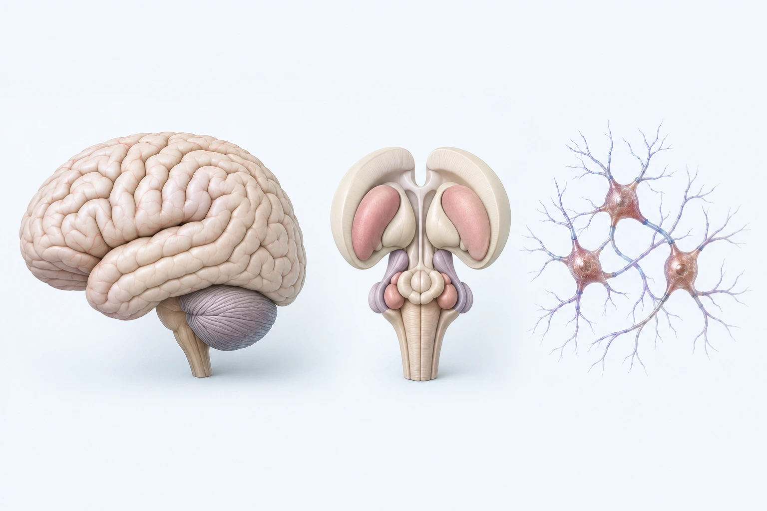
Neutral neuroanatomical orientation for Parkinson disease. The central model is an abstraction, not validated regional anatomy, a scan, histology image, dopamine state, biomarker, mechanism, symptom, or stage.
Scientific media record1 verified identifier
- Subject
- Parkinson Diseasecondition
- Identifiers
- MeSH:D010300
- Review
- Editorial review completeIdentifiers authority-verified · Accessibility validated · · parkinsons-disease|parkinsons-disease-pathology-v1.webp
- Digital source
- Trained-algorithmic mediaCreated with a trained generative algorithm and reviewed by WikiBiome for subject identity, scientific framing, identifiers, provenance, and accessibility.
- Scientific basis
- Parkinson Disease — MeSHParkinson's Disease
- License
- CC BY-SA 4.0Created
The second most common neurodegenerative disease, affecting over 10 million people worldwide.[1]Dorsey 2018 -- The Incidence of Parkinson's Disease: A Systematic Review and Meta-AnalysisE Ray Dorsey, Todd Sherer, Michael S Okun et al. · 2018Open reference 1 ↓ Characterized by progressive loss of dopaminergic neurons in the substantia nigra, alpha-synuclein aggregation (Lewy bodies), and motor symptoms (tremor, rigidity, bradykinesia).[2]Balestrino 2020 -- Parkinson's Disease: A Systematic ReviewRoberta Balestrino, Alberto J Espay · 2020Open reference 2 ↓
From a Metallomics perspective, PD is the disease where ferroptosis is most directly established as a cell death mechanism, manganese-parkinsonism provides the clearest occupational dose-response evidence, and the gut-brain axis offers a compelling metal-microbiome-neurodegeneration framework. Approximately 14 source pages address PD's metallomic dimensions.
Evidence map99 cited passagesInspect provenance +
The second most common neurodegenerative disease, affecting over 10 million people worldwide. Characterized by progressive loss of dopaminergic neurons in the substantia nigra, alpha-synuclein aggregation (Lewy bodies), and motor symptoms (tremor, rigidity, bradykinesia). From a metallomics perspective, PD is the disease where ferroptosis is most directly es
Iron accumulates specifically in the substantia nigra, where it catalyzes Fenton reactions generating hydroxyl radicals that drive lipid peroxidation.
GPX4 (glutathione peroxidase 4) downregulation removes the brake on ferroptotic cell death; GPX4 is the master regulator of ferroptosis.
Neuromelanin normally accumulates iron with age in the substantia nigra; when neuromelanin iron-binding capacity is exceeded, free iron catalyzes lipid peroxidation.
Iron chelation (deferiprone) shows some benefit in PD trials.
Transferrin receptor and ferritin alterations documented in PD.
Pheomelanin (red/yellow pigment dominant in redheads) has weaker metal-binding capacity than eumelanin (brown/black pigment), and may itself generate ROS upon metal exposure.
Neuromelanin in the substantia nigra is a mixed polymer of eumelanin and pheomelanin; its ratio may vary by MC1R genotype.
MC1R loss-of-function variants shift melanogenesis toward pheomelanin in both skin and brain—MC1R is expressed in dopaminergic neurons.
Proposed mechanism: MC1R variants - higher pheomelanin fraction in neuromelanin - reduced iron chelation capacity - greater labile iron pool - enhanced Fenton reactions - ferroptotic neuron death.
Epidemiological support: red hair/fair skin phenotypes and MC1R variants are independent risk factors for PD, with risk increases of approximately 50-100%.
Progression of parkinsonism increases with cumulative Mn exposure: annual UPDRS3 increase of 0.24 points per mg Mn/m3-year (95% CI 0.10-0.38).
Among 886 welders, 15.2% had UPDRS3 = 15 (clinical parkinsonism).
Flux core arc welding in confined spaces showed 6.7x higher progression rate (0.67 vs 0.10 UPDRS3/year per mg Mn/m3-year).
A worker with 20 years of welding exposure would be predicted to have nearly a 7-point increase in UPDRS3 score.
Mn primarily affects the basal ganglia (globus pallidus, striatum) rather than the substantia nigra, producing a phenotype distinct from idiopathic PD.
Mn impairs autophagy at low concentrations; Drp1 inhibition protects against Mn-induced autophagic impairment.
FMT in rats has alleviated Mn-induced neurotoxicity, linking Mn-parkinsonism to the gut microbiome.
Widespread Cu decreases across brain regions in PD dementia, including substantia nigra.
Cu depletion is shared across PD, AD, and DLB, suggesting a common pathway involving disturbed Cu trafficking.
Zinc: Reduced in serum/plasma of PD patients (meta-analysis of 803 PD patients and 796 controls).
Lead: Elevated; Pb mimics Ca in signaling pathways, disrupting neurotransmitter release.
Cadmium: Elevated; BBB disruption and mitochondrial damage.
Alpha-synuclein deposits found in the enteric nervous system years before motor symptoms.
Showing 24 of 99 evidence-bearing passages. Every remaining citation is still indexed in the reference record below.
One disease. Five evidence layers.
A generated systems view of the metals, organisms, host sequestration signals, ecological conditions, and microbial functions indexed for Parkinson's Disease.
Evidence layer
Taxonomic signature
Organisms reported as enriched or depleted, with their indexed functional context kept beside the name.Metal-tolerant pathogens — siderophore-producing, iron piracy, LPS-driven inflammation, Enterobacteriaceae (E. coli-like) express zinc metalloproteases and nickel-dependent urease
Mucus-degrading pathogen — elevated in PD, directly damages intestinal barrier integrity via mucinase enzymes; increases translocation of LPS and bacterial products
Interkingdom functional shielding — biofilm formation, oxygen depletion, metal-acquisition systems; enriched in PD mycobiome; promotes Enterobacteriaceae survival
Curli amyloid producer -- cross-seeds alpha-synuclein aggregation; LPS-driven neuroinflammation
Pro-inflammatory Gram-negative genus enriched in PD gut
Paradoxically enriched in PD (despite being considered beneficial); may reflect compensatory expansion or altered niche availability
nickel (Ni)-dependent urease producer; H. pylori and SIBO are triggering factors for PD
SCFA producer and immune-modulating commensal — consistently depleted across PD cohorts; loss removes barrier protection and neuroprotective metabolites
SCFA producer (butyrate) — crucial for iron regulation via HIF-2alpha and intestinal barrier integrity; depleted in PD
SCFA producers — lost in iron-rich, pro-inflammatory environment; depletion removes colonocyte nutrition and intestinal permeability protection
SCFA producer — depleted in PD; essential for butyrate production and barrier protection
Commensal lactobacilli/lactococci — depleted via bacteriophage predation in PD; produce lactate and other immune-regulating metabolites
Anti-inflammatory SCFA producer; depleted in PD
SCFA producer depleted in PD; associated with reduced fecal butyrate levels
Evidence layer
Nutritional immunity
Host metal-withholding, inflammatory, antioxidant, and microbial-metabolite signals indexed in the signature.Elevated host signals
7Depleted protective signals
6Evidence layer
Ecological state
The environmental conditions that connect the organism-level observations into a system.Evidence layer
Virulence functions
Microbial structures, enzymes, and acquisition systems implicated by the linked evidence.The disease record, in full.
The original WikiBiome disease narrative remains intact beneath the generated signature atlas.
Metallomic Signature#
The Metal-Disease Matrix: A Cross-Source Synthesis identifies PD's profile as: copper (Cu) ↓ (brain), zinc (Zn) ↓ (serum), iron (Fe) ↑ (substantia nigra, ferroptosis), selenium (Se) ↓, manganese (Mn) ↑ (basal ganglia), lead (Pb) ↑, cadmium (Cd) ↑, mercury (Hg) ↑.
Iron: Central to Ferroptosis in the Substantia Nigra#
Iron accumulation in the substantia nigra is the hallmark metallomic finding in PD and the most mechanistically developed metal-disease pathway in this wiki. Iron accumulates specifically in the substantia nigra, where it catalyzes Fenton reactions generating hydroxyl radicals that drive lipid peroxidation.[3]Microbial Metallomics and Parkinson's Disease: A Unified Metal-Driven Framework Linking Ferroptosis, Dysbiosis, and alpha-Synuclein PathologyKaren Pendergrass · 2025Open reference 3 ↓
GPX4 (glutathione peroxidase 4) downregulation removes the brake on ferroptotic cell death; GPX4 is the master regulator of ferroptosis.[3]Microbial Metallomics and Parkinson's Disease: A Unified Metal-Driven Framework Linking Ferroptosis, Dysbiosis, and alpha-Synuclein PathologyKaren Pendergrass · 2025Open reference 3 ↓
Neuromelanin normally accumulates iron with age in the substantia nigra; when neuromelanin iron-binding capacity is exceeded, free iron catalyzes lipid peroxidation.[4]Pheomelanin, Eumelanin, and Neuromelanin: A Metal-Linked Hypothesis for Parkinson's Risk in RedheadsEyer K, Karen Pendergrass · 2025Open reference 4 ↓
Iron chelation (deferiprone) shows some benefit in PD trials.[5]Common and Trace Metals in Alzheimer's and Parkinson's DiseasesDoroszkiewicz J, Farhan JA, Mroczko J et al. · 2023Open reference 5 ↓ Transferrin receptor and ferritin alterations documented in PD.[5]Common and Trace Metals in Alzheimer's and Parkinson's DiseasesDoroszkiewicz J, Farhan JA, Mroczko J et al. · 2023Open reference 5 ↓
The Pheomelanin/Neuromelanin Hypothesis#
A novel hypothesis links MC1R variants (red hair phenotype) to PD risk through differential metal binding. Pheomelanin (red/yellow pigment dominant in redheads) has weaker metal-binding capacity than eumelanin (brown/black pigment), and may itself generate ROS upon metal exposure.[4]Pheomelanin, Eumelanin, and Neuromelanin: A Metal-Linked Hypothesis for Parkinson's Risk in RedheadsEyer K, Karen Pendergrass · 2025Open reference 4 ↓
Neuromelanin in the substantia nigra is a mixed polymer of eumelanin and pheomelanin; its ratio may vary by MC1R genotype.[4]Pheomelanin, Eumelanin, and Neuromelanin: A Metal-Linked Hypothesis for Parkinson's Risk in RedheadsEyer K, Karen Pendergrass · 2025Open reference 4 ↓
MC1R loss-of-function variants shift melanogenesis toward pheomelanin in both skin and brain—MC1R is expressed in dopaminergic neurons.[4]Pheomelanin, Eumelanin, and Neuromelanin: A Metal-Linked Hypothesis for Parkinson's Risk in RedheadsEyer K, Karen Pendergrass · 2025Open reference 4 ↓
Proposed mechanism: MC1R variants -> higher pheomelanin fraction in neuromelanin -> reduced iron chelation capacity -> greater labile iron pool -> enhanced Fenton reactions -> ferroptotic neuron death.[4]Pheomelanin, Eumelanin, and Neuromelanin: A Metal-Linked Hypothesis for Parkinson's Risk in RedheadsEyer K, Karen Pendergrass · 2025Open reference 4 ↓
Epidemiological support: red hair/fair skin phenotypes and MC1R variants are independent risk factors for PD, with risk increases of approximately 50-100%.[4]Pheomelanin, Eumelanin, and Neuromelanin: A Metal-Linked Hypothesis for Parkinson's Risk in RedheadsEyer K, Karen Pendergrass · 2025Open reference 4 ↓
Manganese: Dose-Dependent Parkinsonism in Welders#
Manganese-parkinsonism provides the clearest occupational dose-response evidence for metal-induced neurodegeneration. Progression of parkinsonism increases with cumulative manganese (Mn) exposure: annual UPDRS3 increase of 0.24 points per mg manganese/m3-year (95% CI 0.10-0.38).[6]Dose-Dependent Progression of Parkinsonism in Manganese-Exposed WeldersBrad A. Racette, Susan Searles Nielsen, Susan R. Criswell et al. · 2017Open reference 6 ↓
Among 886 welders, 15.2% had UPDRS3 >= 15 (clinical parkinsonism).[6]Dose-Dependent Progression of Parkinsonism in Manganese-Exposed WeldersBrad A. Racette, Susan Searles Nielsen, Susan R. Criswell et al. · 2017Open reference 6 ↓
Flux core arc welding in confined spaces showed 6.7x higher progression rate (0.67 vs 0.10 UPDRS3/year per mg manganese/m3-year).[6]Dose-Dependent Progression of Parkinsonism in Manganese-Exposed WeldersBrad A. Racette, Susan Searles Nielsen, Susan R. Criswell et al. · 2017Open reference 6 ↓
A worker with 20 years of welding exposure would be predicted to have nearly a 7-point increase in UPDRS3 score.[6]Dose-Dependent Progression of Parkinsonism in Manganese-Exposed WeldersBrad A. Racette, Susan Searles Nielsen, Susan R. Criswell et al. · 2017Open reference 6 ↓
manganese primarily affects the basal ganglia (globus pallidus, striatum) rather than the substantia nigra, producing a phenotype distinct from idiopathic PD.[5]Common and Trace Metals in Alzheimer's and Parkinson's DiseasesDoroszkiewicz J, Farhan JA, Mroczko J et al. · 2023Open reference 5 ↓
manganese impairs autophagy at low concentrations; Drp1 inhibition protects against manganese-induced autophagic impairment.[7]Associations of Environmental Exposure to Arsenic, Manganese, Lead, and Cadmium with Alzheimer's Disease: A Review of Recent Evidence from Mechanistic StudiesGiasuddin Ahmed, Md. Shiblur Rahaman, Enrique Perez et al. · 2025Open reference 7 ↓ FMT in rats has alleviated manganese-induced neurotoxicity, linking manganese-parkinsonism to the Gut Microbiome.[6]Dose-Dependent Progression of Parkinsonism in Manganese-Exposed WeldersBrad A. Racette, Susan Searles Nielsen, Susan R. Criswell et al. · 2017Open reference 6 ↓
Other Metals#
Zinc: Reduced in serum/plasma of PD patients (meta-analysis of 803 PD patients and 796 controls).[5]Common and Trace Metals in Alzheimer's and Parkinson's DiseasesDoroszkiewicz J, Farhan JA, Mroczko J et al. · 2023Open reference 5 ↓ Selenium: Depletion impairs GPx activity, compounding ferroptotic vulnerability. Lead: Elevated; lead (Pb) mimics calcium (Ca) in signaling pathways, disrupting neurotransmitter release.[5]Common and Trace Metals in Alzheimer's and Parkinson's DiseasesDoroszkiewicz J, Farhan JA, Mroczko J et al. · 2023Open reference 5 ↓
Cadmium: Elevated; BBB disruption and mitochondrial damage.[9]Exposure to Cadmium and Its Impacts on Human Health: A Short ReviewPuthiyavalappil Rasin, Ashwathi A V, Sabeel M Basheer et al. · 2025Open reference 9 ↓
Gut-Brain Axis: The Microbial Metallomics Framework#
PD has the strongest evidence for gut-brain axis involvement among neurodegenerative diseases, and a comprehensive metal-microbiome framework has been proposed:
The Braak Hypothesis (Gut-First Origin)#
Alpha-synuclein deposits found in the enteric nervous system years before motor symptoms.[3]Microbial Metallomics and Parkinson's Disease: A Unified Metal-Driven Framework Linking Ferroptosis, Dysbiosis, and alpha-Synuclein PathologyKaren Pendergrass · 2025Open reference 3 ↓[10]Keshavarzian 2020 -- The Gut-Brain Axis and Its Relation to Parkinson's Disease: A ReviewAli Keshavarzian, Phillip Engen, Shohreh Bonvegna et al. · 2020Open reference 10 ↓
The vagus nerve serves as conduit for gut-to-brain propagation of alpha-synuclein pathology (Braak staging).[3]Microbial Metallomics and Parkinson's Disease: A Unified Metal-Driven Framework Linking Ferroptosis, Dysbiosis, and alpha-Synuclein PathologyKaren Pendergrass · 2025Open reference 3 ↓[11]Chiang 2022 -- The Role of the Microbiota-Gut-Brain Axis and Intestinal Microbiome Dysregulation in Parkinson's DiseaseHsiuying Chiang, Jer-An Lin · 2022Open reference 11 ↓
Vagotomy is associated with lower PD risk in epidemiological studies.[12]Diet, Microbiota and Brain Health: Unraveling the Network Intersecting Metabolism and NeurodegenerationGentile F, Doneddu PE, Riva N et al. · 2020Open reference 12 ↓ Bacterial amyloids (curli from Escherichia coli) can cross-seed alpha-synuclein aggregation.[12]Diet, Microbiota and Brain Health: Unraveling the Network Intersecting Metabolism and NeurodegenerationGentile F, Doneddu PE, Riva N et al. · 2020Open reference 12 ↓[13]Gut microbial imbalance and neurodegenerative proteinopathies: from molecular mechanisms to prospects of clinical applicationsAlonso-Garcia P, Martin R, Martinez-Pinilla E · 2021Open reference 13 ↓
Metal-Driven Dysbiosis Cascade#
A multi-step causal framework links metals to PD through the gut:[3]Microbial Metallomics and Parkinson's Disease: A Unified Metal-Driven Framework Linking Ferroptosis, Dysbiosis, and alpha-Synuclein PathologyKaren Pendergrass · 2025Open reference 3 ↓
- Dietary/environmental metal exposure (iron (Fe), manganese (Mn), nickel (Ni), lead (Pb), cadmium (Cd))
- Gut microbial metal stress—selective pressure on microbiome
- Enrichment of metal-resistant pathobionts (Enterobacteriaceae, siderophore producers)
- Loss of SCFA-producing commensals (Prevotellaceae, Lachnospiraceae, Faecalibacterium)
- Impaired gut barrier—reduced tight junction integrity
- LPS/bacterial product translocation—endotoxemia
- Systemic and Neuroinflammation—microglia activation, cytokine cascades
- Alpha-synuclein misfolding in enteric and central nervous system
- Ferroptotic dopaminergic neuron death
Nickel-Dependent Virulence in the PD Gut#
Nickel-dependent bacterial enzymes (Urease, [NiFe] Hydrogenase) in gut pathogens contribute to Dysbiosis and Ammonia-mediated epithelial damage.[3]Microbial Metallomics and Parkinson's Disease: A Unified Metal-Driven Framework Linking Ferroptosis, Dysbiosis, and alpha-Synuclein PathologyKaren Pendergrass · 2025Open reference 3 ↓ This compounds iron-driven ferroptosis by worsening gut barrier breakdown.
PD-Specific Dysbiosis Pattern#
PD patients consistently show reduced Prevotellaceae, Lachnospiraceae, Roseburia, and Faecalibacterium (all SCFA producers) with increased Enterobacteriaceae, Akkermansia, and Verrucomicrobiaceae.[3]Microbial Metallomics and Parkinson's Disease: A Unified Metal-Driven Framework Linking Ferroptosis, Dysbiosis, and alpha-Synuclein PathologyKaren Pendergrass · 2025Open reference 3 ↓[14]Effects of gut microbiota on neurodegenerative diseasesKhatoon S, Kalam N, Rashid S et al. · 2023Open reference 14 ↓[15]Romano 2021 -- Meta-analysis of the Parkinson's Disease Gut Microbiome Suggests Alterations Linked to Intestinal InflammationStefano Romano, George M Savva, Janis R Bedarf et al. · 2021Open reference 15 ↓
Reduced fecal and serum SCFAs (Butyrate, propionate, acetate) correlate with motor symptom severity.[16]Tan 2022 -- The Role of Short-Chain Fatty Acids in Health and DiseaseJing Tan, Craig McKenzie, Maria Potamitis et al. · 2023Open reference 16 ↓[17]Aho 2021 -- Relationships of Gut Microbiota, Short-Chain Fatty Acids, Inflammation, and the Gut Barrier in Parkinson's DiseaseVelma T E Aho, Madelyn C Houser, Pedro A B Pereira et al. · 2021Open reference 17 ↓
Endotoxemia disrupts BBB and increases alpha-synuclein deposition.[12]Diet, Microbiota and Brain Health: Unraveling the Network Intersecting Metabolism and NeurodegenerationGentile F, Doneddu PE, Riva N et al. · 2020Open reference 12 ↓[18]Weis 2022 -- Altered Gut Microbiome in Parkinson's Disease and the Influence of Lipopolysaccharide in a Human Alpha-Synuclein Over-Expressing Mouse ModelSebastian Weis, Marcus J Claus · 2022Open reference 18 ↓
Environmental Metal Exposure Links#
Occupational: Welding (manganese (Mn), iron (Fe), chromium (Cr), nickel (Ni)), mining, battery manufacturing. Welders have the strongest dose-response evidence.[6]Dose-Dependent Progression of Parkinsonism in Manganese-Exposed WeldersBrad A. Racette, Susan Searles Nielsen, Susan R. Criswell et al. · 2017Open reference 6 ↓ Agricultural: Pesticide exposure (many pesticides contain or mobilize metals) associated with PD risk.[19]Environmental pollutants as risk factors for neurodegenerative disorders: Alzheimer and Parkinson diseasesChin-Chan M, Navarro-Yepes J, Quintanilla-Vega B · 2015Open reference 19 ↓
Air pollution: Particulate matter carries metals to the brain via the olfactory pathway.[19]Environmental pollutants as risk factors for neurodegenerative disorders: Alzheimer and Parkinson diseasesChin-Chan M, Navarro-Yepes J, Quintanilla-Vega B · 2015Open reference 19 ↓ Dietary: Iron-rich foods, high-manganese foods (tea, nuts, whole grains), contaminated water.
Developmental Vulnerability#
The pheomelanin/neuromelanin hypothesis implies an inborn vulnerability determined by MC1R genotype—individuals with red hair carry an inherent PD risk modifier.[4]Pheomelanin, Eumelanin, and Neuromelanin: A Metal-Linked Hypothesis for Parkinson's Risk in RedheadsEyer K, Karen Pendergrass · 2025Open reference 4 ↓
Dietary and environmental metal exposure compounds this inherent vulnerability. Early-life metal exposures may set neuroinflammatory trajectories decades before PD onset.
Current Interventions with Metal Relevance#
| Intervention | Evidence | Metal Mechanism |
|---|---|---|
| Iron chelation (deferiprone) | Clinical trials[5]Common and Trace Metals in Alzheimer's and Parkinson's DiseasesDoroszkiewicz J, Farhan JA, Mroczko J et al. · 2023Open reference 5 ↓ | Reduces substantia nigra iron; limits Fenton chemistry |
| Metal-sequestering probiotics | Preclinical[3]Microbial Metallomics and Parkinson's Disease: A Unified Metal-Driven Framework Linking Ferroptosis, Dysbiosis, and alpha-Synuclein PathologyKaren Pendergrass · 2025Open reference 3 ↓ | Reduce gut metal burden; restore SCFA production |
| Dietary metal reduction | Theoretical[3]Microbial Metallomics and Parkinson's Disease: A Unified Metal-Driven Framework Linking Ferroptosis, Dysbiosis, and alpha-Synuclein PathologyKaren Pendergrass · 2025Open reference 3 ↓ | Reduce metal-driven gut dysbiosis cascade |
| SCFA supplementation (butyrate) | Preclinical[3]Microbial Metallomics and Parkinson's Disease: A Unified Metal-Driven Framework Linking Ferroptosis, Dysbiosis, and alpha-Synuclein PathologyKaren Pendergrass · 2025Open reference 3 ↓ | Restore gut barrier; anti-neuroinflammatory |
| FMT | Preclinical (manganese (Mn) neurotoxicity model)[6]Dose-Dependent Progression of Parkinsonism in Manganese-Exposed WeldersBrad A. Racette, Susan Searles Nielsen, Susan R. Criswell et al. · 2017Open reference 6 ↓ | Alleviate manganese-induced neurotoxicity via gut restoration |
| Occupational manganese exposure reduction | Regulatory[6]Dose-Dependent Progression of Parkinsonism in Manganese-Exposed WeldersBrad A. Racette, Susan Searles Nielsen, Susan R. Criswell et al. · 2017Open reference 6 ↓ | Prevention: confined-space ventilation for welders |
| Selenium supplementation | Preliminary[5]Common and Trace Metals in Alzheimer's and Parkinson's DiseasesDoroszkiewicz J, Farhan JA, Mroczko J et al. · 2023Open reference 5 ↓ | Restore GPx4 activity; brake on ferroptosis |
Open Questions#
Unresolved questions identified by the current evidence record.
01Can the gut-brain metal cascade be interrupted at any stage?+
The multi-step framework[3]Microbial Metallomics and Parkinson's Disease: A Unified Metal-Driven Framework Linking Ferroptosis, Dysbiosis, and alpha-Synuclein PathologyKaren Pendergrass · 2025Open reference 3 ↓ predicts that intervention at any point (metal reduction, probiotics, SCFA supplementation, iron chelation) should provide partial benefit—but which stage is most tractable?
02Does neuromelanin composition differ by MC1R genotype?+
The pheomelanin hypothesis[4]Pheomelanin, Eumelanin, and Neuromelanin: A Metal-Linked Hypothesis for Parkinson's Risk in RedheadsEyer K, Karen Pendergrass · 2025Open reference 4 ↓ makes testable predictions about post-mortem neuromelanin composition.
03Are metal-tolerant gut bacteria sufficient to drive PD?+
The microbial metallomics framework proposes that gut metal-stress selects for pathobionts, but direct evidence of causality is lacking.
04Why is manganese (Mn)-parkinsonism distinct from idiopathic PD?+
manganese (Mn) affects globus pallidus while iron (Fe) accumulates in substantia nigra—different metal, different brain region, similar clinical phenotype. What determines the regional specificity?
05Can ferroptosis be specifically blocked in dopaminergic neurons?+
GPX4 activators and iron chelators work systemically; targeted delivery to the substantia nigra would be transformative.
06What is the relative contribution of vagal vs. systemic pathways?+
Alpha-synuclein propagates via vagus nerve; Metal-Driven Inflammation propagates systemically. Which dominates?
07Nickel's role: Is dietary nickel (Ni) exposure via its effects on gut pathogens (urease, hydrogenase) a modifiable PD risk factor?+
The current WikiBiome record identifies this as an unresolved evidence gap.
Comorbidities#
Depression—depression affects 30-40% of PD patients, often preceding motor symptoms by years; shared gut-brain axis disruption via SCFA depletion and tryptophan pathway shifts; serotonergic neuron loss in the raphe nuclei parallels dopaminergic loss in the substantia nigra; L-DOPA treatment can worsen depression by depleting serotonin precursors.
Anxiety Disorders—anxiety disorders in 25-40% of PD patients; often appears in the prodromal phase alongside constipation and anosmia; shared vagal pathway dysfunction and autonomic nervous system degeneration; alpha-synuclein pathology in the amygdala may directly drive anxiety.
Type 2 Diabetes—T2D increases PD risk by 30-40%; shared insulin resistance, mitochondrial dysfunction, and Oxidative Stress pathways; shared gut dysbiosis patterns (Enterobacteriaceae enrichment, SCFA producer depletion); metformin may be protective through AMPK activation and microbiome modulation.
Connections#
- Metals: Iron, Manganese, Copper, Zinc, Lead, Cadmium, Mercury, Selenium, Nickel
- Concepts: Ferroptosis, Alpha-Synuclein, Gut-Brain Axis, oxidative stress, Mis-Metallation
- Analyses: Metal-Disease Matrix: A Cross-Source Synthesis
- Related diseases: Alzheimer's Disease (shared copper (Cu) depletion, ferroptosis, gut-brain axis), Chronic Kidney Disease (shared ferroptosis pathway)
- Pathogens: Escherichia coli (curli cross-seeding alpha-synuclein), Helicobacter pylori (urease/nickel (Ni)-dependent gut pathogen)
- Fecal Microbiota Transplant (FMT)—emerging intervention to restore gut-brain axis signaling; early case series show motor improvement
- Neurodegeneration and Metals—PD is the second most common neurodegenerative disease; ferroptosis as shared mechanism with AD
- Environmental Metal Exposure—occupational manganese (Mn) exposure (welding) and agricultural pesticide-metal mixtures as PD risk factors
- Glutathione (GSH)—GSH depletion in substantia nigra is an early PD feature; enables ferroptotic cascade
- Microbiome-Derived Metabolites—SCFA depletion and TMAO elevation implicated in PD gut-brain signaling
References 38
Numbered by first appearance in the article, then reconciled with its declared source list.
- 1
E Ray Dorsey, Todd Sherer, Michael S Okun et al. (2018). Dorsey 2018 -- The Incidence of Parkinson's Disease: A Systematic Review and Meta-Analysis. Lancet Neurology.
- 2
Roberta Balestrino, Alberto J Espay (2020). Balestrino 2020 -- Parkinson's Disease: A Systematic Review. Basal Ganglia.
- 3
★Karen Pendergrass (2025). Microbial Metallomics and Parkinson's Disease: A Unified Metal-Driven Framework Linking Ferroptosis, Dysbiosis, and alpha-Synuclein Pathology. Conference Presentation.
- 4
★Eyer K, Karen Pendergrass (2025). Pheomelanin, Eumelanin, and Neuromelanin: A Metal-Linked Hypothesis for Parkinson's Risk in Redheads. Conference Presentation.
- 5
Doroszkiewicz J, Farhan JA, Mroczko J et al. (2023). Common and Trace Metals in Alzheimer's and Parkinson's Diseases. International Journal of Molecular Sciences.
- 6
Brad A. Racette, Susan Searles Nielsen, Susan R. Criswell et al. (2017). Dose-Dependent Progression of Parkinsonism in Manganese-Exposed Welders. Neurology.
- 7
★Giasuddin Ahmed, Md. Shiblur Rahaman, Enrique Perez et al. (2025). Associations of Environmental Exposure to Arsenic, Manganese, Lead, and Cadmium with Alzheimer's Disease: A Review of Recent Evidence from Mechanistic Studies. Journal of Xenobiotics.
- 8
★Melissa Scholefield, Stephanie J. Church, Jingshu Xu et al. (2024). Scholefield et al. 2024 — Brain Metallomic Signatures Distinguish DLB from AD and PDD. Frontiers in Neuroscience.
- 9
★Puthiyavalappil Rasin, Ashwathi A V, Sabeel M Basheer et al. (2025). Exposure to Cadmium and Its Impacts on Human Health: A Short Review. Journal of Hazardous Materials Advances.
- 10
Ali Keshavarzian, Phillip Engen, Shohreh Bonvegna et al. (2020). Keshavarzian 2020 -- The Gut-Brain Axis and Its Relation to Parkinson's Disease: A Review. Frontiers in Aging Neuroscience.
- 11
Hsiuying Chiang, Jer-An Lin (2022). Chiang 2022 -- The Role of the Microbiota-Gut-Brain Axis and Intestinal Microbiome Dysregulation in Parkinson's Disease. Frontiers in Neurology.
- 12
Gentile F, Doneddu PE, Riva N et al. (2020). Diet, Microbiota and Brain Health: Unraveling the Network Intersecting Metabolism and Neurodegeneration. International Journal of Molecular Sciences.
- 13
Alonso-Garcia P, Martin R, Martinez-Pinilla E (2021). Gut microbial imbalance and neurodegenerative proteinopathies: from molecular mechanisms to prospects of clinical applications. Exploration of Neuroprotective Therapy.
- 14
Khatoon S, Kalam N, Rashid S et al. (2023). Effects of gut microbiota on neurodegenerative diseases. Frontiers in Aging Neuroscience.
- 15
Stefano Romano, George M Savva, Janis R Bedarf et al. (2021). Romano 2021 -- Meta-analysis of the Parkinson's Disease Gut Microbiome Suggests Alterations Linked to Intestinal Inflammation. npj Parkinson's Disease.
- 16
Jing Tan, Craig McKenzie, Maria Potamitis et al. (2023). Tan 2022 -- The Role of Short-Chain Fatty Acids in Health and Disease. Neuroscience Bulletin.
- 17
Velma T E Aho, Madelyn C Houser, Pedro A B Pereira et al. (2021). Aho 2021 -- Relationships of Gut Microbiota, Short-Chain Fatty Acids, Inflammation, and the Gut Barrier in Parkinson's Disease. Molecular Neurodegeneration.
- 18
Sebastian Weis, Marcus J Claus (2022). Weis 2022 -- Altered Gut Microbiome in Parkinson's Disease and the Influence of Lipopolysaccharide in a Human Alpha-Synuclein Over-Expressing Mouse Model. Frontiers in Neuroscience.
- 19
★Chin-Chan M, Navarro-Yepes J, Quintanilla-Vega B (2015). Environmental pollutants as risk factors for neurodegenerative disorders: Alzheimer and Parkinson diseases. Frontiers in Cellular Neuroscience.
- 20
★Bakulski KM, Seo YA, Hickman RC et al. (2020). Heavy Metals Exposure and Alzheimer's Disease and Related Dementias. Journal of Alzheimer's Disease.
- 21
★Honghong Bao, Yi Wang, Hanlin Xiong et al. (2024). Mechanism of Iron Ion Homeostasis in Intestinal Immunity and Gut Microbiota Remodeling. International Journal of Molecular Sciences.
- 22
Gao C, Jiang J, Tan Y et al. (2023). Microglia in neurodegenerative diseases: mechanism and potential therapeutic targets. Signal Transduction and Targeted Therapy.
- 23
★Federica Giambo, Sebastiano Italia, Michele Teodoro et al. (2021). Influence of Toxic Metal Exposure on the Gut Microbiota (Review). World Academy of Sciences Journal.
- 24
Karen Pendergrass (2025). Pendergrass 2025 — From Dysbiosis to Dyshomeostasis: Why Parkinson's Requires a Metallomic–Microbiome Lens. Zenodo Preprint.
- 25
Yoram Finkelstein, Seth Bhatt, Danielle Bhatt (2022). Finkelstein 2022 -- Lead Exposure, Gut Microbiome, and Parkinson's Disease Risk. npj Parkinson's Disease.
- 26
Jeffrey M Boertien, Pedro A B Pereira, Velma T E Aho et al. (2019). Boertien 2019 -- Changes in Gastrointestinal Microbiome Composition in PD: A Pivotal Role of Covariates. Frontiers in Neurology.
- 27
George Tetz, Victor Tetz (2022). Tetz 2022 -- The Effects of Gut Dysbiosis via Bacteriophages and Its Role in Parkinson's Disease. Pathogens.
- 28
Eduardo De Pablo-Fernandez, Huw R Morris, Andrew J Lees et al. (2024). De Pablo-Fernandez 2024 -- The Faecal Metabolome and Mycobiome in Parkinson's Disease. npj Parkinson's Disease.
- 29
Szu-Ju Chen, Chieh-Chang Chen, Chin-Hsien Lin (2022). Chen 2022 -- Detection of Microbial 16S rRNA Gene in the Blood of Patients with Parkinson's Disease. Frontiers in Aging Neuroscience.
- 30
Szu-Ju Chen, Chin-Hsien Lin (2024). Chen 2024 -- Microbiome-Derived Metabolites and Parkinson's Disease Progression. npj Parkinson's Disease.
- 31
Paula Perez-Pardo, Mitch Hartog, Johan Garssen et al. (2022). Perez-Pardo 2022 -- Mediterranean Diet Adherence in People with Parkinson's Disease Reduces Constipation Symptoms and Changes Fecal Microbiome. Nutrients.
- 32
Roberto Cilia, Emanuele Cereda, Valentina Ferri et al. (2020). Cilia 2020 -- Microbiome-Gut-Brain Axis and Dietary Interventions in Parkinson's Disease. CNS & Neurological Disorders - Drug Targets.
- 33
Filippo Rosario, Almut Heinken, Ines Thiele (2019). Rosario 2019 -- Constraint-Based Modelling of Host-Microbiome Co-Metabolism in Alzheimer's and Parkinson's Disease. Microbiome.
- 34
Hiroshi Nishiwaki, Miklos Ito, Kenichi Hamaguchi (2020). Nishiwaki 2020 -- Exploring Gut Microbiota Alterations in Parkinson's Disease: Insights from a 16S Amplicon Sequencing Study. BMC Microbiology.
- 35
Abigail Jackson, Amrutha Bagavathi, Kyle Schmitt et al. (2022). Jackson 2022 -- Outside In: Unraveling the Role of Neuroinflammation in the Progression of Parkinson's Disease. Current Neurology and Neuroscience Reports.
- 36
Tuomas H Mertsalmi, Filip Scheperjans (2021). Mertsalmi 2021 -- Viral Gut Microbiota and Neuroinflammation in Parkinson's Disease. Molecular Neurobiology.
- 37
Polina Novikova (2025). Novikova 2025 -- Microbiome-Derived Metabolites in Parkinson's Disease (Thesis). PhD Thesis.
- 38
★Sweta Ghosh, Syam P. Nukavarpu, Venkatakrishna Rao Jala (2024). Effects of Heavy Metals on Gut Barrier Integrity and Gut Microbiota. Microbiota and Host.
Article network
Mentioned here 18
Pages linking here 62
Connect the evidence
Publicly readable discussion by ORCID-authenticated researchers. Questions, interpretation, methods, corrections, and new evidence stay attached to this record.
No discussion yet. Start with a precise question or a source-backed observation.
Activity and accepted changes
Accepted researcher context, editorial status, public discussion, and upstream Git revisions are shown together. Pending, declined, and withdrawn proposals remain private.
- published revision
Add reviewed neuroinflammation coverage batch
Karen Pendergrass · +1 −1
Inspect exact Git diff ↗ - published revision
Strengthen Metallomics and link high-leverage contexts
Karen Pendergrass · +1 −1
Inspect exact Git diff ↗ - published revision
Backfill oxidative stress concept links
Karen Pendergrass · +2 −2
Inspect exact Git diff ↗ - published revision
Backfill gut microbiome concept links
Karen Pendergrass · +1 −1
Inspect exact Git diff ↗ - published revision
Backfill inflammation concept links
Karen Pendergrass · +1 −1
Inspect exact Git diff ↗ - published revision
Complete corpus-wide Dysbiosis linking
Karen Pendergrass · +1 −1
Inspect exact Git diff ↗ - published revision
Complete Ammonia contextual coverage
Karen Pendergrass · +1 −1
Inspect exact Git diff ↗ - published revision
Complete Hydrogenase contextual coverage
Karen Pendergrass · +1 −1
Inspect exact Git diff ↗ - published revision
Complete Tight junctions contextual coverage
Karen Pendergrass · +1 −1
Inspect exact Git diff ↗ - published revision
Complete reviewed Urease contextual coverage
Karen Pendergrass · +1 −1
Inspect exact Git diff ↗ - published revision
massive wiki expansion: 149 stubs fixed, 100+ new pages, Rule 15 scan, keystone papers
WikiBiome Deploy Bot · +37 −37
Inspect exact Git diff ↗ - published revision
nightly maintenance: 94 stub demotions, 181 source_count fixes, 22 auto-discovered stubs, 5 adversarial audits, 3 boundary fixes, 3 evidence-level corrections
WikiBiome Deploy Bot · +1 −0
Inspect exact Git diff ↗ - published revision
cycle 1: health check + lint fixes + 8 ingests + 2 stubs + gestational-diabetes signature
WikiBiome Deploy Bot · +1 −0
Inspect exact Git diff ↗ - published revision
Deep citation pass on 10 disease entities + expand 3 thin entities
WikiBiome Deploy Bot · +6 −5
Inspect exact Git diff ↗ - published revision
Deepen metal/concept entities + 8 new sources for T1D/schizophrenia
WikiBiome Deploy Bot · +313 −0
Inspect exact Git diff ↗