
 metals · microbes · host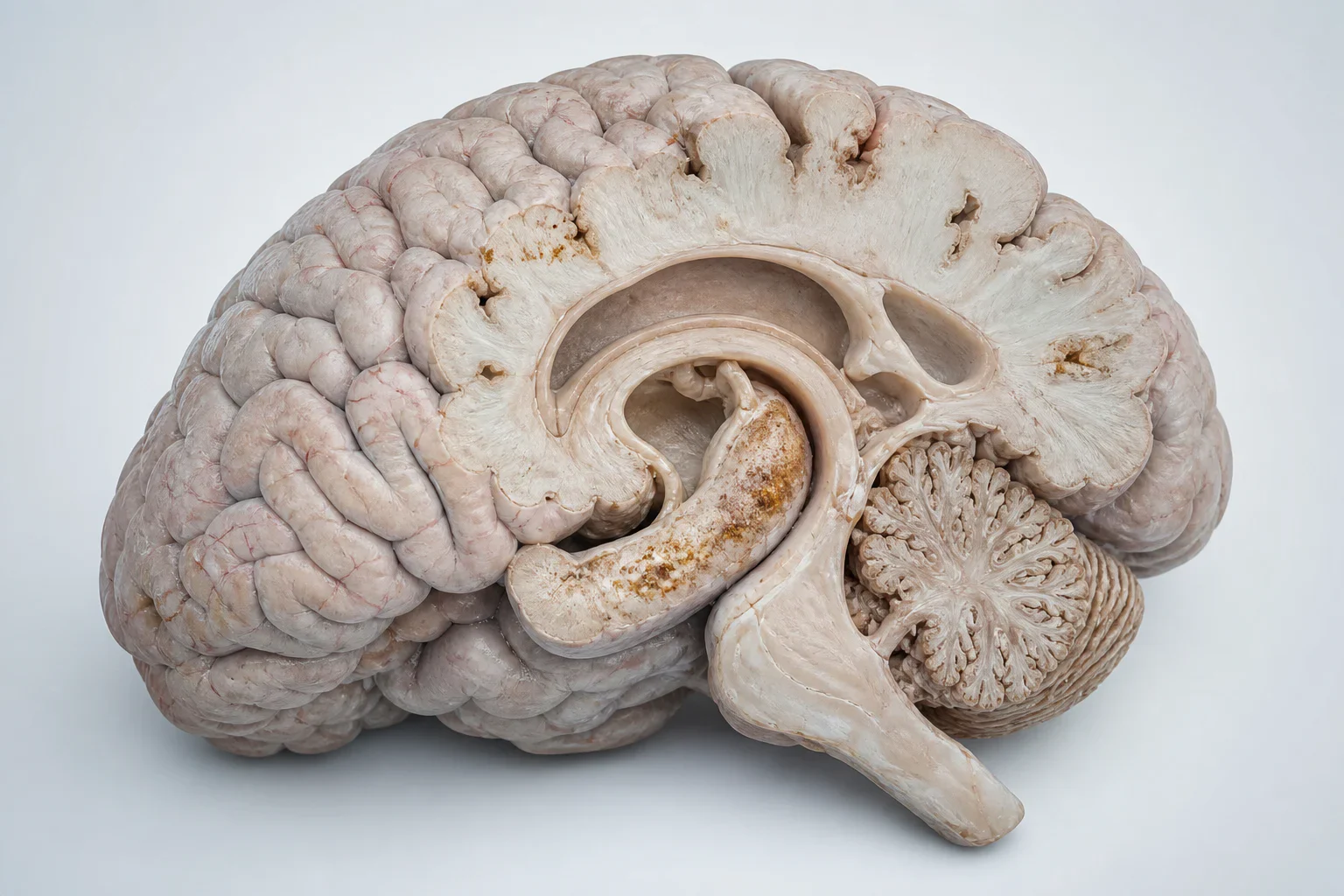
Pathology-informed reconstruction of a sagittal human brain specimen, with restrained emphasis on structures commonly discussed in Alzheimer’s disease. This is an educational illustration, not a diagnostic image.
Scientific media record1 verified identifier
- Subject
- Alzheimer Diseasecondition
- Identifiers
- MeSH:D000544
- Review
- Editorial review completeIdentifiers authority-verified · Accessibility validated · · alzheimers-disease|alzheimers-disease-pathology-v2.webp
- Digital source
- Trained-algorithmic mediaCreated with a trained generative algorithm and reviewed by WikiBiome for subject identity, scientific framing, identifiers, provenance, and accessibility.
- Scientific basis
- Alzheimer Disease — Medical Subject HeadingsWhat Happens to the Brain in Alzheimer's Disease?
- License
- CC BY-SA 4.0Created
The most common form of dementia, affecting an estimated 55 million people worldwide and projected to triple by 2050.[1]Recent advances in Alzheimer's disease: mechanisms, clinical trials and new drug development strategiesZhang Y, et al. · 2024Open reference 1 ↓ Characterized by progressive memory loss, cognitive decline, amyloid-beta plaques, and neurofibrillary tau tangles.[2]Recent Advances in Therapeutics for the Treatment of Alzheimer's DiseasePasseri E, et al. · 2024Open reference 2 ↓
From a Metallomics perspective, Alzheimer's disease presents one of the most complex metal signatures of any disease—featuring a central paradox of brain copper depletion alongside peripheral copper elevation, iron accumulation in plaques, and the strongest epidemiological evidence for lead as a neurodegenerative risk factor.
Approximately 23 source pages address AD's metallomic dimensions.
Evidence map121 cited passagesInspect provenance +
The most common form of dementia, affecting an estimated 55 million people worldwide and projected to triple by 2050. Characterized by progressive memory loss, cognitive decline, amyloid-beta plaques, and neurofibrillary tau tangles. From a metallomics perspective, Alzheimer's disease presents one of the most complex metal signatures of any disease—featur
Post-mortem brain metallomics reveals widespread Cu decreases across multiple brain regions in AD (also in DLB and PDD), with Cu changes contributing most to the multivariate separation between dementia types.
Cu depletion spans hippocampus, cingulate gyrus, middle temporal gyrus, substantia nigra, primary visual cortex, and putamen.
Ceruloplasmin dysfunction is a candidate mechanism: it both elevates circulating Cu and fails to deliver Cu to the brain.
Cu-amyloid-beta interactions promote toxic oligomer formation—Cu binds A-beta at histidine residues, catalyzing ROS production and accelerating aggregation.
Iron accumulates in hippocampus and cortex in AD.
Transferrin receptor and ferritin alterations documented in AD brain.
Pb is the most extensively studied metal in relation to AD; cumulative bone lead levels provide better exposure estimates than blood lead.
Higher tibia lead associated with cognitive decline in older adults.
Early-life Pb exposure produces latent effects on AD-related gene expression through epigenetic mechanisms that manifest decades later—the "developmental origins" hypothesis.
Pb disrupts calcium signaling (mimics Ca2+ in signaling pathways), promotes oxidative stress, and affects protein phosphorylation.
Pb induces BBB disruption, enabling further metal and toxin entry to the brain.
21 of 46 mechanistic studies reviewed focused on Pb—the most of any metal.
Zinc accumulates in amyloid plaques, where it induces A-beta aggregation; 100 uM Zn2+ produced 5-14 fold increases in A-beta aggregation rates in vitro.
Ni2+ enhances A-beta-40 aggregation 5.7-fold at 100 uM.
Commercial recombinant A-beta-40 preparations contain 1,005 ug Ni per gram of peptide, reflecting intrinsic Ni binding.
The nickel chelator dimethylglyoxime (DMG) inhibits A-beta-40 aggregation 40-85% in a dose-dependent manner.
Ni chelation represents a dual therapeutic strategy against both the "metal hypothesis" and the "infection hypothesis" of AD by disabling Ni-dependent pathogen enzymes (hydrogenase, urease).
Continues to be debated as a risk factor; some studies show increased Al in AD brain regions.
Arsenic: Increases A-beta(1-42) production and BACE1 activity; RAGE levels up 220-fold in animal models; dose-dependent tau phosphorylation via GSK3-beta and ERK1/2.
Cadmium: BBB disruption, calcium signaling disruption, mitochondrial dysfunction; blood Cd associated with lower cognitive scores,.
Mercury: Both inorganic and methylmercury are neurotoxic; can increase A-beta production and tau phosphorylation; fish consumption confounds epidemiology (source of MeHg but also neuroprotective omega-3).
Manganese: Impairs autophagy; Drp1 inhibition protective against Mn-induced autophagic impairment; acute Mn exposure increases seizure susceptibility in AD mouse models.
Se deficiency associated with increased neurodegeneration risk via impaired selenoproteins (glutathione peroxidases, thioredoxin reductases).
Showing 24 of 121 evidence-bearing passages. Every remaining citation is still indexed in the reference record below.
One disease. Five evidence layers.
A generated systems view of the metals, organisms, host sequestration signals, ecological conditions, and microbial functions indexed for Alzheimer's Disease.
Evidence layer
Taxonomic signature
Organisms reported as enriched or depleted, with their indexed functional context kept beside the name.Gram-negative pathogen associated with AD; neuroinflammatory LPS stimulus; gastric barrier disruption enabling translocation
Primary Gram-negative driver; amyloid-producing curli fibers cross-seed host amyloid-beta aggregation; iron siderophore competition
LPS-producing Gram-negative; neuroinflammatory stimulus; iron piracy; siderophore production
Enriched in AD; strict anaerobe indicating hypoxia; LPS endotoxin driver of neuroinflammation
Specific biomarker upregulated by cadmium in ApoE4 mice; pro-inflammatory phenotype; associated with neuroinflammation
Paradoxically upregulated in AD and cadmium (Cd)-exposed mice; mucin-degrading; barrier disruption; pro-inflammatory in disease context
Fungal dysbiosis in AD; functional shielding of bacterial pathogens; oxygen depletion creating anaerobic niches; metal-dependent biofilm formation
DEPLETED in AD; loss of probiotic properties and metal-binding capacity
SCFA producers — lost in metal-rich pro-inflammatory environment; colonocyte dysfunction when depleted
SCFA producers — depleted in dysbiotic AD microbiota; loss of butyrate support for BBB integrity
Butyrate producer — depleted in AD; protective against neuroinflammation via histone deacetylase inhibition
Depleted in dysbiotic AD signature; SCFA producer loss compromises intestinal barrier
A cited review reports the historical species label as decreased in AD; it does not establish causation or a species-specific mechanism
Commensal species depleted in AD; reduced diversity in Bacteroidetes phylum
Anti-inflammatory SCFA producer; depletion contributes to systemic inflammation and neuroinflammation
Evidence layer
Nutritional immunity
Host metal-withholding, inflammatory, antioxidant, and microbial-metabolite signals indexed in the signature.Elevated host signals
7Depleted protective signals
7Evidence layer
Ecological state
The environmental conditions that connect the organism-level observations into a system.Evidence layer
Virulence functions
Microbial structures, enzymes, and acquisition systems implicated by the linked evidence.The disease record, in full.
The original WikiBiome disease narrative remains intact beneath the generated signature atlas.
Metallomic Signature#
The Metal-Disease Matrix: A Cross-Source Synthesis identifies AD's profile as: copper (Cu) ↓ (brain), zinc (Zn) ↑↓ (plaques ↑, serum ↓), iron (Fe) ↑ (brain accumulation), selenium (Se) ↓, manganese (Mn) ↑↓, lead (Pb) ↑ (epigenetic), cadmium (Cd) ↑, mercury (Hg) ↑, As ↑, aluminum (Al) ↑ (brain).
The Copper Paradox: Brain Depletion, Peripheral Excess#
This is the defining metallomic feature of Alzheimer's disease. Post-mortem brain metallomics reveals widespread copper (Cu) decreases across multiple brain regions in AD (also in DLB and PDD), with copper changes contributing most to the multivariate separation between dementia types.[3]Scholefield et al. 2024 — Brain Metallomic Signatures Distinguish DLB from AD and PDDMelissa Scholefield, Stephanie J. Church, Jingshu Xu et al. · 2024Open reference 3 ↓
copper depletion spans hippocampus, cingulate gyrus, middle temporal gyrus, substantia nigra, primary visual cortex, and putamen.[3]Scholefield et al. 2024 — Brain Metallomic Signatures Distinguish DLB from AD and PDDMelissa Scholefield, Stephanie J. Church, Jingshu Xu et al. · 2024Open reference 3 ↓ Yet peripherally, copper is often normal or elevated—the paradox suggests disturbed copper trafficking rather than simple depletion.
Ceruloplasmin dysfunction is a candidate mechanism: it both elevates circulating copper and fails to deliver copper to the brain.[4]Common and Trace Metals in Alzheimer's and Parkinson's DiseasesDoroszkiewicz J, Farhan JA, Mroczko J et al. · 2023Open reference 4 ↓
copper-amyloid-beta interactions promote toxic oligomer formation—copper binds A-beta at Histidine residues, catalyzing ROS production and accelerating aggregation.[4]Common and Trace Metals in Alzheimer's and Parkinson's DiseasesDoroszkiewicz J, Farhan JA, Mroczko J et al. · 2023Open reference 4 ↓
copper depletion impairs cytochrome c oxidase (mitochondrial respiration), copper/zinc superoxide dismutase (Cu/Zn-SOD) (antioxidant defense), and ceruloplasmin (iron homeostasis) in brain tissue.
Iron: Accumulation and Ferroptosis#
Iron accumulates in hippocampus and cortex in AD.[4]Common and Trace Metals in Alzheimer's and Parkinson's DiseasesDoroszkiewicz J, Farhan JA, Mroczko J et al. · 2023Open reference 4 ↓ Iron-catalyzed Fenton reactions generate hydroxyl radicals driving lipid peroxidation. Ferroptosis (iron-dependent lipid peroxidation cell death) is an emerging cell death pathway in AD, parallel to its established role in Parkinson's Disease.
Transferrin receptor and ferritin alterations documented in AD brain.[4]Common and Trace Metals in Alzheimer's and Parkinson's DiseasesDoroszkiewicz J, Farhan JA, Mroczko J et al. · 2023Open reference 4 ↓ Iron chelation (deferiprone) is under investigation as a therapeutic strategy.
Lead: Strongest Epidemiological Evidence#
lead (Pb) is the most extensively studied metal in relation to AD; cumulative bone lead levels provide better exposure estimates than blood lead.[5]Heavy Metals Exposure and Alzheimer's Disease and Related DementiasBakulski KM, Seo YA, Hickman RC et al. · 2020Open reference 5 ↓
Higher tibia lead associated with cognitive decline in older adults.[5]Heavy Metals Exposure and Alzheimer's Disease and Related DementiasBakulski KM, Seo YA, Hickman RC et al. · 2020Open reference 5 ↓
Early-life lead exposure produces latent effects on AD-related gene expression through epigenetic mechanisms that manifest decades later—the "developmental origins" hypothesis.[5]Heavy Metals Exposure and Alzheimer's Disease and Related DementiasBakulski KM, Seo YA, Hickman RC et al. · 2020Open reference 5 ↓
lead disrupts calcium signaling (mimics calcium(II) (Ca2+) in signaling pathways), promotes Oxidative Stress, and affects protein phosphorylation.[4]Common and Trace Metals in Alzheimer's and Parkinson's DiseasesDoroszkiewicz J, Farhan JA, Mroczko J et al. · 2023Open reference 4 ↓ lead induces BBB disruption, enabling further metal and toxin entry to the brain.[6]Associations of Environmental Exposure to Arsenic, Manganese, Lead, and Cadmium with Alzheimer's Disease: A Review of Recent Evidence from Mechanistic StudiesGiasuddin Ahmed, Md. Shiblur Rahaman, Enrique Perez et al. · 2025Open reference 6 ↓
21 of 46 mechanistic studies reviewed focused on lead—the most of any metal.[6]Associations of Environmental Exposure to Arsenic, Manganese, Lead, and Cadmium with Alzheimer's Disease: A Review of Recent Evidence from Mechanistic StudiesGiasuddin Ahmed, Md. Shiblur Rahaman, Enrique Perez et al. · 2025Open reference 6 ↓
Zinc: Enriched in Plaques, Depleted Systemically#
Zinc accumulates in amyloid plaques, where it induces A-beta aggregation;[4]Common and Trace Metals in Alzheimer's and Parkinson's DiseasesDoroszkiewicz J, Farhan JA, Mroczko J et al. · 2023Open reference 4 ↓ 100 uM zinc(II) (Zn2+) produced 5-14 fold increases in A-beta aggregation rates in vitro.[7]Benoit & Maier 2021 — Nickel Chelator Inhibits Amyloid-Beta AggregationBenoit, S.L., Bhatt et al. · 2021Open reference 7 ↓
Serum/plasma zinc is reduced in AD patients. Zinc transporter dysfunction documented in AD brains.
The dual role of zinc—protective at physiological levels (antioxidant, synaptic function) but pathological when trapped in plaques—mirrors the copper (Cu) paradox.
Nickel: Newly Recognized A-beta Aggregation Driver#
nickel(II) (Ni2+) enhances A-beta-40 aggregation 5.7-fold at 100 uM.[7]Benoit & Maier 2021 — Nickel Chelator Inhibits Amyloid-Beta AggregationBenoit, S.L., Bhatt et al. · 2021Open reference 7 ↓ Commercial recombinant A-beta-40 preparations contain 1,005 ug nickel per gram of peptide, reflecting intrinsic nickel binding.[7]Benoit & Maier 2021 — Nickel Chelator Inhibits Amyloid-Beta AggregationBenoit, S.L., Bhatt et al. · 2021Open reference 7 ↓
The nickel chelator dimethylglyoxime (DMG) inhibits A-beta-40 aggregation 40-85% in a dose-dependent manner.[8]The nickel-chelator dimethylglyoxime inhibits human amyloid beta peptide in vitro aggregationBenoit SL, Bhatt RJ, Maier RJ · 2021Open reference 8 ↓
nickel chelation represents a dual therapeutic strategy against both the "metal hypothesis" and the "infection hypothesis" of AD by disabling nickel-dependent pathogen enzymes (Hydrogenase, Urease).[8]The nickel-chelator dimethylglyoxime inhibits human amyloid beta peptide in vitro aggregationBenoit SL, Bhatt RJ, Maier RJ · 2021Open reference 8 ↓
Aluminum: Controversial but Persistent#
aluminum (Al) accumulates in brain tissue in AD Metal-Disease Matrix: A Cross-Source Synthesis. Continues to be debated as a risk factor; some studies show increased aluminum in AD brain regions.[9]The Health Effects of Aluminum ExposureKatrin Klotz, Wobbeke Weistenhofer, Frauke Neff et al. · 2017Open reference 9 ↓ aluminum's neurotoxicity mechanisms include oxidative stress, inflammatory cytokine induction, and interference with iron homeostasis.
Other Toxic Metals#
Arsenic: Increases A-beta(1-42) production and BACE1 activity; RAGE levels up 220-fold in animal models; dose-dependent tau phosphorylation via GSK3-beta and ERK1/2.[6]Associations of Environmental Exposure to Arsenic, Manganese, Lead, and Cadmium with Alzheimer's Disease: A Review of Recent Evidence from Mechanistic StudiesGiasuddin Ahmed, Md. Shiblur Rahaman, Enrique Perez et al. · 2025Open reference 6 ↓
Cadmium: BBB disruption, calcium signaling disruption, mitochondrial dysfunction; blood cadmium (Cd) associated with lower cognitive scores.[5]Heavy Metals Exposure and Alzheimer's Disease and Related DementiasBakulski KM, Seo YA, Hickman RC et al. · 2020Open reference 5 ↓[6]Associations of Environmental Exposure to Arsenic, Manganese, Lead, and Cadmium with Alzheimer's Disease: A Review of Recent Evidence from Mechanistic StudiesGiasuddin Ahmed, Md. Shiblur Rahaman, Enrique Perez et al. · 2025Open reference 6 ↓
Mercury: Both inorganic and methylmercury are neurotoxic; can increase A-beta production and tau phosphorylation; fish consumption confounds epidemiology (source of methylmercury (MeHg) but also neuroprotective omega-3).[5]Heavy Metals Exposure and Alzheimer's Disease and Related DementiasBakulski KM, Seo YA, Hickman RC et al. · 2020Open reference 5 ↓
Manganese: Impairs autophagy; Drp1 inhibition protective against manganese (Mn)-induced autophagic impairment; acute manganese exposure increases seizure susceptibility in AD mouse models.[6]Associations of Environmental Exposure to Arsenic, Manganese, Lead, and Cadmium with Alzheimer's Disease: A Review of Recent Evidence from Mechanistic StudiesGiasuddin Ahmed, Md. Shiblur Rahaman, Enrique Perez et al. · 2025Open reference 6 ↓
Selenium Depletion#
selenium (Se) deficiency associated with increased neurodegeneration risk via impaired selenoproteins (glutathione peroxidases, thioredoxin reductases).[4]Common and Trace Metals in Alzheimer's and Parkinson's DiseasesDoroszkiewicz J, Farhan JA, Mroczko J et al. · 2023Open reference 4 ↓ selenium supplementation may have neuroprotective effects, though evidence is preliminary.
Gut Microbiome Connection#
The gut-brain axis is a major emerging pathway in AD metallomic research. AD patients show reduced Bacteroidetes diversity, decreased Firmicutes, and increased Gram-negative bacteria.[12]Diet, Microbiota and Brain Health: Unraveling the Network Intersecting Metabolism and NeurodegenerationGentile F, Doneddu PE, Riva N et al. · 2020Open reference 12 ↓ Increased H. pylori, Escherichia/Shigella, K. pneumoniae, B. fragilis; decreased Eubacterium rectale and Bacteroides in AD.[13]Effects of gut microbiota on neurodegenerative diseasesKhatoon S, Kalam N, Rashid S et al. · 2023Open reference 13 ↓
LPS enhances amyloid-beta fibrillization and triggers NF-kB signaling.[12]Diet, Microbiota and Brain Health: Unraveling the Network Intersecting Metabolism and NeurodegenerationGentile F, Doneddu PE, Riva N et al. · 2020Open reference 12 ↓
Bacterial amyloids (curli from Escherichia coli) can cross-seed with cerebral amyloid-beta, providing a direct microbial-to-neurodegeneration pathway.[12]Diet, Microbiota and Brain Health: Unraveling the Network Intersecting Metabolism and NeurodegenerationGentile F, Doneddu PE, Riva N et al. · 2020Open reference 12 ↓[14]Gut microbial imbalance and neurodegenerative proteinopathies: from molecular mechanisms to prospects of clinical applicationsAlonso-Garcia P, Martin R, Martinez-Pinilla E · 2021Open reference 14 ↓
TMAO (trimethylamine N-oxide) from gut bacteria traverses the BBB and is found at increased levels in CSF of cognitively impaired AD patients.[13]Effects of gut microbiota on neurodegenerative diseasesKhatoon S, Kalam N, Rashid S et al. · 2023Open reference 13 ↓
FMT from wild-type to AD mice alleviated cognitive impairment and reduced A-beta plaque burden.[12]Diet, Microbiota and Brain Health: Unraveling the Network Intersecting Metabolism and NeurodegenerationGentile F, Doneddu PE, Riva N et al. · 2020Open reference 12 ↓ SCFAs are reduced in AD; sodium Butyrate curbs cholesterol-induced neuronal amyloidosis.[12]Diet, Microbiota and Brain Health: Unraveling the Network Intersecting Metabolism and NeurodegenerationGentile F, Doneddu PE, Riva N et al. · 2020Open reference 12 ↓
The metal-microbiome connection: Heavy Metals (lead (Pb), cadmium (Cd), mercury (Hg), arsenic (As)) disrupt gut barrier integrity,[15]Effects of Heavy Metals on Gut Barrier Integrity and Gut MicrobiotaSweta Ghosh, Syam P. Nukavarpu, Venkatakrishna Rao Jala · 2024Open reference 15 ↓ reshaping the microbiome toward metal-tolerant, LPS-producing Gram-negative species. This gut-derived Metal-Driven Inflammation reaches the brain via the vagus nerve and systemic circulation, amplifying metal-induced neuroinflammation.
Environmental Metal Exposure Links#
Occupational exposure (welding, mining, battery manufacturing) provides high-dose lead (Pb) and manganese (Mn) exposure.[5]Heavy Metals Exposure and Alzheimer's Disease and Related DementiasBakulski KM, Seo YA, Hickman RC et al. · 2020Open reference 5 ↓ Drinking water contamination: arsenic (As) in water linked to cognitive deficits in some populations.[5]Heavy Metals Exposure and Alzheimer's Disease and Related DementiasBakulski KM, Seo YA, Hickman RC et al. · 2020Open reference 5 ↓
Air pollution: particulate matter carries metals (lead, manganese, nickel (Ni)) to the brain via olfactory pathway.[16]Environmental pollutants as risk factors for neurodegenerative disorders: Alzheimer and Parkinson diseasesChin-Chan M, Navarro-Yepes J, Quintanilla-Vega B · 2015Open reference 16 ↓ Dietary metals: fish (methylmercury (MeHg) but also omega-3), contaminated rice (arsenic, cadmium (Cd)), processed foods.[17]Heavy metals in the diet: unraveling the molecular pathways linked to neurodegenerative disease riskGuevara-Ramirez P, Tamayo-Trujillo R, Cadena-Ullauri S et al. · 2024Open reference 17 ↓
Cigarette smoking: major non-dietary cadmium source.[5]Heavy Metals Exposure and Alzheimer's Disease and Related DementiasBakulski KM, Seo YA, Hickman RC et al. · 2020Open reference 5 ↓
Developmental Vulnerability#
AD may have its origins decades before symptom onset.
Early-life lead (Pb) exposure alters epigenetic programming of AD-related genes, manifesting as disease 40-60 years later.[5]Heavy Metals Exposure and Alzheimer's Disease and Related DementiasBakulski KM, Seo YA, Hickman RC et al. · 2020Open reference 5 ↓ Obesity and T2D at midlife increase AD risk by up to 74%; metals contribute to both conditions.[12]Diet, Microbiota and Brain Health: Unraveling the Network Intersecting Metabolism and NeurodegenerationGentile F, Doneddu PE, Riva N et al. · 2020Open reference 12 ↓
The APOE4 genotype may modify metal-AD associations, creating gene-environment interactions.[5]Heavy Metals Exposure and Alzheimer's Disease and Related DementiasBakulski KM, Seo YA, Hickman RC et al. · 2020Open reference 5 ↓ Mixed metal exposures during development remain critically understudied.[5]Heavy Metals Exposure and Alzheimer's Disease and Related DementiasBakulski KM, Seo YA, Hickman RC et al. · 2020Open reference 5 ↓
Dietary Metal Paradoxes#
Full evidence is maintained on the canonical paradox articles.
Current Interventions with Metal Relevance#
| Intervention | Evidence | Metal Mechanism |
|---|---|---|
| Iron chelation (deferiprone) | Clinical trials ongoing[4]Common and Trace Metals in Alzheimer's and Parkinson's DiseasesDoroszkiewicz J, Farhan JA, Mroczko J et al. · 2023Open reference 4 ↓ | Reduces brain iron, limits Fenton chemistry and ferroptosis |
| Mediterranean diet | Moderate-strong[12]Diet, Microbiota and Brain Health: Unraveling the Network Intersecting Metabolism and NeurodegenerationGentile F, Doneddu PE, Riva N et al. · 2020Open reference 12 ↓ | Anti-inflammatory; antioxidant; but mixed metal exposure profile |
| Selenium supplementation | Preliminary[4]Common and Trace Metals in Alzheimer's and Parkinson's DiseasesDoroszkiewicz J, Farhan JA, Mroczko J et al. · 2023Open reference 4 ↓ | Restores GPx and thioredoxin reductase activity |
| Probiotics (Bifidobacterium breve A1) | Moderate[13]Effects of gut microbiota on neurodegenerative diseasesKhatoon S, Kalam N, Rashid S et al. · 2023Open reference 13 ↓ | Significant RBANS/MMSE improvement; reduces neuroinflammation |
| FMT | Preclinical[12]Diet, Microbiota and Brain Health: Unraveling the Network Intersecting Metabolism and NeurodegenerationGentile F, Doneddu PE, Riva N et al. · 2020Open reference 12 ↓ | Reduces A-beta plaques in AD mice; restores SCFA production |
| Lead exposure reduction | Public health[5]Heavy Metals Exposure and Alzheimer's Disease and Related DementiasBakulski KM, Seo YA, Hickman RC et al. · 2020Open reference 5 ↓ | Prevention: reducing cumulative lifetime lead (Pb) burden |
| Copper-targeted therapy | Theoretical | Must address the paradox: restore brain copper (Cu) while not worsening peripheral excess |
Open Questions#
Unresolved questions identified by the current evidence record.
01Can brain copper (Cu) be restored without raising peripheral levels?+
The copper (Cu) paradox demands compartment-specific therapeutics—a major pharmacological challenge.
02What is the critical window for lead (Pb) exposure?+
Epigenetic evidence points to early life, but cumulative bone lead (Pb) suggests lifelong accumulation matters.
03Is the Gut Microbiome a viable therapeutic target for AD?+
FMT results in mice are promising; human trials are needed.
04How do metal mixtures interact in AD risk?+
Nearly all studies examine single metals, but real-world exposure involves complex mixtures.
05Does aluminum genuinely contribute to AD, or is brain aluminum (Al) accumulation an epiphenomenon?+
Decades of debate remain unresolved.
06Can metallomic brain profiling become an in vivo diagnostic?+
Post-mortem data is extensive;[3]Scholefield et al. 2024 — Brain Metallomic Signatures Distinguish DLB from AD and PDDMelissa Scholefield, Stephanie J. Church, Jingshu Xu et al. · 2024Open reference 3 ↓ translating to MRI-based or biofluid-based diagnostics is the challenge.
07APOE4 gene-metal interactions: Does APOE genotype modify susceptibility to metal-driven AD pathways?+
The current WikiBiome record identifies this as an unresolved evidence gap.
Connections#
- Metals: Copper, Iron, Lead, Zinc, Aluminum, Cadmium, Mercury, Arsenic, Manganese, Selenium
- Concepts: Ferroptosis, Amyloid-Beta, Blood-Brain Barrier, oxidative stress, Epigenetic Modifications, Mis-Metallation, Gut-Brain Axis
- Analyses: Metal-Disease Matrix: A Cross-Source Synthesis, Dietary Metal Paradoxes: When Healthy Foods and Good Intentions Backfire
- Related diseases: Parkinson's Disease (shared iron (Fe)/copper (Cu) brain dysregulation, ferroptosis, gut-brain axis), Chronic Kidney Disease (shared ferroptosis pathway), Type 2 Diabetes ("type-3 diabetes" hypothesis)
- Pathogens: Helicobacter pylori (increased in AD gut), Escherichia coli (curli cross-seeding with A-beta)
- Interventions: Mediterranean Diet, Probiotics, Selenium Supplementation
- Neurodegeneration and Metals—AD is the most common neurodegenerative disease; ferroptosis, amyloid, and tau as convergent pathways
- Environmental Metal Exposure—lifetime cumulative lead (Pb), aluminum (Al), and occupational metal exposure as AD risk factors
- Glutathione (GSH)—GSH depletion in AD brain enables iron-driven oxidative damage
- Microbiome-Derived Metabolites—SCFA depletion and tryptophan shunting implicated in AD gut-brain signaling
- Biomarkers—brain metal accumulation (iron, copper, zinc (Zn)), plasma amyloid, and metallomic panels for AD
References 31
Numbered by first appearance in the article, then reconciled with its declared source list.
- 1
Zhang Y, et al. (2024). Recent advances in Alzheimer's disease: mechanisms, clinical trials and new drug development strategies. (Signal Transduction and Targeted Therapy / related journal).
- 2
Passeri E, et al. (2024). Recent Advances in Therapeutics for the Treatment of Alzheimer's Disease. (Brain Sciences / related journal).
- 3
★Melissa Scholefield, Stephanie J. Church, Jingshu Xu et al. (2024). Scholefield et al. 2024 — Brain Metallomic Signatures Distinguish DLB from AD and PDD. Frontiers in Neuroscience.
- 4
Doroszkiewicz J, Farhan JA, Mroczko J et al. (2023). Common and Trace Metals in Alzheimer's and Parkinson's Diseases. International Journal of Molecular Sciences.
- 5
★Bakulski KM, Seo YA, Hickman RC et al. (2020). Heavy Metals Exposure and Alzheimer's Disease and Related Dementias. Journal of Alzheimer's Disease.
- 6
★Giasuddin Ahmed, Md. Shiblur Rahaman, Enrique Perez et al. (2025). Associations of Environmental Exposure to Arsenic, Manganese, Lead, and Cadmium with Alzheimer's Disease: A Review of Recent Evidence from Mechanistic Studies. Journal of Xenobiotics.
- 7
Benoit, S.L., Bhatt et al. (2021). Benoit & Maier 2021 — Nickel Chelator Inhibits Amyloid-Beta Aggregation. Scientific Reports.
- 8
Benoit SL, Bhatt RJ, Maier RJ (2021). The nickel-chelator dimethylglyoxime inhibits human amyloid beta peptide in vitro aggregation. Scientific Reports.
- 9
Katrin Klotz, Wobbeke Weistenhofer, Frauke Neff et al. (2017). The Health Effects of Aluminum Exposure. Deutsches Arzteblatt International.
- 10
Gao C, Jiang J, Tan Y et al. (2023). Microglia in neurodegenerative diseases: mechanism and potential therapeutic targets. Signal Transduction and Targeted Therapy.
- 11
Bakulski KM, et al. (2025). Contributions of heavy metal exposure to late-onset Alzheimer's disease. (Behavioural Brain Research / related journal).
- 12
Gentile F, Doneddu PE, Riva N et al. (2020). Diet, Microbiota and Brain Health: Unraveling the Network Intersecting Metabolism and Neurodegeneration. International Journal of Molecular Sciences.
- 13
Khatoon S, Kalam N, Rashid S et al. (2023). Effects of gut microbiota on neurodegenerative diseases. Frontiers in Aging Neuroscience.
- 14
Alonso-Garcia P, Martin R, Martinez-Pinilla E (2021). Gut microbial imbalance and neurodegenerative proteinopathies: from molecular mechanisms to prospects of clinical applications. Exploration of Neuroprotective Therapy.
- 15
★Sweta Ghosh, Syam P. Nukavarpu, Venkatakrishna Rao Jala (2024). Effects of Heavy Metals on Gut Barrier Integrity and Gut Microbiota. Microbiota and Host.
- 16
★Chin-Chan M, Navarro-Yepes J, Quintanilla-Vega B (2015). Environmental pollutants as risk factors for neurodegenerative disorders: Alzheimer and Parkinson diseases. Frontiers in Cellular Neuroscience.
- 17
★Guevara-Ramirez P, Tamayo-Trujillo R, Cadena-Ullauri S et al. (2024). Heavy metals in the diet: unraveling the molecular pathways linked to neurodegenerative disease risk. Food and Agricultural Immunology.
- 18
Islam F, Shohag S, Akhter S et al. (2022). Exposure of metal toxicity in Alzheimer's disease: An extensive review. Frontiers in Pharmacology.
- 19
Jakubowska E, Hoppe-Mitera E, Sionek I et al. (2024). Metal toxicity exposure in Alzheimer's disease - literature review. Journal of Education, Health and Sport.
- 20
Armstrong RA, et al. (2024). Alzheimer's disease: the role of extrinsic factors in its development, an investigation of the evidence. (Journal of Alzheimer's Disease / related journal).
- 21
Althomali RH, Abbood MA, Saleh EAM et al. (2024). Exposure to heavy metals and neurocognitive function in adults: a systematic review. Environmental Sciences Europe.
- 22
★Monisha Jaishankar, Tenzin Tseten, Naresh Anbalagan et al. (2014). Toxicity, Mechanism and Health Effects of Some Heavy Metals. Interdisciplinary Toxicology.
- 23
★Puthiyavalappil Rasin, Ashwathi A V, Sabeel M Basheer et al. (2025). Exposure to Cadmium and Its Impacts on Human Health: A Short Review. Journal of Hazardous Materials Advances.
- 24
Stefano Romano, George M Savva, Janis R Bedarf (2021). Romano 2021 -- The Role of Microbiome-Host Interactions in the Development of Alzheimer's Disease. Frontiers in Pharmacology.
- 25
Pengya Zhang, Huizhen Zheng, Guangbo Qu (2021). Zhang 2021 -- Lead Exposure and Gut Microbiome Alterations in Neurodegeneration. Microbiome.
- 26
Angela Zhang, Megumi Matsushita, Liang Zhang et al. (2021). Cadmium exposure modulates the gut-liver axis in an Alzheimer's disease mouse model. Communications Biology.
- 27
Zhi Ling, Yiwen Cheng, Feng Yan et al. (2020). Ling 2020 -- Fecal Fungal Dysbiosis in Chinese Patients with Alzheimer's Disease. Frontiers in Cell and Developmental Biology.
- 28
Borghini R, Porpora MG, Casale R et al. (2020). Irritable Bowel Syndrome-Like Disorders in Endometriosis: Prevalence of Nickel Sensitivity and Effects of a Low-Nickel Diet. An Open-Label Pilot Study. Nutrients.
- 29
Yi Zhao, Haotian Zhao, Mengqi Li et al. (2023). Zhao 2023 -- Efficacy and Safety of Probiotics for the Treatment of Alzheimer's Disease, Mild Cognitive Impairment, and Parkinson's Disease. npj Parkinson's Disease.
- 30
Zara Siu Wa Chui, Lily Man Lee Chan, Esther Wan Hei Zhang et al. (2024). Effects of microbiome-based interventions on neurodegenerative diseases: a systematic review and meta-analysis. Scientific Reports.
- 31
Dorines Rosario, Saeed Shoaie (2025). Rosario 2025 -- Constraint-Based Modelling of Host-Microbiome Co-Metabolism in Alzheimer's and Parkinson's Disease. Microbiome.
Article network
Mentioned here 18
Pages linking here 44
Connect the evidence
Publicly readable discussion by ORCID-authenticated researchers. Questions, interpretation, methods, corrections, and new evidence stay attached to this record.
No discussion yet. Start with a precise question or a source-backed observation.
Activity and accepted changes
Accepted researcher context, editorial status, public discussion, and upstream Git revisions are shown together. Pending, declined, and withdrawn proposals remain private.
- published revision
Add reviewed neuroinflammation coverage batch
Karen Pendergrass · +1 −1
Inspect exact Git diff ↗ - published revision
Strengthen Metallomics and link high-leverage contexts
Karen Pendergrass · +1 −1
Inspect exact Git diff ↗ - published revision
Backfill heavy metals concept links
Karen Pendergrass · +1 −1
Inspect exact Git diff ↗ - published revision
Backfill oxidative stress concept links
Karen Pendergrass · +2 −2
Inspect exact Git diff ↗ - published revision
Backfill gut microbiome concept links
Karen Pendergrass · +1 −1
Inspect exact Git diff ↗ - published revision
Backfill inflammation concept links
Karen Pendergrass · +1 −1
Inspect exact Git diff ↗ - published revision
Add reviewed Histidine concept coverage
Karen Pendergrass · +1 −1
Inspect exact Git diff ↗ - published revision
Complete Hydrogenase contextual coverage
Karen Pendergrass · +1 −1
Inspect exact Git diff ↗ - published revision
Complete reviewed Urease contextual coverage
Karen Pendergrass · +1 −1
Inspect exact Git diff ↗ - published revision
massive wiki expansion: 149 stubs fixed, 100+ new pages, Rule 15 scan, keystone papers
WikiBiome Deploy Bot · +31 −31
Inspect exact Git diff ↗ - published revision
nightly maintenance: 94 stub demotions, 181 source_count fixes, 22 auto-discovered stubs, 5 adversarial audits, 3 boundary fixes, 3 evidence-level corrections
WikiBiome Deploy Bot · +1 −0
Inspect exact Git diff ↗ - published revision
cycle 1: health check + lint fixes + 8 ingests + 2 stubs + gestational-diabetes signature
WikiBiome Deploy Bot · +1 −0
Inspect exact Git diff ↗ - published revision
Deep citation pass on 10 disease entities + expand 3 thin entities
WikiBiome Deploy Bot · +13 −6
Inspect exact Git diff ↗ - published revision
Deepen metal/concept entities + 8 new sources for T1D/schizophrenia
WikiBiome Deploy Bot · +301 −0
Inspect exact Git diff ↗ - published revision
Citation integrity pass: 38 citations added, 24 parenthesized citations fixed
WikiBiome Deploy Bot · +1 −1
Inspect exact Git diff ↗