

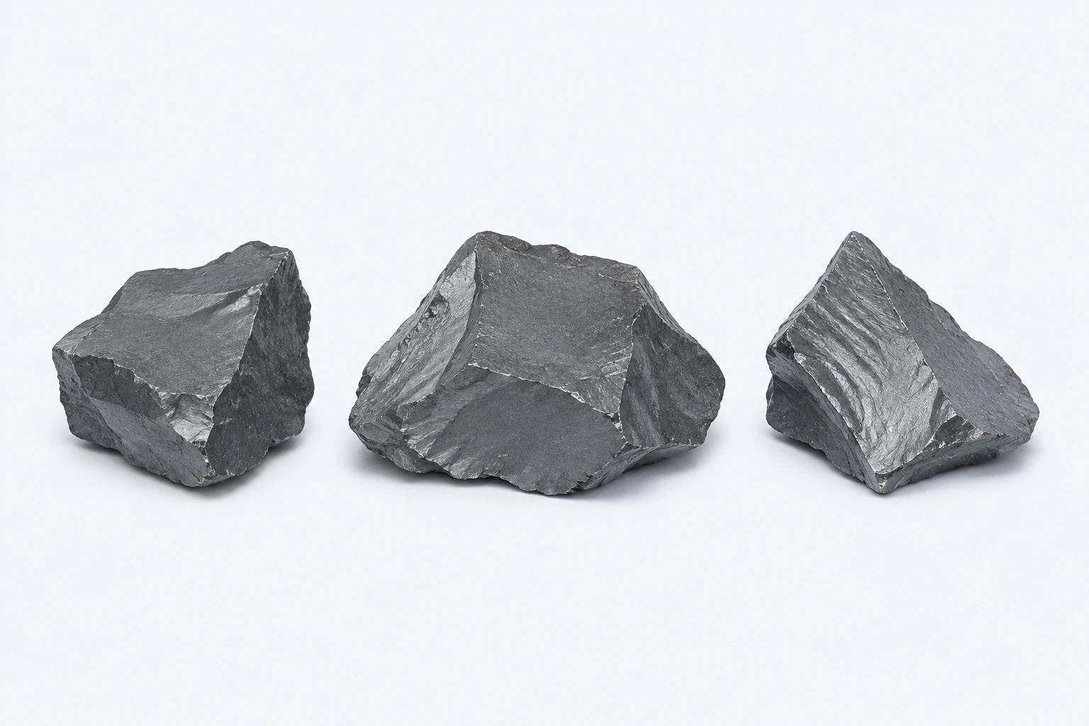
Gray elemental selenium (Se), shown as three representative dark steel-gray brittle-looking specimens. Selenium has multiple allotropes; this plate represents the gray form and is not powder, a supplement, analytical reference material, or a photograph.
Scientific media record2 verified identifiers
- Subject
- Seleniumelement
- Identifiers
- Atomic number 34PubChem CID:6326970
- Review
- Editorial review completeIdentifiers authority-verified · Accessibility validated · · selenium|selenium-technical-specimen-v1.webp
- Digital source
- Trained-algorithmic mediaCreated with a trained generative algorithm and reviewed by WikiBiome for subject identity, scientific framing, identifiers, provenance, and accessibility.
- Scientific basis
- Selenium — PubChem ElementSelenium, PubChem CID 6326970
- License
- CC BY-SA 4.0Created
An essential trace element that is primarily protective in its biological roles. Unlike most metals in this wiki, selenium's toxicological significance lies more in the consequences of its deficiency than its excess.
Selenium is the backbone of the selenoproteome—a family of ~25 selenoproteins including glutathione peroxidases, thioredoxin reductases, and iodothyronine deiodinases—that collectively defend against Oxidative Stress, regulate thyroid hormone metabolism, and modulate immune function.
Selenium depletion is among the most consistent metallomic findings across cancer, cardiovascular disease, inflammatory bowel disease, and neurodegeneration, positioning it as a critical node in the metal-microbe-disease landscape.
Evidence map89 cited passagesInspect provenance +
The thyroid gland contains the highest concentration of selenium per gram of any organ in the body.
IARC classification: Group 3 (not classifiable as carcinogenic to humans)—selenium and selenium compounds.
| Selenoprotein | Function | Disease Relevance | |---|---|---| | GPX1-4 (Glutathione peroxidases) | Reduce H2O2 and lipid hydroperoxides | Cancer, neurodegeneration, CKD | | GPX4 | Specifically prevents membrane lipid peroxidation | ferroptosis—loss of GPX4 is the key trigger | | GPX3 | Plasma-phase extracellular antioxidant | Functional Se status marker;
Antioxidant defense: GPX enzymes reduce H2O2 and lipid hydroperoxides; TrxR enzymes maintain the thioredoxin system—together they constitute the primary enzymatic antioxidant network.
Anti-ferroptotic activity: GPX4 specifically prevents lipid peroxidation in membranes; its loss triggers ferroptotic cell death. This mechanism links selenium deficiency directly to iron-dependent cellular damage.
Thyroid hormone activation: DIO1/2 convert inactive T4 to active T3; DIO3 inactivates T3—Se status directly controls thyroid hormone bioavailability,.
Immunomodulation: Se supports regulatory T cells and suppresses Th1/Th17 responses. Supplementation increases GPx3 and selenoprotein P1; decreases MDA (oxidative stress marker); modulates anti-TPO antibody production,.
H2O2 clearance in thyroid: Thyroglobulin iodination by thyroid peroxidase (TPO) generates substantial H2O2 via DUOX1/2. GPx1, GPx3, and GPx4 neutralize this H2O2, protecting thyrocytes from oxidative damage. Without adequate selenium, thyroid tissue accumulates oxidative damage during normal hormone synthesis.
Cereal selenium content varies geographically—soils in parts of China, Finland, New Zealand, and central Europe are Se-poor, creating population-level deficiency zones.
Fruit juices tested in Pakistan contained measurable selenium across all categories, though the primary concern in those samples was other metals (Ni, Pb, Fe) exceeding WHO limits.
Infant formula selenium concentration measured at 130 ug/kg in German market samples, well below the health-based guidance value.
Selenium in human milk decreases during SARS-CoV-2 infection: 0.5-fold reduction compared to prepandemic controls (p=0.0001), alongside reductions in iron, copper, and nickel.
Human milk selenium is part of the micronutrient layer of milk-mediated neonatal protection, complementing zinc, lactoferrin-iron handling, and HMOs.
200 ug/day selenomethionine is the most commonly used dose in clinical trials.
Combined selenomethionine + myoinositol has been tested in autoimmune thyroid disease trials.
Tampons: Se detected in 98.3% of samples tested, representing an underrecognized mucosal exposure route.
Infant gut diversity: In a prospective cohort of 146 mother-infant pairs, Shannon diversity in 3-month-old infants correlated positively with maternal selenium exposure (measured in hair) and negatively with copper—suggesting selenium promotes a more diverse early-life microbial ecosystem,.
This stands in contrast to toxic metals such as copper and arsenic, which at high levels enriched antibiotic resistance genes and drove dysbiosis in the same cohort.
Mercury antagonism: Selenium directly antagonizes mercury toxicity and protects gut microbiota from mercury-induced compositional disruption. The Se-Hg binding complex is biologically inert, and the Se:Hg molar ratio determines net toxicity,.
Selenoprotein protection: Selenium-dependent selenoproteins (GPX, TrxR) protect against the oxidative stress cascade generated by toxic metals (cadmium, lead, arsenic, mercury), which deplete glutathione through direct thiol binding. Selenium supplementation can partially restore antioxidant capacity in metal-exposed populations.
Cadmium antagonism: Selenium binds cadmium and facilitates biliary excretion, reducing cadmium's half-life in tissues. The cadmium-selenium antagonistic interaction in pregnant women is a developing research area with implications for developmental protection,.
The selenium-mercury interaction deserves special attention because mercury toxicity operates substantially through selenium biology. Mercury (Hg2+ and methylmercury) binds selenocysteine active sites in GPX and TrxR with higher affinity than these enzymes bind their native selenium, effectively displacing selenium and disabling the entire selenoproteome. Th
Gut bacteria participate in selenium absorption, biotransformation, and recycling. The bidirectional relationship between gut microbiota and essential metals including selenium is well established—gut bacteria modulate pH, oxidative balance, and detoxification enzyme activity, all of which influence selenium bioavailability.
In the thyroid-gut axis, gut microbiota influence selenium utilization for thyroid hormone synthesis. Bacterial dysbiosis may impair selenium absorption, contributing to the selenium and other micronutrient deficiencies observed in hashimotos thyroiditis,,.
Showing 24 of 89 evidence-bearing passages. Every remaining citation is still indexed in the reference record below.
Contents
1. Chemical Properties and Forms2. Biological Roles3. Dietary and Environmental Sources4. Microbiome Interactions5. Nutritional Immunity6. Conditions Associated7. Interactions with Other Metals8. Biomarkers9. Key Studies10. Open Questions11. Cross-ReferencesChemical Properties and Forms#
Metalloid (Group 16), incorporated into proteins as the amino acid selenocysteine (the "21st amino acid"). Key organic forms: selenomethionine (dietary, supplement), selenocysteine (in selenoproteins), methylselenocysteine. Inorganic forms: selenite (SeO3 2-), selenate (SeO4 2-)—used in some supplements.
Narrow therapeutic window: deficiency below ~40 ug/day; toxicity (selenosis) above ~400 ug/day. The thyroid gland contains the highest concentration of selenium per gram of any organ in the body.[1]McGregor Brock 2015 — The Role of Selenium in Thyroid Autoimmunity: A ReviewMcGregor Brock · 2015Open reference 1 ↓
IARC classification: Group 3 (not classifiable as carcinogenic to humans)—selenium and selenium compounds.[2]Heavy Metal Pollution in the Environment and Their Toxicological Effects on HumansBriffa J, Sinagra E, Blundell R · 2020Open reference 2 ↓
Biological Roles#
Selenium's biological significance is mediated almost entirely through its incorporation into selenoproteins as selenocysteine. These proteins form the backbone of multiple defense and regulatory systems.
The Selenoproteome#
| Selenoprotein | Function | Disease Relevance | |
|---|---|---|---|
| GPX1-4 (Glutathione peroxidases) | Reduce H2O2 and lipid hydroperoxides | Cancer, neurodegeneration, CKD | |
| GPX4 | Specifically prevents membrane lipid peroxidation | Ferroptosis—loss of GPX4 is the key trigger | |
| GPX3 | Plasma-phase extracellular antioxidant | Functional selenium (Se) status marker; decreased in fibromyalgia ([3]Duran-Gonzalez 2025 — Multi-Omics Diagnosis of FibromyalgiaDuran-Gonzalez et al. · 2025Open reference 3 ↓) | |
| TXNRD1/2 (Thioredoxin reductases) | Maintain thioredoxin in reduced state | Redox signaling, cancer | |
| DIO1/2/3 (Iodothyronine deiodinases) | Convert T4 to T3 (DIO1/2) or inactivate T3 (DIO3) | [[gut-thyroid-axis | thyroid-hormone-metabolism]] |
| Selenoprotein P (SELENOP) | selenium transport and delivery; antioxidant | Primary selenium status biomarker | |
| Selenoprotein S (SELENOS) | Anti-inflammatory; ER stress response | Genetic variants implicated in Hashimoto's susceptibility[4]Hu & Rayman 2017 — Multiple nutritional factors and the risk of Hashimoto's thyroiditisShiqian Hu, Margaret P Rayman · 2017Open reference 4 ↓ |
Core Biological Functions#
- Antioxidant defense: GPX enzymes reduce H2O2 and lipid hydroperoxides; TrxR enzymes maintain the thioredoxin system—together they constitute the primary enzymatic antioxidant network.[5]Toxicity, Mechanism and Health Effects of Some Heavy MetalsMonisha Jaishankar, Tenzin Tseten, Naresh Anbalagan et al. · 2014Open reference 5 ↓
- Anti-ferroptotic activity: GPX4 specifically prevents lipid peroxidation in membranes; its loss triggers ferroptotic cell death. This mechanism links selenium deficiency directly to iron-dependent cellular damage.[6]Microbial Metallomics and Parkinson's Disease: A Unified Metal-Driven Framework Linking Ferroptosis, Dysbiosis, and alpha-Synuclein PathologyKaren Pendergrass · 2025Open reference 6 ↓
- Thyroid hormone activation: DIO1/2 convert inactive T4 to active T3; DIO3 inactivates T3—selenium (Se) status directly controls thyroid hormone bioavailability.[1]McGregor Brock 2015 — The Role of Selenium in Thyroid Autoimmunity: A ReviewMcGregor Brock · 2015Open reference 1 ↓[7]Effects of Trace Elements on Endocrine Function and Pathogenesis of Thyroid Diseases — A Literature ReviewBrylinski L, Kostelecka K, Wolinski F et al. · 2025Open reference 7 ↓
- Immunomodulation: selenium supports regulatory T cells and suppresses Th1/Th17 responses. Supplementation increases GPx3 and selenoprotein P1; decreases MDA (oxidative stress marker); modulates anti-TPO antibody production.[8]Kravchenko 2023 — Thyroid hormones and minerals in immunocorrection of disorders in autoimmune thyroid diseasesKravchenko V, Zakharchenko T · 2023Open reference 8 ↓[9]Recent advances of trace elements in autoimmune thyroid diseaseLi S, Xu Q, Wang S et al. · 2025Open reference 9 ↓
- H2O2 clearance in thyroid: Thyroglobulin iodination by thyroid peroxidase (TPO) generates substantial H2O2 via DUOX1/2. GPx1, GPx3, and GPx4 neutralize this H2O2, protecting thyrocytes from oxidative damage. Without adequate selenium, thyroid tissue accumulates oxidative damage during normal hormone synthesis.[4]Hu & Rayman 2017 — Multiple nutritional factors and the risk of Hashimoto's thyroiditisShiqian Hu, Margaret P Rayman · 2017Open reference 4 ↓
Dietary and Environmental Sources#
Dietary Sources#
Brazil nuts are the highest natural source of selenium (ranges vary widely with soil selenium, but can deliver >50 ug per nut). Seafood, organ meats, eggs, and dairy products are reliable dietary sources.
Cereal selenium content varies geographically—soils in parts of China, Finland, New Zealand, and central Europe are selenium (Se)-poor, creating population-level deficiency zones.[1]McGregor Brock 2015 — The Role of Selenium in Thyroid Autoimmunity: A ReviewMcGregor Brock · 2015Open reference 1 ↓
Fruit juices tested in Pakistan contained measurable selenium across all categories, though the primary concern in those samples was other metals (nickel (Ni), lead (Pb), iron (Fe)) exceeding WHO limits.[10]Evaluation of Heavy Metals Content in the Canned/Packed Fruit Juices from Local and Imported Origin in Lahore, PakistanNazeefa Fatima, Munazza Khan, Muhammad Shuaib Kabeer · 2020Open reference 10 ↓
Infant Exposure#
Infant formula selenium concentration measured at 130 ug/kg in German market samples, well below the health-based guidance value.[11]The contribution of infant formula to the food survey-based dietary exposure of nine selected elementsHopfner T, Wollenberg M, Jager A et al. · 2025Open reference 11 ↓
Selenium in human milk decreases during SARS-CoV-2 infection: 0.5-fold reduction compared to prepandemic controls (p=0.0001), alongside reductions in iron, copper, and nickel.[12]Metallomic and Untargeted Metabolomic Signatures of Human Milk from SARS-CoV-2 Positive MothersArias-Borrego A, Soto Cruz FJ, Selma-Royo M et al. · 2022Open reference 12 ↓
Human milk selenium is part of the micronutrient layer of milk-mediated neonatal protection, complementing zinc, lactoferrin-iron handling, and HMOs.[13]Sami 2023 — Human Milk Nutrients Preventing NECSami et al. · 2023Open reference 13 ↓
Supplementation Forms#
200 ug/day selenomethionine is the most commonly used dose in clinical trials.[1]McGregor Brock 2015 — The Role of Selenium in Thyroid Autoimmunity: A ReviewMcGregor Brock · 2015Open reference 1 ↓ Combined selenomethionine + myoinositol has been tested in autoimmune thyroid disease trials.[8]Kravchenko 2023 — Thyroid hormones and minerals in immunocorrection of disorders in autoimmune thyroid diseasesKravchenko V, Zakharchenko T · 2023Open reference 8 ↓
The optimal selenium form (selenomethionine vs. sodium selenite vs. selenium (Se)-enriched yeast) remains an open question across disease contexts.
Other Exposure Routes#
- Tampons: selenium (Se) detected in 98.3% of samples tested, representing an underrecognized mucosal exposure route.[14]Tampons as a Source of Exposure to Metal(loid)sJenni A. Shearston, Kristen Upson, Milo Gordon et al. · 2024Open reference 14 ↓
Microbiome Interactions#
This is where WikiBiome's coverage departs most significantly from conventional selenium literature. The selenium-microbiome axis is bidirectional: selenium status shapes microbial communities, and gut bacteria influence selenium bioavailability and metabolism.
Selenium Shapes Gut Microbial Diversity#
Infant gut diversity: In a prospective cohort of 146 mother-infant pairs, Shannon diversity in 3-month-old infants correlated positively with maternal selenium exposure (measured in hair) and negatively with copper—suggesting selenium promotes a more diverse early-life microbial ecosystem.[15]Xiong 2025 — Prenatal Exposure to Trace Elements Impacts Mother-Infant Gut Microbiome, Metabolome and Resistome During the First Year of LifeShimao Xiong, Bing Xie, Naiyi Yin et al. · 2025Open reference 15 ↓[16]Xiong 2025 — Prenatal Exposure to Trace Elements Impacts Mother-Infant Gut Microbiome, Metabolome and ResistomeShimao Xiong, Bing Xie, Naiyi Yin et al. · 2025Open reference 16 ↓
This stands in contrast to toxic metals such as copper and arsenic, which at high levels enriched antibiotic resistance genes and drove Dysbiosis in the same cohort.[15]Xiong 2025 — Prenatal Exposure to Trace Elements Impacts Mother-Infant Gut Microbiome, Metabolome and Resistome During the First Year of LifeShimao Xiong, Bing Xie, Naiyi Yin et al. · 2025Open reference 15 ↓
Selenium Protects Against Metal-Induced Dysbiosis#
Mercury antagonism: Selenium directly antagonizes mercury toxicity and protects gut microbiota from mercury-induced compositional disruption. The selenium (Se)-mercury (Hg) binding complex is biologically inert, and the selenium:mercury molar ratio determines net toxicity.[17]Rezazadegan 2025 — Major Heavy Metals and Human Gut Microbiota Composition: A Systematic Review with Nutritional ApproachMahsa Rezazadegan, Bita Forootani, Yeganeh Hoveyda et al. · 2025Open reference 17 ↓[1]McGregor Brock 2015 — The Role of Selenium in Thyroid Autoimmunity: A ReviewMcGregor Brock · 2015Open reference 1 ↓
Selenoprotein protection: Selenium-dependent selenoproteins (GPX, TrxR) protect against the oxidative stress cascade generated by toxic metals (cadmium, lead, arsenic, mercury), which deplete glutathione through direct thiol binding.[5]Toxicity, Mechanism and Health Effects of Some Heavy MetalsMonisha Jaishankar, Tenzin Tseten, Naresh Anbalagan et al. · 2014Open reference 5 ↓ Selenium supplementation can partially restore antioxidant capacity in metal-exposed populations.[18]The Effects of Cadmium ToxicityGiuseppe Genchi, Maria Stefania Sinicropi, Graziantonio Lauria et al. · 2020Open reference 18 ↓
Cadmium antagonism: Selenium binds cadmium and facilitates biliary excretion, reducing cadmium's half-life in tissues. The cadmium-selenium antagonistic interaction in pregnant women is a developing research area with implications for developmental protection.[19]Developmental Toxicity of Cadmium in Infants and Children: A ReviewChandravanshi L, Shiv K, Kumar S · 2021Open reference 19 ↓[1]McGregor Brock 2015 — The Role of Selenium in Thyroid Autoimmunity: A ReviewMcGregor Brock · 2015Open reference 1 ↓
Mercury Disrupts Selenoprotein Function#
The selenium-mercury interaction deserves special attention because mercury toxicity operates substantially through selenium biology. Mercury (mercury(II) (Hg2+) and methylmercury) binds selenocysteine active sites in GPX and TrxR with higher affinity than these enzymes bind their native selenium, effectively displacing selenium and disabling the entire selenoproteome.[5]Toxicity, Mechanism and Health Effects of Some Heavy MetalsMonisha Jaishankar, Tenzin Tseten, Naresh Anbalagan et al. · 2014Open reference 5 ↓
This means mercury toxicity is not merely additive with selenium deficiency—it is mechanistically dependent on selenoprotein disruption. An individual with adequate selenium may tolerate higher mercury exposure because the selenoprotein reserve buffers against displacement, while a selenium-deficient individual is acutely vulnerable.
Gut Microbiota Influence Selenium Metabolism#
Gut bacteria participate in selenium absorption, biotransformation, and recycling. The bidirectional relationship between gut microbiota and essential metals including selenium is well established—gut bacteria modulate pH, oxidative balance, and detoxification enzyme activity, all of which influence selenium bioavailability.[20]Zhu 2024 — Toxic and Essential Metals: Metabolic Interactions with the Gut Microbiota and Health ImplicationsQinheng Zhu, Boyan Chen, Fu Zhang et al. · 2024Open reference 20 ↓
In the thyroid-gut axis, gut microbiota influence selenium utilization for thyroid hormone synthesis.
Bacterial dysbiosis may impair selenium absorption, contributing to the selenium and other micronutrient deficiencies observed in Hashimoto's Thyroiditis.[21]Identifying the metabolic profile of Hashimoto's thyroiditis from the METHAP clinical studySarandi E, Tsoukalas D, Rudofsky G et al. · 2025Open reference 21 ↓[22]The relationship between thyroid and human-associated microbiota: A systematic review of reviewsVirili C, Stramazzo I, Bagaglini MF et al. · 2024Open reference 22 ↓[23]Gut Microbiota and Thyroid Diseases: A Comprehensive Review of Mechanisms and Clinical ImplicationsBao K, Lin H, Guo S · 2025Open reference 23 ↓
Probiotic-Selenium Synergy#
- In an RCT of 152 occupational metal workers, probiotic yogurt containing Pediococcus acidilactici GR-1 reduced blood copper (34.45%) and nickel (38.34%) after 12 weeks. Enriched Blautia and Bifidobacterium abundance correlated positively with antioxidant capacity, demonstrating that selenium-probiotic co-supplementation could further enhance protection against heavy metal-driven dysbiosis.[24]Feng 2022 — Human Supplementation with Pediococcus acidilactici GR-1 Decreases Heavy Metals Levels through Modifying Gut Microbiota and MetabolomePengya Feng, Jinfeng Yang, Shuai Zhao et al. · 2022Open reference 24 ↓
Nutritional Immunity#
Selenium occupies an unusual position in the nutritional immunity framework. Unlike iron, zinc, manganese, and nickel—which are actively sequestered by the host to starve pathogens—selenium does not appear to function as a direct antimicrobial withholding metal. Instead, selenium supports immune function indirectly through selenoprotein-dependent pathways.
Th1/Th2/Treg balance: Selenium deficiency skews immune responses. Mice on high-selenium (Se) diets showed increased T cell receptor signaling in CD4+ T cells and shifted balance toward Th1 phenotype. In humans, selenium supplementation increases Tregs and reduces Foxp3 deficiency.[9]Recent advances of trace elements in autoimmune thyroid diseaseLi S, Xu Q, Wang S et al. · 2025Open reference 9 ↓
Anti-inflammatory selenoproteins: Selenoprotein S (SELENOS) is a key anti-inflammatory protein in the ER stress response. Genetic variants in SELENOS are implicated in Hashimoto's thyroiditis susceptibility.[4]Hu & Rayman 2017 — Multiple nutritional factors and the risk of Hashimoto's thyroiditisShiqian Hu, Margaret P Rayman · 2017Open reference 4 ↓
Magnesium-selenium interaction: Magnesium modulates selenium bioavailability and tissue distribution, indirectly supporting T4-to-T3 conversion as a cofactor for deiodinases. Serum magnesium (Mg) <0.55 mmol/L is associated with higher TgAb positivity.[9]Recent advances of trace elements in autoimmune thyroid diseaseLi S, Xu Q, Wang S et al. · 2025Open reference 9 ↓
Conditions Associated#
Thyroid Autoimmunity#
The selenium-thyroid axis is among the most clinically significant and well-evidenced associations in trace element medicine.
Hashimoto's thyroiditis. selenium (Se) levels are significantly lower in HT patients (104.36 ug/l) and GD patients (97.68 ug/l) compared to controls (122.63 ug/l, P<0.001).[9]Recent advances of trace elements in autoimmune thyroid diseaseLi S, Xu Q, Wang S et al. · 2025Open reference 9 ↓
Network meta-analysis of 7 RCTs: Selenium supplementation (200 ug/day selenomethionine) significantly reduced TPOAb (SMD -2.44, 95% CI -4.19 to -0.69) and TgAb (SMD -2.76, 95% CI -4.50 to -1.02) in euthyroid Hashimoto's patients.
A 6-month treatment duration was required for detectable effects; 3-month studies were insufficient.[25]Peng 2024 — Effects of different supplements on Hashimoto's thyroiditis: network meta-analysisBingcong Peng, Weiwei Wang, Qingling Gu et al. · 2024Open reference 25 ↓
A separate meta-analysis confirmed selenium supplementation reduced FT3 (MD=-0.40, P=0.009), FT4 (MD=-0.76, P=0.07), and TPOAb (MD=-150.25, P<0.00001).[9]Recent advances of trace elements in autoimmune thyroid diseaseLi S, Xu Q, Wang S et al. · 2025Open reference 9 ↓
selenium monotherapy (83 ug/day selenomethionine, 196 patients, 4 months) normalized TSH in 17.2% of subjects.[9]Recent advances of trace elements in autoimmune thyroid diseaseLi S, Xu Q, Wang S et al. · 2025Open reference 9 ↓ Combined selenomethionine + myoinositol reduced TSH, TPOAb, and TgAb after 6 and 12 months.[8]Kravchenko 2023 — Thyroid hormones and minerals in immunocorrection of disorders in autoimmune thyroid diseasesKravchenko V, Zakharchenko T · 2023Open reference 8 ↓
selenium is essential for GPx-mediated H2O2 clearance during thyroglobulin iodination; selenoprotein S genetic variants are implicated in HT risk.[4]Hu & Rayman 2017 — Multiple nutritional factors and the risk of Hashimoto's thyroiditisShiqian Hu, Margaret P Rayman · 2017Open reference 4 ↓
Greatest antibody reduction (40%) seen in those with anti-TPO >1200 IU/mL.[1]McGregor Brock 2015 — The Role of Selenium in Thyroid Autoimmunity: A ReviewMcGregor Brock · 2015Open reference 1 ↓
Graves' disease and ophthalmopathy. GD patients with ophthalmopathy have even lower selenium (96.82 ug/dL) than GD patients without (94.53 ug/dL) vs. controls (102.55 ug/dL).[9]Recent advances of trace elements in autoimmune thyroid diseaseLi S, Xu Q, Wang S et al. · 2025Open reference 9 ↓
Double-blind RCT: 200 ug selenium/day for 6 months significantly decreased GO severity, improved quality of life; benefits persisted after withdrawal.[1]McGregor Brock 2015 — The Role of Selenium in Thyroid Autoimmunity: A ReviewMcGregor Brock · 2015Open reference 1 ↓
selenium deficiency is an independent risk factor for Graves' ophthalmopathy.[1]McGregor Brock 2015 — The Role of Selenium in Thyroid Autoimmunity: A ReviewMcGregor Brock · 2015Open reference 1 ↓
Pregnancy. Selenomethionine 200 ug/day during pregnancy and postpartum decreased risk of permanent hypothyroidism and postpartum thyroiditis.[1]McGregor Brock 2015 — The Role of Selenium in Thyroid Autoimmunity: A ReviewMcGregor Brock · 2015Open reference 1 ↓
The iodine-selenium interaction is clinically important: in combined selenium and iodine deficiency, normalizing selenium without iodine worsens hypothyroidism; selenium co-administration maintains GPx function during iodine repletion.[1]McGregor Brock 2015 — The Role of Selenium in Thyroid Autoimmunity: A ReviewMcGregor Brock · 2015Open reference 1 ↓
Hashimoto's and thyroid cancer. In a meta-analysis of 65 studies (n=47,237), coexisting HT in papillary thyroid carcinoma (PTC) patients was associated with lower risk of lymph node metastasis (OR 0.787), distant metastasis (OR 0.435), and better 20-year survival (OR 1.396).
Selenium status is a candidate mediator of this protective effect, alongside the immune microenvironment created by HT lymphocytic infiltration.[26]Tang 2022 — Hashimoto Thyroiditis and Clinical Outcomes of Papillary Thyroid Carcinoma: A Meta-AnalysisQizhi Tang, Weiyu Pan, Liangyue Peng · 2022Open reference 26 ↓
Cancer#
Selenium depletion is one of the most consistent findings across cancer metallomic studies.
Pan-cancer metallomic pattern: selenium (Se) tends to decrease across cancer types in blood/serum/plasma, alongside copper (Cu) elevation and zinc (Zn) depletion. Selenoprotein gene variants (GPX1, GPX4, TXNRD1/2) are associated with cancer risk and susceptibility.[27]Recent advances in the application of metallomics in diagnosis and prognosis of human cancerYan Zhang, Jie He, Jiao Jin et al. · 2022Open reference 27 ↓
Prostate cancer: selenium significantly decreased (0.07 vs 0.13 ug/ml, p<0.005); low selenium correlates with carcinogenesis risk through impaired antioxidant defense.[28]Serum Levels of Selenium, Zinc, Copper, Manganese, and Iron in Prostate Cancer PatientsSaleh A. K. Saleh, Heba M. Adly, Altaf A. Abdelkhaliq et al. · 2020Open reference 28 ↓
Hepatocellular carcinoma: In non-cirrhotic HCC, tumor tissue shows metal depletion relative to non-tumoral liver for most elements including selenium, suggesting cancer cells systematically modulate their metal metabolism.
selenium concentration in tumor tissue was associated with extended survival only in the Peruvian cohort (above 1.48 ug/g: mean survival 323 weeks vs. 50 weeks below threshold, p=0.048).[29]Metallomic profile in non-cirrhotic hepatocellular carcinoma supports a phenomenon of metal metabolism adaptation in tumor cellsCano L, Bertani S, Island ML et al. · 2021Open reference 29 ↓
Lung cancer/COPD: selenium elevated 1.23-fold in the COPD-to-lung-cancer transition group vs. healthy controls; the normally strong zinc-selenium correlation (rho=0.69) is disrupted in disease states.[30]Metallomic Signatures of Lung Cancer and Chronic Obstructive Pulmonary DiseaseBelen Callejon-Leblic, Saida Sanchez Espirilla, Carolina Gotera-Rivera et al. · 2023Open reference 30 ↓
Breast cancer: Decreased selenium reported in multiple metallomic studies alongside elevated copper.[27]Recent advances in the application of metallomics in diagnosis and prognosis of human cancerYan Zhang, Jie He, Jiao Jin et al. · 2022Open reference 27 ↓
However, a large prospective study (Sister Study, n=1,495 cases) using toenail biomarkers found little evidence for individual metal-breast cancer associations; the authors note toenail selenium reflects longer-term exposure (6-12 months) which may explain discrepancies with blood/serum studies.[31]Metals and Breast Cancer Risk: A Prospective Study Using Toenail BiomarkersNiehoff NM, O'Brien KM, Keil AP et al. · 2021Open reference 31 ↓
Cadmium-selenium interaction in breast cancer: Selenium may have a modified protective effect against cadmium-driven breast carcinogenesis, where cadmium acts as a metalloestrogen by binding ERa (Kd = 4.5x10^-10 M).[32]Cadmium and breast cancer - Current state and research gaps in the underlying mechanismsTarhonska K, Lesicka M, Janasik B et al. · 2022Open reference 32 ↓
Cardiovascular Disease#
selenium (Se) decreased in AMI patients (90.31 vs. 99.98 ng/mL, p<0.01), with persistent depression at 6 months post-event.[33]Plasma metallomics reveals potential biomarkers and insights into the ambivalent associations of elements with acute myocardial infarctionSi Ying Lim, Hiranya Dayal, Song Jie Seah et al. · 2023Open reference 33 ↓
copper (Cu)/selenium ratio increased in AMI and showed significant longitudinal trajectory; identified as one of the most specific AMI biomarkers. A 10-feature random forest model incorporating metallomic ratios (copper/selenium, iron (Fe)/copper) with traditional risk factors achieved AUC 0.942.[33]Plasma metallomics reveals potential biomarkers and insights into the ambivalent associations of elements with acute myocardial infarctionSi Ying Lim, Hiranya Dayal, Song Jie Seah et al. · 2023Open reference 33 ↓
copper elevated and selenium decreased represents a consistent cardiovascular risk metallomic signature.[33]Plasma metallomics reveals potential biomarkers and insights into the ambivalent associations of elements with acute myocardial infarctionSi Ying Lim, Hiranya Dayal, Song Jie Seah et al. · 2023Open reference 33 ↓
Inflammatory Bowel Disease#
In a Greek cross-sectional study (76 CD + 39 UC + 38 HC), plasma selenium was significantly lower in both Crohn's disease (50 ug/L) and ulcerative colitis (44 ug/L) compared to healthy controls (77 ug/L, p=0.009).[34]Clinical and inflammatory biomarkers of inflammatory bowel diseases are linked to plasma trace elements and toxic metals; new insights into an old conceptAmerikanou C, Karavoltsos S, Gioxari A et al. · 2022Open reference 34 ↓
Manganese, zinc, and strontium were also depleted in IBD, while nickel was elevated in active Crohn's disease.[34]Clinical and inflammatory biomarkers of inflammatory bowel diseases are linked to plasma trace elements and toxic metals; new insights into an old conceptAmerikanou C, Karavoltsos S, Gioxari A et al. · 2022Open reference 34 ↓
The selenium depletion in IBD parallels findings across autoimmune conditions and may reflect both malabsorption from inflamed gut epithelium and increased antioxidant demand from chronic Metal-Driven Inflammation.
Neurodegeneration#
Selenium deficiency is associated with increased neurodegeneration risk; some studies suggest selenium (Se) supplementation may have neuroprotective effects.[35]Common and Trace Metals in Alzheimer's and Parkinson's DiseasesDoroszkiewicz J, Farhan JA, Mroczko J et al. · 2023Open reference 35 ↓
In dementia with Lewy bodies (DLB), selenium deficiency in primary visual cortex may compound effects of copper (Cu) loss by impairing GPx and TrxR activity.[36]Scholefield et al. 2024 — Brain Metallomic Signatures Distinguish DLB from AD and PDDMelissa Scholefield, Stephanie J. Church, Jingshu Xu et al. · 2024Open reference 36 ↓
selenium's role in GPX4 directly connects to Ferroptosis resistance in neurons—the same pathway implicated in Parkinson's disease dopaminergic neuron death. Iron accumulation in the substantia nigra catalyzes Fenton reactions driving lipid peroxidation; GPX4 downregulation removes the brake on ferroptotic cell death.[6]Microbial Metallomics and Parkinson's Disease: A Unified Metal-Driven Framework Linking Ferroptosis, Dysbiosis, and alpha-Synuclein PathologyKaren Pendergrass · 2025Open reference 6 ↓
A systematic review of 8 studies (n=1,828,126) found significant correlation between cadmium and mercury exposure and deleterious neurocognitive outcomes in adults.
The selenium-mercury interaction is relevant here: mercury (Hg) displaces selenium from selenocysteine active sites, and adequate selenium status may buffer against mercury-driven cognitive decline.[37]Exposure to heavy metals and neurocognitive function in adults: a systematic reviewAlthomali RH, Abbood MA, Saleh EAM et al. · 2024Open reference 37 ↓
Kidney Function#
- In a prospective Swiss cohort (n=4,704, mean follow-up 12.5 years), urinary selenium was associated with increased risk of incident impaired kidney function or CKD (HR 1.48). This paradoxical finding—an essential protective element associated with kidney risk—likely reflects that urinary selenium (Se) is a marker of renal filtration dynamics rather than selenium toxicity per se.[38]Association between urinary heavy metal/trace element concentrations and kidney function: a prospective studyXie S, Perrais M, Golshayan D et al. · 2025Open reference 38 ↓
PCOS#
A systematic review of 15 studies found that selenium (Se) supplementation shows potential benefits for reducing oxidative stress and improving endocrinological parameters in PCOS women.[39]Heavy Metals and Essential Elements in Association with Oxidative Stress in Women with Polycystic Ovary Syndrome -- A Systematic ReviewSmovrsnik T, Virant-Klun I, Pinter B · 2023Open reference 39 ↓
However, a Slovenian case-control study (n=70) found no significant differences in selenium between PCOS and control groups, contrasting with the consistent copper (Cu) elevation and novel molybdenum (Mo) depletion observed in the same cohort.[40]Smovršnik 2025 — Association of Trace Elements with Polycystic Ovary Syndrome in Women — A Case-Control StudyTinkara Smovršnik, Bojana Pinter, Milena Horvat et al. · 2025Open reference 40 ↓
Fibromyalgia#
In a multi-omics study (199 FM patients + 43 controls), GPX3 (selenium-dependent glutathione peroxidase-3) was decreased to 0.85-fold in fibromyalgia plasma, alongside 2.05-fold elevation of zinc-alpha-2-glycoprotein (ZA2G). This links FM to impaired selenium-dependent antioxidant capacity.[3]Duran-Gonzalez 2025 — Multi-Omics Diagnosis of FibromyalgiaDuran-Gonzalez et al. · 2025Open reference 3 ↓
A Mendelian randomization study (n>400,000) found no significant causal association for selenium and FM risk, though copper was positively associated and iron inversely associated.[41]Zeng 2025 — Copper, Iron and Trace Elements in Fibromyalgia (Mendelian Randomization)Zeng et al. · 2025Open reference 41 ↓
COVID-19 and Long COVID#
SARS-CoV-2 infection reduces selenium in human milk by approximately 50% compared to prepandemic controls (p=0.0001). The selenium (Se)/zinc (Zn) ratio was also significantly reduced, contrasting with the unique increase in zinc.[12]Metallomic and Untargeted Metabolomic Signatures of Human Milk from SARS-CoV-2 Positive MothersArias-Borrego A, Soto Cruz FJ, Selma-Royo M et al. · 2022Open reference 12 ↓
Higher anti-SARS-CoV-2 IgA levels were associated with lower selenium and cobalt (Co) in milk.[12]Metallomic and Untargeted Metabolomic Signatures of Human Milk from SARS-CoV-2 Positive MothersArias-Borrego A, Soto Cruz FJ, Selma-Royo M et al. · 2022Open reference 12 ↓
Long COVID with ME/CFS phenotype shows metabolomic signatures consistent with mitochondrial dysfunction and glutathione depletion—patterns that implicate selenium-dependent antioxidant defenses.[42]Saito et al 2024 — Metabolomic and Immune Alterations in Long COVID Patients with Chronic Fatigue SyndromeSuguru Saito, Shima Shahbaz, Xian Luo et al. · 2024Open reference 42 ↓
Postpartum Depression#
Selenium deficiency is among the micronutrient deficiencies associated with PPD through psychoneuroimmunological mechanisms.[43]Ellsworth-Bowers 2012 — Nutrition and the Psychoneuroimmunology of Postpartum DepressionE. R. Ellsworth-Bowers, E. J. Corwin · 2012Open reference 43 ↓[44]Lee 2023 — How Are Micronutrient Deficiencies Associated with Outcomes of Postpartum Anxiety and Depression in Women?Destiny Lee · 2023Open reference 44 ↓
In a prospective cohort (Project Viva, n=1,226), erythrocyte selenium showed weak evidence of association with elevated depressive symptoms (OR 2.09 for elevated-then-decreasing trajectory, 95% CI: 0.74-5.94), though the overall metal mixture was not significantly associated.[45]Early pregnancy essential and non-essential metal mixtures and maternal antepartum and postpartum depressive symptomsRokoff LB, Cardenas A, Lin PI et al. · 2023Open reference 45 ↓
Autism Spectrum Disorder#
In a hair element study of 181 children (57 controls, 53 mild-moderate ASD, 71 severe ASD), no significant differences in selenium, zinc, iron, or magnesium were found between groups—though Heavy Metals (vanadium (V), cobalt (Co), nickel (Ni), arsenic (As), cadmium (Cd), lead (Pb)) were significantly elevated in severe ASD.[46]Zhou 2025 — Evaluation of Heavy Metals and Essential Minerals in the Hair of Children with ASD and Their Association with Symptom SeverityXulan Zhou, Xiaochun Xia, Liming Li et al. · 2025Open reference 46 ↓
In a meta-analysis of two enriched-risk pregnancy cohorts (n=401), prenatal selenium was not consistently associated with ASD risk, though cadmium above detection level was associated with 1.69x higher ASD risk.[47]Dou 2024 — Exposure to Heavy Metals in Utero and Autism Spectrum Disorder at Age 3: A Meta-Analysis of Two Longitudinal CohortsJohn F. Dou, Rebecca J. Schmidt, Heather E. Volk et al. · 2024Open reference 47 ↓
Environmental and Thyroid Disruption#
Heavy metals (arsenic (As), cadmium (Cd), chromium (Cr), mercury (Hg), nickel (Ni), lead (Pb)) have never been systematically tested as potential human thyroid carcinogens, despite growing circumstantial evidence. Links between thyroid volume and chromium, selenium (Se), zinc (Zn) in hair samples of children have been documented.[48]Street et al. 2024 — The Impact of Environmental Factors and Contaminants on Thyroid Function and Disease from Fetal to Adult LifeStreet ME, Shulhai A, Petraroli M et al. · 2024Open reference 48 ↓
The 1.1% annual increase in thyroid disorders may reflect cumulative environmental metal exposure alongside endocrine disruptors.[48]Street et al. 2024 — The Impact of Environmental Factors and Contaminants on Thyroid Function and Disease from Fetal to Adult LifeStreet ME, Shulhai A, Petraroli M et al. · 2024Open reference 48 ↓
Interactions with Other Metals#
Mercury (Se-Hg Antagonism)#
The selenium-mercury interaction is the most mechanistically characterized protective metal-metal relationship in human biology. Mercury binds selenocysteine active sites with higher affinity than the native selenium-protein bond, effectively hijacking the entire selenoproteome. This means.
mercury (Hg) displaces selenium (Se) from GPX and TrxR, disabling antioxidant defense.[5]Toxicity, Mechanism and Health Effects of Some Heavy MetalsMonisha Jaishankar, Tenzin Tseten, Naresh Anbalagan et al. · 2014Open reference 5 ↓ selenium-mercury complexes are biologically inert and facilitate mercury excretion.[1]McGregor Brock 2015 — The Role of Selenium in Thyroid Autoimmunity: A ReviewMcGregor Brock · 2015Open reference 1 ↓ The selenium:mercury molar ratio determines net toxicity—adequate selenium creates a buffer zone.
No effective chelator exists for methylmercury in the brain; selenium supplementation may be partially protective by forming inert mercury-selenium complexes.[5]Toxicity, Mechanism and Health Effects of Some Heavy MetalsMonisha Jaishankar, Tenzin Tseten, Naresh Anbalagan et al. · 2014Open reference 5 ↓
Iron (Se-Fe Cooperation via GPX4)#
Selenium and iron cooperate through GPX4: selenium (Se) provides the enzyme, iron (Fe) homeostasis determines the substrate (lipid peroxides from Fenton chemistry). selenium deficiency amplifies iron-driven ferroptosis.
In Parkinson's disease, this axis is critical—iron accumulates in the substantia nigra while GPX4 is downregulated, creating a convergent vulnerability.[6]Microbial Metallomics and Parkinson's Disease: A Unified Metal-Driven Framework Linking Ferroptosis, Dysbiosis, and alpha-Synuclein PathologyKaren Pendergrass · 2025Open reference 6 ↓
Copper (Cu/Se Ratio as Biomarker)#
copper (Cu)/selenium (Se) ratio is emerging as a multi-disease biomarker. copper elevation + selenium depletion is a consistent pathological signature. In AMI: copper/selenium ratio increased with significant longitudinal trajectory.[33]Plasma metallomics reveals potential biomarkers and insights into the ambivalent associations of elements with acute myocardial infarctionSi Ying Lim, Hiranya Dayal, Song Jie Seah et al. · 2023Open reference 33 ↓
In cancer: copper elevated and selenium decreased is a pan-cancer metallomic pattern.[27]Recent advances in the application of metallomics in diagnosis and prognosis of human cancerYan Zhang, Jie He, Jiao Jin et al. · 2022Open reference 27 ↓
In COVID-19 milk: selenium/zinc (Zn) ratio significantly reduced while copper decreased separately—tissue-specific metal regulation during infection.[12]Metallomic and Untargeted Metabolomic Signatures of Human Milk from SARS-CoV-2 Positive MothersArias-Borrego A, Soto Cruz FJ, Selma-Royo M et al. · 2022Open reference 12 ↓
copper/selenium ratio may be more clinically relevant than absolute copper concentration in autoimmune thyroid disease.[9]Recent advances of trace elements in autoimmune thyroid diseaseLi S, Xu Q, Wang S et al. · 2025Open reference 9 ↓
Zinc (Zn-Se Correlation)#
zinc (Zn)-selenium (Se) correlation (rho=0.69) in healthy subjects is disrupted in lung cancer and COPD-LC, reflecting systemic metal dyshomeostasis.[30]Metallomic Signatures of Lung Cancer and Chronic Obstructive Pulmonary DiseaseBelen Callejon-Leblic, Saida Sanchez Espirilla, Carolina Gotera-Rivera et al. · 2023Open reference 30 ↓ Both elements are typically co-depleted in IBD and autoimmune disease.
Cadmium (Se-Cd Protection)#
selenium (Se) binding facilitates cadmium (Cd) biliary excretion, reducing tissue cadmium burden.[1]McGregor Brock 2015 — The Role of Selenium in Thyroid Autoimmunity: A ReviewMcGregor Brock · 2015Open reference 1 ↓ The antagonistic interaction is particularly relevant in pregnancy, where cadmium crosses the placenta and selenium may provide developmental protection.[19]Developmental Toxicity of Cadmium in Infants and Children: A ReviewChandravanshi L, Shiv K, Kumar S · 2021Open reference 19 ↓
Selenium may also modify the protective effect against cadmium's metalloestrogen activity in breast tissue.[32]Cadmium and breast cancer - Current state and research gaps in the underlying mechanismsTarhonska K, Lesicka M, Janasik B et al. · 2022Open reference 32 ↓
Iodine (Two-Way Relationship)#
selenium (Se) protects against both iodine excess and deficiency in the thyroid. Combined selenium+I deficiency requires careful sequential repletion—normalizing selenium without iodine worsens hypothyroidism.[1]McGregor Brock 2015 — The Role of Selenium in Thyroid Autoimmunity: A ReviewMcGregor Brock · 2015Open reference 1 ↓
Iodine has a U-shaped dose-response relationship with autoimmune thyroid disease: excess iodine directly inhibits TPO activity, induces ROS, and promotes Th17 proliferation—all effects that adequate selenium buffers against.[9]Recent advances of trace elements in autoimmune thyroid diseaseLi S, Xu Q, Wang S et al. · 2025Open reference 9 ↓[4]Hu & Rayman 2017 — Multiple nutritional factors and the risk of Hashimoto's thyroiditisShiqian Hu, Margaret P Rayman · 2017Open reference 4 ↓
Magnesium#
magnesium (Mg) modulates selenium (Se) bioavailability and tissue distribution; indirectly supports T4-to-T3 conversion as a cofactor for deiodinases.[9]Recent advances of trace elements in autoimmune thyroid diseaseLi S, Xu Q, Wang S et al. · 2025Open reference 9 ↓
Biomarkers#
| Biomarker | What it measures | Clinical context |
|---|---|---|
| Plasma/serum selenium (Se) | Total circulating selenium | Decreased in AMI (persistent at 6 months), prostate cancer, IBD, and other malignancies[33]Plasma metallomics reveals potential biomarkers and insights into the ambivalent associations of elements with acute myocardial infarctionSi Ying Lim, Hiranya Dayal, Song Jie Seah et al. · 2023Open reference 33 ↓[28]Serum Levels of Selenium, Zinc, Copper, Manganese, and Iron in Prostate Cancer PatientsSaleh A. K. Saleh, Heba M. Adly, Altaf A. Abdelkhaliq et al. · 2020Open reference 28 ↓[34]Clinical and inflammatory biomarkers of inflammatory bowel diseases are linked to plasma trace elements and toxic metals; new insights into an old conceptAmerikanou C, Karavoltsos S, Gioxari A et al. · 2022Open reference 34 ↓ |
| copper (Cu)/selenium ratio | Oxidative stress balance | Elevated in AMI; tracks longitudinally; incorporated into a 10-feature random forest model achieving AUC 0.942[33]Plasma metallomics reveals potential biomarkers and insights into the ambivalent associations of elements with acute myocardial infarctionSi Ying Lim, Hiranya Dayal, Song Jie Seah et al. · 2023Open reference 33 ↓ |
| Selenoprotein P | Functional selenium status | Reflects selenium available for selenoprotein synthesis rather than total selenium |
| GPX3 activity | selenium-dependent extracellular antioxidant capacity | Decreased to 0.85-fold in fibromyalgia;[3]Duran-Gonzalez 2025 — Multi-Omics Diagnosis of FibromyalgiaDuran-Gonzalez et al. · 2025Open reference 3 ↓ functional marker in AITD[8]Kravchenko 2023 — Thyroid hormones and minerals in immunocorrection of disorders in autoimmune thyroid diseasesKravchenko V, Zakharchenko T · 2023Open reference 8 ↓ |
| Anti-TPO antibodies | Thyroid autoimmune response | Response to selenium supplementation serves as clinical endpoint in thyroid autoimmunity trials (SMD -2.44 reduction)[25]Peng 2024 — Effects of different supplements on Hashimoto's thyroiditis: network meta-analysisBingcong Peng, Weiwei Wang, Qingling Gu et al. · 2024Open reference 25 ↓ |
| Urinary selenium | Renal selenium handling | Associated with kidney function decline in prospective studies (HR 1.48), likely reflecting filtration dynamics[38]Association between urinary heavy metal/trace element concentrations and kidney function: a prospective studyXie S, Perrais M, Golshayan D et al. · 2025Open reference 38 ↓ |
| Hair selenium | Chronic selenium exposure | Used in prenatal/maternal studies; correlates with infant gut microbial diversity[15]Xiong 2025 — Prenatal Exposure to Trace Elements Impacts Mother-Infant Gut Microbiome, Metabolome and Resistome During the First Year of LifeShimao Xiong, Bing Xie, Naiyi Yin et al. · 2025Open reference 15 ↓ |
Key Studies#
Peng 2024: Network meta-analysis of 10 RCTs establishing selenium (Se) as the most effective supplement for reducing TPOAb and TgAb in euthyroid Hashimoto's patients, requiring 6-month treatment duration.[25]Peng 2024 — Effects of different supplements on Hashimoto's thyroiditis: network meta-analysisBingcong Peng, Weiwei Wang, Qingling Gu et al. · 2024Open reference 25 ↓
Lim 2023: Prospective plasma metallomics study identifying copper (Cu)/selenium ratio as a longitudinal AMI biomarker within a 10-feature model achieving AUC 0.942.[33]Plasma metallomics reveals potential biomarkers and insights into the ambivalent associations of elements with acute myocardial infarctionSi Ying Lim, Hiranya Dayal, Song Jie Seah et al. · 2023Open reference 33 ↓
Cano 2021: Tissue metallomics of HCC demonstrating tumor selenium concentration predicts survival in Peruvian cohort (323 vs. 50 weeks).[29]Metallomic profile in non-cirrhotic hepatocellular carcinoma supports a phenomenon of metal metabolism adaptation in tumor cellsCano L, Bertani S, Island ML et al. · 2021Open reference 29 ↓
Xiong 2025: Nature Communications study demonstrating maternal selenium exposure positively correlates with infant gut microbial diversity—first direct evidence linking selenium to early-life microbiome development.[15]Xiong 2025 — Prenatal Exposure to Trace Elements Impacts Mother-Infant Gut Microbiome, Metabolome and Resistome During the First Year of LifeShimao Xiong, Bing Xie, Naiyi Yin et al. · 2025Open reference 15 ↓
Amerikanou 2022: Cross-sectional study quantifying selenium depletion in both CD (50 ug/L) and UC (44 ug/L) vs. controls (77 ug/L, p=0.009).[34]Clinical and inflammatory biomarkers of inflammatory bowel diseases are linked to plasma trace elements and toxic metals; new insights into an old conceptAmerikanou C, Karavoltsos S, Gioxari A et al. · 2022Open reference 34 ↓
Duran-Gonzalez 2025: Multi-omics fibromyalgia study identifying GPX3 (selenium-dependent) as a depressed protein biomarker alongside zinc-alpha-2-glycoprotein.[3]Duran-Gonzalez 2025 — Multi-Omics Diagnosis of FibromyalgiaDuran-Gonzalez et al. · 2025Open reference 3 ↓
Open Questions#
Unresolved questions identified by the current evidence record.
01Whether population-level selenium (Se) supplementation in selenium-poor regions could reduce thyroid autoimmunity, cardiovascular events, and cancer incidence simultaneously.+
The current WikiBiome record identifies this as an unresolved evidence gap.
02The optimal form and dose of selenium (Se) supplementation across different disease contexts (selenomethionine vs. selenite vs. selenium-enriched yeast).+
The current WikiBiome record identifies this as an unresolved evidence gap.
03Why selenium (Se) depletion is so consistently observed across cancer types—is it cause (impaired antioxidant defense enabling carcinogenesis) or consequence (tumor selenium consumption)?+
The current WikiBiome record identifies this as an unresolved evidence gap.
04How selenoprotein gene polymorphisms (GPX1, GPX4, TXNRD) modify individual cancer susceptibility and treatment response.+
The current WikiBiome record identifies this as an unresolved evidence gap.
05Whether selenium (Se) supplementation could enhance ferroptosis-based cancer therapies by selectively protecting normal cells while allowing tumor cell death.+
The current WikiBiome record identifies this as an unresolved evidence gap.
06The mechanism by which selenium positively correlates with infant gut microbial diversity—is it through maternal selenoprotein activity, direct luminal effects, or breast milk composition?+
The current WikiBiome record identifies this as an unresolved evidence gap.
07Whether selenium-microbiome-thyroid interactions represent a single causal pathway or parallel independent effects.+
The current WikiBiome record identifies this as an unresolved evidence gap.
08How selenium status interacts with cadmium body burden during pregnancy to influence both developmental outcomes and infant microbiome colonization.+
The current WikiBiome record identifies this as an unresolved evidence gap.
09Whether the copper (Cu)/selenium (Se) ratio should be standardized as a clinical biomarker across cardiovascular, oncological, and autoimmune contexts.+
The current WikiBiome record identifies this as an unresolved evidence gap.
Cross-References#
Ferroptosis—GPX4 is the master regulator; selenium (Se) deficiency enables ferroptotic cell death. Hashimoto's Thyroiditis—200 ug selenium reduces anti-TPO antibodies (meta-analysis: SMD -2.44). Graves' Disease—selenium deficiency as independent risk factor for ophthalmopathy.
Iron—selenium-iron (Fe) cooperation through GPX4; selenium deficiency amplifies iron-driven oxidative damage. Copper—copper (Cu)/selenium ratio as cardiovascular, cancer, and autoimmune biomarker. Cadmium—selenium facilitates cadmium (Cd) detoxification via biliary excretion; antagonistic interaction in pregnancy.
Mercury—selenium-mercury (Hg) binding provides protective effects; mercury hijacks selenoprotein active sites. Zinc—zinc (Zn)-selenium correlation disrupted in disease states; co-depletion in IBD and autoimmunity. Iodine—Two-way thyroid relationship; combined selenium+I deficiency requires careful repletion.
oxidative stress—selenoproteins are the primary enzymatic antioxidant network. Metallomics—selenium as key element in metallomic profiling of cancer, CVD, IBD, and neurodegeneration. Nutritional Immunity (Metal Sequestration)—selenium supports immune function via selenoproteins rather than direct metal withholding.
Gut-Metal-Microbiome Interactions—bidirectional selenium-microbiome interactions shape both microbial ecology and selenium bioavailability. Inflammatory Bowel Disease (IBD)—selenium depletion in both CD and UC. Postpartum Depression—selenium deficiency as modifiable PPD risk factor.
Fibromyalgia—GPX3 depletion as metallomic biomarker.
References 50
Numbered by first appearance in the article, then reconciled with its declared source list.
- 1
McGregor Brock (2015). McGregor Brock 2015 — The Role of Selenium in Thyroid Autoimmunity: A Review. Journal of Restorative Medicine.
- 2
Briffa J, Sinagra E, Blundell R (2020). Heavy Metal Pollution in the Environment and Their Toxicological Effects on Humans. Heliyon.
- 3
Duran-Gonzalez et al. (2025). Duran-Gonzalez 2025 — Multi-Omics Diagnosis of Fibromyalgia. Frontiers in Microbiology.
- 4
Shiqian Hu, Margaret P Rayman (2017). Hu & Rayman 2017 — Multiple nutritional factors and the risk of Hashimoto's thyroiditis. Thyroid.
- 5
★Monisha Jaishankar, Tenzin Tseten, Naresh Anbalagan et al. (2014). Toxicity, Mechanism and Health Effects of Some Heavy Metals. Interdisciplinary Toxicology.
- 6
★Karen Pendergrass (2025). Microbial Metallomics and Parkinson's Disease: A Unified Metal-Driven Framework Linking Ferroptosis, Dysbiosis, and alpha-Synuclein Pathology. Conference Presentation.
- 7
★Brylinski L, Kostelecka K, Wolinski F et al. (2025). Effects of Trace Elements on Endocrine Function and Pathogenesis of Thyroid Diseases — A Literature Review. Nutrients.
- 8
★Kravchenko V, Zakharchenko T (2023). Kravchenko 2023 — Thyroid hormones and minerals in immunocorrection of disorders in autoimmune thyroid diseases. Frontiers in Endocrinology.
- 9
Li S, Xu Q, Wang S et al. (2025). Recent advances of trace elements in autoimmune thyroid disease. Frontiers in Immunology.
- 10
Nazeefa Fatima, Munazza Khan, Muhammad Shuaib Kabeer (2020). Evaluation of Heavy Metals Content in the Canned/Packed Fruit Juices from Local and Imported Origin in Lahore, Pakistan. Journal of Food Safety and Hygiene.
- 11
Hopfner T, Wollenberg M, Jager A et al. (2025). The contribution of infant formula to the food survey-based dietary exposure of nine selected elements. Journal of Environmental Exposure Assessment.
- 12
Arias-Borrego A, Soto Cruz FJ, Selma-Royo M et al. (2022). Metallomic and Untargeted Metabolomic Signatures of Human Milk from SARS-CoV-2 Positive Mothers. Molecular Nutrition and Food Research.
- 13
Sami et al. (2023). Sami 2023 — Human Milk Nutrients Preventing NEC. Frontiers in Pediatrics.
- 14
Jenni A. Shearston, Kristen Upson, Milo Gordon et al. (2024). Tampons as a Source of Exposure to Metal(loid)s. Environment International.
- 15
Shimao Xiong, Bing Xie, Naiyi Yin et al. (2025). Xiong 2025 — Prenatal Exposure to Trace Elements Impacts Mother-Infant Gut Microbiome, Metabolome and Resistome During the First Year of Life. Nature Communications.
- 16
Shimao Xiong, Bing Xie, Naiyi Yin et al. (2025). Xiong 2025 — Prenatal Exposure to Trace Elements Impacts Mother-Infant Gut Microbiome, Metabolome and Resistome. Nature Communications.
- 17
Mahsa Rezazadegan, Bita Forootani, Yeganeh Hoveyda et al. (2025). Rezazadegan 2025 — Major Heavy Metals and Human Gut Microbiota Composition: A Systematic Review with Nutritional Approach. Journal of Health, Population and Nutrition.
- 18
Giuseppe Genchi, Maria Stefania Sinicropi, Graziantonio Lauria et al. (2020). The Effects of Cadmium Toxicity. International Journal of Environmental Research and Public Health.
- 19
Chandravanshi L, Shiv K, Kumar S (2021). Developmental Toxicity of Cadmium in Infants and Children: A Review. Environmental Analysis Health and Toxicology.
- 20
Qinheng Zhu, Boyan Chen, Fu Zhang et al. (2024). Zhu 2024 — Toxic and Essential Metals: Metabolic Interactions with the Gut Microbiota and Health Implications. Frontiers in Nutrition.
- 21
Sarandi E, Tsoukalas D, Rudofsky G et al. (2025). Identifying the metabolic profile of Hashimoto's thyroiditis from the METHAP clinical study. Scientific Reports.
- 22
Virili C, Stramazzo I, Bagaglini MF et al. (2024). The relationship between thyroid and human-associated microbiota: A systematic review of reviews. Reviews in Endocrine and Metabolic Disorders.
- 23
Bao K, Lin H, Guo S (2025). Gut Microbiota and Thyroid Diseases: A Comprehensive Review of Mechanisms and Clinical Implications. X-Disciplinarity.
- 24
Pengya Feng, Jinfeng Yang, Shuai Zhao et al. (2022). Feng 2022 — Human Supplementation with Pediococcus acidilactici GR-1 Decreases Heavy Metals Levels through Modifying Gut Microbiota and Metabolome. npj Biofilms and Microbiomes.
- 25
Bingcong Peng, Weiwei Wang, Qingling Gu et al. (2024). Peng 2024 — Effects of different supplements on Hashimoto's thyroiditis: network meta-analysis. Frontiers in Endocrinology.
- 26
Qizhi Tang, Weiyu Pan, Liangyue Peng (2022). Tang 2022 — Hashimoto Thyroiditis and Clinical Outcomes of Papillary Thyroid Carcinoma: A Meta-Analysis. PLoS ONE.
- 27
Yan Zhang, Jie He, Jiao Jin et al. (2022). Recent advances in the application of metallomics in diagnosis and prognosis of human cancer. Metallomics.
- 28
★Saleh A. K. Saleh, Heba M. Adly, Altaf A. Abdelkhaliq et al. (2020). Serum Levels of Selenium, Zinc, Copper, Manganese, and Iron in Prostate Cancer Patients. Current Urology.
- 29
Cano L, Bertani S, Island ML et al. (2021). Metallomic profile in non-cirrhotic hepatocellular carcinoma supports a phenomenon of metal metabolism adaptation in tumor cells. Scientific Reports.
- 30
Belen Callejon-Leblic, Saida Sanchez Espirilla, Carolina Gotera-Rivera et al. (2023). Metallomic Signatures of Lung Cancer and Chronic Obstructive Pulmonary Disease. International Journal of Molecular Sciences.
- 31
Niehoff NM, O'Brien KM, Keil AP et al. (2021). Metals and Breast Cancer Risk: A Prospective Study Using Toenail Biomarkers. American Journal of Epidemiology.
- 32
Tarhonska K, Lesicka M, Janasik B et al. (2022). Cadmium and breast cancer - Current state and research gaps in the underlying mechanisms. Toxicology Letters.
- 33
Si Ying Lim, Hiranya Dayal, Song Jie Seah et al. (2023). Plasma metallomics reveals potential biomarkers and insights into the ambivalent associations of elements with acute myocardial infarction. Journal of Trace Elements in Medicine and Biology.
- 34
Amerikanou C, Karavoltsos S, Gioxari A et al. (2022). Clinical and inflammatory biomarkers of inflammatory bowel diseases are linked to plasma trace elements and toxic metals; new insights into an old concept. Frontiers in Nutrition.
- 35
Doroszkiewicz J, Farhan JA, Mroczko J et al. (2023). Common and Trace Metals in Alzheimer's and Parkinson's Diseases. International Journal of Molecular Sciences.
- 36
★Melissa Scholefield, Stephanie J. Church, Jingshu Xu et al. (2024). Scholefield et al. 2024 — Brain Metallomic Signatures Distinguish DLB from AD and PDD. Frontiers in Neuroscience.
- 37
Althomali RH, Abbood MA, Saleh EAM et al. (2024). Exposure to heavy metals and neurocognitive function in adults: a systematic review. Environmental Sciences Europe.
- 38
Xie S, Perrais M, Golshayan D et al. (2025). Association between urinary heavy metal/trace element concentrations and kidney function: a prospective study. Clinical Kidney Journal.
- 39
Smovrsnik T, Virant-Klun I, Pinter B (2023). Heavy Metals and Essential Elements in Association with Oxidative Stress in Women with Polycystic Ovary Syndrome -- A Systematic Review. Antioxidants.
- 40
Tinkara Smovršnik, Bojana Pinter, Milena Horvat et al. (2025). Smovršnik 2025 — Association of Trace Elements with Polycystic Ovary Syndrome in Women — A Case-Control Study. Metabolites.
- 41
Zeng et al. (2025). Zeng 2025 — Copper, Iron and Trace Elements in Fibromyalgia (Mendelian Randomization). Scientific Reports.
- 42
Suguru Saito, Shima Shahbaz, Xian Luo et al. (2024). Saito et al 2024 — Metabolomic and Immune Alterations in Long COVID Patients with Chronic Fatigue Syndrome. Frontiers in Immunology.
- 43
E. R. Ellsworth-Bowers, E. J. Corwin (2012). Ellsworth-Bowers 2012 — Nutrition and the Psychoneuroimmunology of Postpartum Depression. Nutrition Research Reviews.
- 44
Destiny Lee (2023). Lee 2023 — How Are Micronutrient Deficiencies Associated with Outcomes of Postpartum Anxiety and Depression in Women?. Tennessee Tech University (poster/review).
- 45
Rokoff LB, Cardenas A, Lin PI et al. (2023). Early pregnancy essential and non-essential metal mixtures and maternal antepartum and postpartum depressive symptoms. Neurotoxicology.
- 46
Xulan Zhou, Xiaochun Xia, Liming Li et al. (2025). Zhou 2025 — Evaluation of Heavy Metals and Essential Minerals in the Hair of Children with ASD and Their Association with Symptom Severity. Biological Trace Element Research.
- 47
John F. Dou, Rebecca J. Schmidt, Heather E. Volk et al. (2024). Dou 2024 — Exposure to Heavy Metals in Utero and Autism Spectrum Disorder at Age 3: A Meta-Analysis of Two Longitudinal Cohorts. Environmental Health.
- 48
★Street ME, Shulhai A, Petraroli M et al. (2024). Street et al. 2024 — The Impact of Environmental Factors and Contaminants on Thyroid Function and Disease from Fetal to Adult Life. Frontiers in Endocrinology.
- 49
Smovrsnik T, Pinter B, Horvat M et al. (2025). Association of Trace Elements with Polycystic Ovary Syndrome in Women -- A Case-Control Study. Metabolites.
- 50
Xing Yan, Jun Qiu, Ruiwen Huang et al. (2025). Yan 2025 — Association Between Infants' Serum Levels of 26 Metals and Gut Microbiota: A Hospital-Based Cross-Sectional Study in China. Frontiers in Microbiology.
Article network
Mentioned here 18
Pages linking here 47
Connect the evidence
Publicly readable discussion by ORCID-authenticated researchers. Questions, interpretation, methods, corrections, and new evidence stay attached to this record.
No discussion yet. Start with a precise question or a source-backed observation.
Activity and accepted changes
Accepted researcher context, editorial status, public discussion, and upstream Git revisions are shown together. Pending, declined, and withdrawn proposals remain private.
- published revision
Backfill heavy metals concept links
Karen Pendergrass · +1 −1
Inspect exact Git diff ↗ - published revision
Backfill oxidative stress concept links
Karen Pendergrass · +2 −2
Inspect exact Git diff ↗ - published revision
Backfill inflammation concept links
Karen Pendergrass · +1 −1
Inspect exact Git diff ↗ - published revision
Complete corpus-wide Dysbiosis linking
Karen Pendergrass · +1 −1
Inspect exact Git diff ↗ - published revision
massive wiki expansion: 149 stubs fixed, 100+ new pages, Rule 15 scan, keystone papers
WikiBiome Deploy Bot · +55 −55
Inspect exact Git diff ↗ - published revision
ops: token-saving levers (batch 5, audit 3, alt-night maintenance, sonnet) + accumulated ingest work
WikiBiome Deploy Bot · +232 −78
Inspect exact Git diff ↗ - published revision
pre-overnight checkpoint 2026-04-18
WikiBiome Deploy Bot · +5 −5
Inspect exact Git diff ↗ - published revision
WikiBiome update — 2026-04-15 17:23
WikiBiome Deploy Bot · +4 −4
Inspect exact Git diff ↗ - published revision
wiki: bulk entity upgrades, new article pages, and site regeneration
WikiBiome Deploy Bot · +6 −0
Inspect exact Git diff ↗ - published revision
WikiBiome v2 migration: signature pages + safety fixes + gap analysis
WikiBiome Deploy Bot · +2 −1
Inspect exact Git diff ↗ - published revision
WikiBiome update — integrity fixes, metallomic diet pages, cross-condition analyses
WikiBiome Deploy Bot · +42 −40
Inspect exact Git diff ↗ - published revision
WikiBiome v7 — interactive microbiome metallomics encyclopedia
Karen Pendergrass · +134 −0
Inspect exact Git diff ↗