

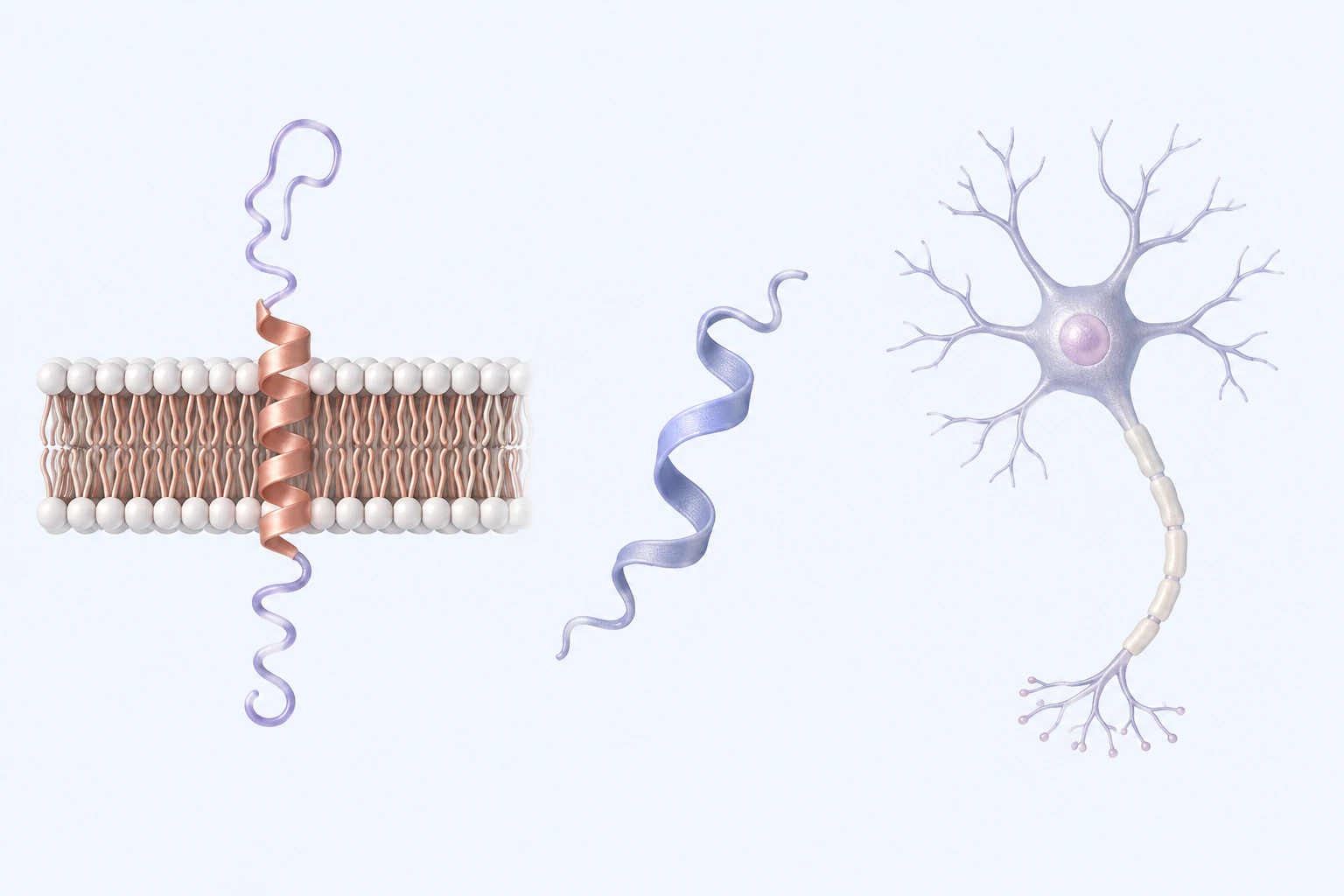
Amyloid-beta peptide context. The forms do not show literal APP structure, peptide sequence or length, cleavage, aggregation, plaque, toxicity, mechanism, biomarker, or diagnosis.
Scientific media record3 verified identifiers
- Subject
- Amyloid beta-Peptidesbiological-process
- Review
- Editorial review completeIdentifiers authority-verified · Accessibility validated · · amyloid-beta|amyloid-beta-mechanism-v1.webp
- Digital source
- Trained-algorithmic mediaCreated with a trained generative algorithm and reviewed by WikiBiome for subject identity, scientific framing, identifiers, provenance, and accessibility.
- License
- CC BY-SA 4.0Created
Amyloid-beta (Aβ) is a 36–43 amino acid peptide derived from the proteolytic cleavage of amyloid precursor protein (APP) by beta-secretase (BACE1) and gamma-secretase. Its aggregation from soluble monomers into insoluble fibrillar plaques in the brain is the defining neuropathological feature of Alzheimer's Disease.
However, the story of amyloid-beta is more complex than a simple pathological protein—it is a normal physiological peptide with antimicrobial functions whose relationship to infection and the Gut Microbiome has become mechanistically central to understanding Alzheimer's pathogenesis.
Evidence map11 cited passagesInspect provenance +
An antimicrobial peptide (AMP): Aβ42 has documented antimicrobial activity against bacteria, fungi (Candida albicans), and viruses (HSV-1, HIV). It traps pathogens in fibrillar nets structurally similar to neutrophil extracellular traps (NETs). In mouse models, Aβ deposition accelerates in response to bacterial or viral brain infection—and transgenic mice
A growing body of mechanistic evidence repositions amyloid-beta plaque formation as a misdirected innate immune response rather than a primary neurodegenerative event:
The metallomic signature of Alzheimer's disease—elevated iron, copper, and zinc in plaques and affected brain regions—converges directly on amyloid-beta biochemistry:
Arsenic exposure—which disrupts iron regulatory protein activity and increases BACE1 activity—increases Aβ(1-42) production in 3xTg-AD mouse models; 10 ppm chronic arsenic exposure elevates amyloid plaques and RAGE expression 220-fold
Arsenic uniquely disrupts nitric oxide (S-nitrosylation) signaling in the hippocampus and striatum, adding a neurochemical dimension to its Aβ-promoting effects
Curli-mediated cross-seeding: E. coli strains expressing curli fibers (functional bacterial amyloids structurally analogous to mammalian amyloids) can cross-seed mammalian amyloid aggregation. In mouse models, oral gavage with curli-expressing E. coli accelerates brain Aβ and alpha-synuclein pathology compared to curli-negative strains. This is the most dire
LPS has been detected in amyloid plaques at concentrations 3× higher than in age-matched controls without AD.
FMT reduces brain amyloid: Fecal microbiota transplant from healthy donors into AD mouse models reduced tau phosphorylation and brain Aβ levels, and improved synaptic plasticity. This causal experiment establishes the gut microbiome as a functional upstream modulator of brain amyloid pathology—not merely a correlate.
heavy metals (lead, cadmium) directly disrupt BBB tight junctions—providing the gateway for microbial products to reach the brain at concentrations that drive chronic Aβ overproduction
A notably specific connection: a nickel chelator (DMG-H) was shown to inhibit amyloid-beta aggregation in vitro, suggesting that nickel occupancy of Aβ metal-binding sites contributes to pathological aggregation. This connects nickel dyshomeostasis (a distinctive WikiBiome focus) to Alzheimer's pathology through the mis-metallation mechanism.
| Source | Evidence Level | Key Contribution | |--------|---------------|-----------------| | (2021) | Expert opinion (review) | LPS in plaques; curli cross-seeding; FMT reduces amyloid in AD models | | (2023) | Expert opinion (review) | Zinc, copper, iron profiles in AD plaques and brain regions | | (2025) | Systematic review (preprint) | 46 mechanistic stu
Contents
1. Normal Function: An Antimicrobial Peptide2. The Infection Hypothesis3. Metal-Driven Aggregation4. Gut Microbiome Modulation of Aβ Biology5. Blood-Brain Barrier Failure as the Gateway6. Nickel-Chelator Evidence7. Key Studies8. Cross-ReferencesNormal Function: An Antimicrobial Peptide#
Amyloid-beta is not intrinsically pathological. At physiological concentrations, Aβ42 functions as:
An antimicrobial peptide (AMP): Aβ42 has documented antimicrobial activity against bacteria, fungi (Candida albicans), and viruses (HSV-1, HIV). It traps pathogens in fibrillar nets structurally similar to neutrophil extracellular traps (NETs).
In mouse models, Aβ deposition accelerates in response to bacterial or viral brain infection—and transgenic mice overproducing Aβ survive Salmonella meningitis at higher rates than wild-type mice.[1]Romano 2021 -- The Role of Microbiome-Host Interactions in the Development of Alzheimer's DiseaseStefano Romano, George M Savva, Janis R Bedarf · 2021Open reference 1 ↓
A metal-binding peptide: Aβ binds zinc and copper with high affinity at specific N-terminal and C-terminal sites. This may serve a protective role in sequestering redox-active metals from pathogens—fitting the Nutritional Immunity (Metal Sequestration) framework. Under normal conditions, Aβ-metal binding is reversible and transient.
A synaptic regulator: At low concentrations, Aβ monomers modulate synaptic transmission and facilitate memory consolidation. Only at pathological oligomeric concentrations does Aβ become synaptotoxic.
The Infection Hypothesis#
A growing body of mechanistic evidence repositions amyloid-beta plaque formation as a misdirected innate immune response rather than a primary neurodegenerative event.[1]Romano 2021 -- The Role of Microbiome-Host Interactions in the Development of Alzheimer's DiseaseStefano Romano, George M Savva, Janis R Bedarf · 2021Open reference 1 ↓
Aβ production is upregulated in response to bacterial LPS, viral infection, and fungal exposure. Brain infections (HSV-1, H. pylori bacteremia, bacterial translocation across a disrupted blood-brain barrier) may trigger Aβ overproduction as a first-line antimicrobial defense.
The gut-brain axis connects gut Dysbiosis to brain Aβ burden—systemic LPS from Gram-negative pathobionts stimulates neuroinflammation and may drive chronic Aβ overproduction.
Germ-free mice show dramatically reduced Aβ plaque burden; colonization with human AD-patient microbiota increases brain amyloid.
This framework positions amyloid-beta accumulation not as the cause of AD but as a chronic innate immune response to gut-derived pathogenic stimuli that becomes pathological through overactivation and metal-catalyzed aggregation.
Metal-Driven Aggregation#
The metallomic signature of Alzheimer's disease—elevated iron, copper, and zinc in plaques and affected brain regions—converges directly on amyloid-beta biochemistry:[2]Common and Trace Metals in Alzheimer's and Parkinson's DiseasesDoroszkiewicz J, Farhan JA, Mroczko J et al. · 2023Open reference 2 ↓
Zinc (zinc (Zn)) Zinc binds to Aβ at His6, His13, His14, and Glu11, promoting aggregation from soluble monomers to insoluble oligomers and fibrils at physiological concentrations. Zinc-Aβ aggregates are structurally distinct from unmetallated Aβ fibrils—they may form faster and be less amenable to disaggregation.
Zinc accumulation in amyloid plaques is 2–3× higher than in adjacent tissue; intraneuronal zinc release during synaptic transmission may contribute to local Aβ aggregation at synaptic clefts.
Copper (copper (Cu))
Copper binds Aβ at His6, His13, His14 (same sites as zinc) with higher affinity. copper(I/II) cycling at the Aβ surface catalyzes hydrogen peroxide and hydroxyl radical generation—Fenton-like chemistry that oxidatively damages surrounding neurons. Copper-Aβ complexes are more neurotoxic than Aβ alone; soluble copper-Aβ oligomers show elevated pro-apoptotic activity.
Ceruloplasmin (the major circulating copper protein) shows reduced activity in AD, impairing ferroxidase function and promoting iron accumulation.
Iron (iron (Fe)) Iron does not directly aggregate Aβ but accumulates at plaque surfaces and catalyzes oxidative damage to surrounding tissue via Fenton chemistry. Elevated ferritin and reduced transferrin saturation in CSF are early biomarkers of AD progression. Iron accumulation in the hippocampus tracks with cognitive decline progression.
Arsenic exposure—which disrupts iron regulatory protein activity and increases BACE1 activity—increases Aβ(1-42) production in 3xTg-AD mouse models; 10 ppm chronic arsenic exposure elevates amyloid plaques and RAGE expression 220-fold.[3]Associations of Environmental Exposure to Arsenic, Manganese, Lead, and Cadmium with Alzheimer's Disease: A Review of Recent Evidence from Mechanistic StudiesGiasuddin Ahmed, Md. Shiblur Rahaman, Enrique Perez et al. · 2025Open reference 3 ↓
Mis-metallation When toxic metals (lead, cadmium, nickel) occupy the copper- and zinc-binding sites on Aβ, they alter its aggregation kinetics. Lead- and cadmium-Aβ complexes show different structural properties and disaggregation profiles than zinc-Aβ or copper-Aβ, potentially accelerating pathological accumulation Mis-Metallation.
Arsenic uniquely disrupts nitric oxide (S-nitrosylation) signaling in the hippocampus and striatum, adding a neurochemical dimension to its Aβ-promoting effects.[3]Associations of Environmental Exposure to Arsenic, Manganese, Lead, and Cadmium with Alzheimer's Disease: A Review of Recent Evidence from Mechanistic StudiesGiasuddin Ahmed, Md. Shiblur Rahaman, Enrique Perez et al. · 2025Open reference 3 ↓
Gut Microbiome Modulation of Aβ Biology#
Curli-mediated cross-seeding: E. coli strains expressing curli fibers (functional bacterial amyloids structurally analogous to mammalian amyloids) can cross-seed mammalian amyloid aggregation. In mouse models, oral gavage with curli-expressing E. coli accelerates brain Aβ and alpha-synuclein pathology compared to curli-negative strains.[1]Romano 2021 -- The Role of Microbiome-Host Interactions in the Development of Alzheimer's DiseaseStefano Romano, George M Savva, Janis R Bedarf · 2021Open reference 1 ↓
This is the most direct mechanism linking gut bacterial products to brain amyloid accumulation.
LPS-driven neuroinflammation: Gram-negative gut pathobionts (E. coli, Klebsiella, H. pylori) release LPS. Systemic LPS activates TLR4 receptors on brain microglia, inducing pro-inflammatory cytokine release (TNF-α, IL-1β, IL-6). This neuroinflammatory state.
Upregulates BACE1 expression (increasing Aβ production). Impairs microglial phagocytosis of Aβ plaques (reducing clearance). Activates NLRP3 inflammasome in microglia, amplifying IL-1β production.
LPS has been detected in amyloid plaques at concentrations 3× higher than in age-matched controls without AD.[1]Romano 2021 -- The Role of Microbiome-Host Interactions in the Development of Alzheimer's DiseaseStefano Romano, George M Savva, Janis R Bedarf · 2021Open reference 1 ↓
H. pylori and Aβ: Helicobacter pylori infection is associated with elevated serum amyloid and increased AD risk in epidemiological studies. H. pylori produces Ammonia (via nickel-dependent Urease), vacuolating toxin VacA, and CagA protein—all of which trigger mucosal and systemic inflammatory responses that chronically stimulate Aβ production.
FMT reduces brain amyloid: Fecal microbiota transplant from healthy donors into AD mouse models reduced tau phosphorylation and brain Aβ levels, and improved synaptic plasticity.[4]Gut microbial imbalance and neurodegenerative proteinopathies: from molecular mechanisms to prospects of clinical applicationsAlonso-Garcia P, Martin R, Martinez-Pinilla E · 2021Open reference 4 ↓
This causal experiment establishes the gut microbiome as a functional upstream modulator of brain amyloid pathology—not merely a correlate.
SCFA depletion increases amyloid burden: SCFA-producing bacteria (Faecalibacterium prausnitzii, Roseburia, Bifidobacterium) are depleted in AD microbiomes. Butyrate inhibits HDAC activity in brain tissue, maintains blood-brain barrier tight junctions, and suppresses neuroinflammatory gene expression. Loss of these bacteria removes multiple layers of protection against Aβ accumulation and aggregation.
Blood-Brain Barrier Failure as the Gateway#
The blood-brain barrier (BBB) normally prevents LPS, microbial products, and amyloid-seeding proteins from reaching brain parenchyma. Gut dysbiosis compromises the BBB through multiple mechanisms. Decreased butyrate production reduces tight junction protein expression in BBB endothelial cells.
Systemic LPS directly increases BBB permeability via TLR4 signaling.
Heavy Metals (lead, cadmium) directly disrupt BBB tight junctions—providing the gateway for microbial products to reach the brain at concentrations that drive chronic Aβ overproduction.[3]Associations of Environmental Exposure to Arsenic, Manganese, Lead, and Cadmium with Alzheimer's Disease: A Review of Recent Evidence from Mechanistic StudiesGiasuddin Ahmed, Md. Shiblur Rahaman, Enrique Perez et al. · 2025Open reference 3 ↓
Once the BBB is compromised, the loop closes: microbial products drive more Aβ production, Aβ aggregation drives neuroinflammation, neuroinflammation increases BBB permeability, and more microbial products enter.
Nickel-Chelator Evidence#
A notably specific connection: a nickel chelator (DMG-H) was shown to inhibit amyloid-beta aggregation in vitro, suggesting that nickel occupancy of Aβ metal-binding sites contributes to pathological aggregation.[5]Benoit & Maier 2021 — Nickel Chelator Inhibits Amyloid-Beta AggregationBenoit, S.L., Bhatt et al. · 2021Open reference 5 ↓ This connects nickel dyshomeostasis (a distinctive WikiBiome focus) to Alzheimer's pathology through the mis-metallation mechanism.
Key Studies#
| Source | Evidence Level | Key Contribution |
|---|---|---|
| [1]Romano 2021 -- The Role of Microbiome-Host Interactions in the Development of Alzheimer's DiseaseStefano Romano, George M Savva, Janis R Bedarf · 2021Open reference 1 ↓ (2021) | Expert opinion (review) | LPS in plaques; curli cross-seeding; FMT reduces amyloid in AD models |
| [2]Common and Trace Metals in Alzheimer's and Parkinson's DiseasesDoroszkiewicz J, Farhan JA, Mroczko J et al. · 2023Open reference 2 ↓ (2023) | Expert opinion (review) | Zinc, copper, iron profiles in AD plaques and brain regions |
| [3]Associations of Environmental Exposure to Arsenic, Manganese, Lead, and Cadmium with Alzheimer's Disease: A Review of Recent Evidence from Mechanistic StudiesGiasuddin Ahmed, Md. Shiblur Rahaman, Enrique Perez et al. · 2025Open reference 3 ↓ (2025) | Systematic review (preprint) | 46 mechanistic studies; arsenic increases BACE1/RAGE, Aβ(1-42) production |
| [4]Gut microbial imbalance and neurodegenerative proteinopathies: from molecular mechanisms to prospects of clinical applicationsAlonso-Garcia P, Martin R, Martinez-Pinilla E · 2021Open reference 4 ↓ (2021) | Expert opinion (review) | FMT reduces tau/amyloid; Lactobacillus/Bifidobacterium improve cognition |
Cross-References#
Alzheimer's Disease—the primary disease context. Mis-Metallation—toxic metal displacement of zinc/copper in Aβ binding sites. Iron—catalyzes oxidative damage at plaque surfaces; elevated in AD hippocampus.
Zinc—promotes Aβ aggregation; accumulated 2–3× higher in plaques. Copper—copper (Cu)-Aβ complexes generate reactive oxygen species via Fenton-like chemistry. Gut-Brain Axis—the route by which gut dysbiosis reaches brain Aβ pathology.
Neuroinflammation—the chronic inflammatory state that drives BACE1 upregulation and Aβ overproduction. Blood-Brain Barrier—barrier failure allows LPS and microbial amyloids to reach the brain. Nutritional Immunity (Metal Sequestration)—normal Aβ antimicrobial function fits the nutritional immunity framework.
Helicobacter pylori—enriched in AD signatures; triggers mucosal Metal-Driven Inflammation that reaches brain. dysbiosis—the upstream disruption that initiates chronic Aβ overproduction. Alpha-Synuclein—parallel proteinopathy with overlapping metal and microbiome interactions.
References 8
Numbered by first appearance in the article, then reconciled with its declared source list.
- 1
Stefano Romano, George M Savva, Janis R Bedarf (2021). Romano 2021 -- The Role of Microbiome-Host Interactions in the Development of Alzheimer's Disease. Frontiers in Pharmacology.
- 2
Doroszkiewicz J, Farhan JA, Mroczko J et al. (2023). Common and Trace Metals in Alzheimer's and Parkinson's Diseases. International Journal of Molecular Sciences.
- 3
★Giasuddin Ahmed, Md. Shiblur Rahaman, Enrique Perez et al. (2025). Associations of Environmental Exposure to Arsenic, Manganese, Lead, and Cadmium with Alzheimer's Disease: A Review of Recent Evidence from Mechanistic Studies. Journal of Xenobiotics.
- 4
Alonso-Garcia P, Martin R, Martinez-Pinilla E (2021). Gut microbial imbalance and neurodegenerative proteinopathies: from molecular mechanisms to prospects of clinical applications. Exploration of Neuroprotective Therapy.
- 5
Benoit, S.L., Bhatt et al. (2021). Benoit & Maier 2021 — Nickel Chelator Inhibits Amyloid-Beta Aggregation. Scientific Reports.
- 6
Jakubowska E, Hoppe-Mitera E, Sionek I et al. (2024). Metal toxicity exposure in Alzheimer's disease - literature review. Journal of Education, Health and Sport.
- 7
★Bakulski KM, Seo YA, Hickman RC et al. (2020). Heavy Metals Exposure and Alzheimer's Disease and Related Dementias. Journal of Alzheimer's Disease.
- 8
Islam F, Shohag S, Akhter S et al. (2022). Exposure of metal toxicity in Alzheimer's disease: An extensive review. Frontiers in Pharmacology.
Article network
Mentioned here 18
Pages linking here 14
Connect the evidence
Publicly readable discussion by ORCID-authenticated researchers. Questions, interpretation, methods, corrections, and new evidence stay attached to this record.
No discussion yet. Start with a precise question or a source-backed observation.
Activity and accepted changes
Accepted researcher context, editorial status, public discussion, and upstream Git revisions are shown together. Pending, declined, and withdrawn proposals remain private.
- published revision
Backfill heavy metals concept links
Karen Pendergrass · +1 −1
Inspect exact Git diff ↗ - published revision
Backfill gut microbiome concept links
Karen Pendergrass · +1 −1
Inspect exact Git diff ↗ - published revision
Backfill inflammation concept links
Karen Pendergrass · +1 −1
Inspect exact Git diff ↗ - published revision
Complete corpus-wide Dysbiosis linking
Karen Pendergrass · +2 −2
Inspect exact Git diff ↗ - published revision
Complete Ammonia contextual coverage
Karen Pendergrass · +1 −1
Inspect exact Git diff ↗ - published revision
Complete Tight junctions contextual coverage
Karen Pendergrass · +1 −1
Inspect exact Git diff ↗ - published revision
Complete reviewed Urease contextual coverage
Karen Pendergrass · +1 −1
Inspect exact Git diff ↗ - published revision
massive wiki expansion: 149 stubs fixed, 100+ new pages, Rule 15 scan, keystone papers
WikiBiome Deploy Bot · +15 −15
Inspect exact Git diff ↗ - published revision
Deepen 8 concept pages + 2 CP source pages (co-selection, AMR, alpha-synuclein, metabolites, gut-microbiome, propionic-acid, amyloid-beta, dyshomeostasis)
WikiBiome Deploy Bot · +91 −27
Inspect exact Git diff ↗ - published revision
Deep content + citation pass: 10 entities enriched, 7 new sources, DOI integrity
WikiBiome Deploy Bot · +1 −1
Inspect exact Git diff ↗ - published revision
WikiBiome v2 migration: signature pages + safety fixes + gap analysis
WikiBiome Deploy Bot · +53 −0
Inspect exact Git diff ↗