
 metals · microbes · host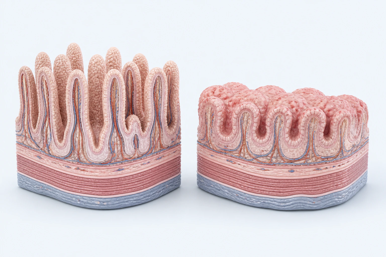
Representative small-intestinal mucosal architecture for celiac disease, comparing tall villi with shortened, broadened projections. This reconstruction is not a biopsy, Marsh grade, treatment response, or diagnosis.
Scientific media record1 verified identifier
- Subject
- Celiac Diseasecondition
- Identifiers
- MeSH:D002446
- Review
- Editorial review completeIdentifiers authority-verified · Accessibility validated · · celiac-disease|celiac-disease-pathology-v1.webp
- Digital source
- Trained-algorithmic mediaCreated with a trained generative algorithm and reviewed by WikiBiome for subject identity, scientific framing, identifiers, provenance, and accessibility.
- Scientific basis
- Celiac Disease — MeSHCeliac Disease
- License
- CC BY-SA 4.0Created
Celiac disease is a chronic autoimmune condition triggered by dietary gluten (gliadin proteins from wheat, barley, and rye) in genetically susceptible individuals (HLA-DQ2/DQ8, carried by ~30-40% of Europeans but penetrant in only ~1% of carriers).
Gluten exposure causes immune-mediated destruction of small intestinal villi (Marsh grade 3 villous atrophy at diagnosis), resulting in malabsorption of iron, zinc, selenium, copper, calcium, magnesium, folate, and fat-soluble vitamins. Global prevalence is ~0.7-1.4% by serology, with clinical diagnosis lagging considerably (Singh et al. 2018 meta-analysis; Lebwohl & Rubio-Tapia 2021).
The pathogenesis proceeds in three coupled steps: (1) intestinal permeability allows gliadin to reach the lamina propria; (2) tissue transglutaminase 2 (tTG2) deamidates gliadin peptides, enhancing HLA-DQ2/DQ8 binding affinity; (3) CD4+ T cells activate against deamidated gliadin, and intraepithelial lymphocytes driven by IL-15 lyse enterocytes.
Each step is modifiable by the Gut Microbiome and by metal status.
Evidence map4 cited passagesInspect provenance +
The gluten-free diet (GFD) paradox with nickel: The standard treatment for celiac disease is strict adherence to a gluten-free diet. However, many gluten-free substitute foods (rice, corn, oats, soy, pseudocereals like quinoa and amaranth) are high-nickel foods. Patients who switch to a GFD to manage celiac symptoms may simultaneously increase their dietary
The combined gluten-free + low-nickel diet (GFD+LNiD) has been studied specifically in celiac patients who also have Nickel Allergic Contact Mucositis. A combined elimination diet produces superior symptom resolution compared to GFD alone in patients with both conditions, establishing that nickel and gluten represent independent but co-occurring triggers in
Nickel—Dietary nickel intake increases paradoxically on a gluten-free diet because GFD substitute foods (rice, corn, oats, soy, quinoa, amaranth, legumes) are high-nickel foods. In patients with nickel allergic contact mucositis (NACM), this triggers gastrointestinal inflammation that mimics celiac symptoms. Estimated prevalence of Ni ACM exceeds 30% in th
Dietary nickel is the primary environmental metal exposure in celiac disease. Average dietary nickel intake ranges from 100-600 ug/day depending on food choices. The gluten-free diet paradox means patients replacing wheat with corn, rice, buckwheat, and legume-based products inadvertently increase their daily nickel intake substantially. Plant-based and whol
One disease. Five evidence layers.
A generated systems view of the metals, organisms, host sequestration signals, ecological conditions, and microbial functions indexed for Celiac Disease.
Evidence layer
Taxonomic signature
Organisms reported as enriched or depleted, with their indexed functional context kept beside the name.Proteolytic activity generates residual immunogenic gliadin peptides that enhance HLA-DQ2 presentation
Elevated proteolytic strains deamidate gliadin peptides similarly to tTG2, priming immune activation
Phylum-level bloom — consistent dysbiosis marker in both active and GFD-treated celiac patients
Enriched in celiac gut; functional role unclear
Consistently reduced (B. longum, B. adolescentis) — loss impairs barrier support, immune tolerance, and gluten peptide degradation
Reduced luminal and mucosal populations; healthy Lactobacillus strains fully degrade the immunogenic 33-mer gliadin peptide
Butyrate producer whose loss compounds epithelial energy deficit in villous atrophy
SCFA producers — loss worsens barrier dysfunction and mucosal healing
Depleted in active celiac disease
Evidence layer
Nutritional immunity
Host metal-withholding, inflammatory, antioxidant, and microbial-metabolite signals indexed in the signature.Elevated host signals
1Depleted protective signals
1Evidence layer
Ecological state
The environmental conditions that connect the organism-level observations into a system.Evidence layer
Virulence functions
Microbial structures, enzymes, and acquisition systems implicated by the linked evidence.The disease record, in full.
The original WikiBiome disease narrative remains intact beneath the generated signature atlas.
Metallomic Angle#
Celiac disease is notable in this knowledge base for a paradoxical metal intersection with nickel:
The gluten-free diet (GFD) paradox with nickel: The standard treatment for celiac disease is strict adherence to a gluten-free diet. However, many gluten-free substitute foods (rice, corn, oats, soy, pseudocereals like quinoa and amaranth) are high-nickel foods.
Patients who switch to a GFD to manage celiac symptoms may simultaneously increase their dietary nickel intake, triggering Nickel Allergic Contact Mucositis (NACM)—a systemic allergic response to nickel that causes gastrointestinal symptoms overlapping significantly with celiac symptoms (bloating, diarrhea, pain).
This creates diagnostic confusion and can lead to inadequate management of both conditions simultaneously.[1]Beneficial Effects of a Low-Nickel Diet on Relapsing IBS-Like and Extraintestinal Symptoms of Celiac Patients during a Proper Gluten-Free DietBorghini R, De Amicis N, Bella A et al. · 2020Open reference 1 ↓
Iron and zinc malabsorption: Villous atrophy in celiac disease severely impairs absorption of iron and zinc from the upper small intestine, causing deficiencies that mirror the metallomic pattern of nutritional immunity but arise from absorptive failure rather than host sequestration.
Distinguishing true absorptive iron deficiency (celiac-driven) from functional anemia requires celiac antibody testing and duodenal biopsy.
Microbiome Signature#
Celiac disease is associated with a characteristic gut Dysbiosis documented in both active disease and treated (GFD-adherent) patients:
Depleted. Bifidobacterium (especially B. longum and B. adolescentis)—consistently reduced across paediatric and adult cohorts (Collado et al. 2009; De Palma et al. 2010; Golfetto et al.
2014).
Lactobacillus—reduced luminal and mucosal populations, correlating with impaired gluten peptide degradation. Faecalibacterium prausnitzii and other Lachnospiraceae—Butyrate producers whose loss worsens epithelial energy deficit in the setting of villous atrophy. Prevotella spp.—depleted in active disease.
Enriched. Proteobacteria (Pseudomonadota) at phylum level (a consistent dysbiosis marker). Escherichia coli—some strains express proteolytic activity that enhances rather than reduces gliadin immunogenicity (Caminero et al. 2019, Gastroenterology). Staphylococcus spp.
Bacteroides fragilis—elevated proteolytic strains that deamidate gliadin peptides similarly to tTG2, priming HLA-DQ2 presentation.
Mechanistic bridge: Caminero et al. (2016, Gut) showed that lactobacilli from healthy subjects fully degrade the immunogenic 33-mer gliadin peptide, while pseudomonal and E. coli proteases from celiac patients only partially cleave it, generating residual epitopes that remain antigenic.
This establishes the microbiome as a checkpoint on gluten immunogenicity rather than a passive observer. Wheat-associated Pseudomonas aeruginosa specifically produces elastase-like proteases that generate more, not fewer, HLA-DQ2-binding peptides.
This dysbiosis pattern overlaps substantially with other conditions in this knowledge base, suggesting shared metal-microbiome mechanisms may operate in celiac disease beyond the primary gluten-immune axis.
Metabolomic Signature#
Short-chain fatty acids (butyrate, propionate, acetate)—reduced due to depletion of SCFA-producing Faecalibacterium and Lachnospiraceae; compounds epithelial repair deficit.
Tryptophan metabolites—altered indole/AhR ligand production due to loss of Lactobacillus reuteri and related species, impairing AhR (Aryl Hydrocarbon Receptor) signalling and Treg induction (Lamas et al. 2016).
Bile acids—secondary bile acid profile shifted in treated celiac patients (Wacklin et al. 2014), with implications for FXR-mediated intestinal barrier regulation. Zonulin—elevated serum levels signal active tight-junction disassembly; a biomarker of the "leaky gut" phase of celiac pathogenesis.
Relationship to [[low-nickel-diet]]#
The combined gluten-free + low-nickel diet (GFD+LNiD) has been studied specifically in celiac patients who also have Nickel Allergic Contact Mucositis.
A combined elimination diet produces superior symptom resolution compared to GFD alone in patients with both conditions, establishing that nickel and gluten represent independent but co-occurring triggers in a subset of celiac patients.[1]Beneficial Effects of a Low-Nickel Diet on Relapsing IBS-Like and Extraintestinal Symptoms of Celiac Patients during a Proper Gluten-Free DietBorghini R, De Amicis N, Bella A et al. · 2020Open reference 1 ↓
Cross-References#
- Low-Nickel Diet—the intervention with documented benefit in celiac + NACM overlap
- Nickel Allergy and Allergic Contact Dermatitis—Nickel Allergic Contact Mucositis (NACM); the overlapping condition
- Dietary Nickel Exposure—gluten-free foods as a high-nickel exposure pathway
- Iron—malabsorption as a mechanism distinct from nutritional immunity
- Zinc—malabsorption in untreated celiac disease
- gut microbiome—dysbiosis co-occurring with celiac
- Type 1 Diabetes—shared autoimmune HLA-DQ associations and Bifidobacterium depletion
- Hashimoto's Thyroiditis—autoimmune comorbidity with shared metal malabsorption (iron, zinc, selenium)
- Intestinal Permeability—villous atrophy and tight junction disruption drive nutrient malabsorption
- Zonulin—gluten-triggered zonulin release increases intestinal permeability in celiac disease
- Bifidobacterium—consistently depleted in celiac gut; loss impairs barrier support and immune tolerance
References 8
Numbered by first appearance in the article, then reconciled with its declared source list.
- 1
Borghini R, De Amicis N, Bella A et al. (2020). Beneficial Effects of a Low-Nickel Diet on Relapsing IBS-Like and Extraintestinal Symptoms of Celiac Patients during a Proper Gluten-Free Diet. Nutrients.
- 2
de Vos WM, de Vos EAJ (2012). Role of the Intestinal Microbiome in Health and Disease: From Correlation to Causation. Nutrition Reviews.
- 3
Xu Q, Ni JJ, Han BX et al. (2022). Causal Relationship Between Gut Microbiota and Autoimmune Diseases: A Two-Sample Mendelian Randomization Study. Frontiers in Immunology.
- 4
Rodziewicz A, Szewczyk A, Bryl E (2024). Rodziewicz et al. 2024 — Gluten-Free Diet Alters the Gut Microbiome in Women with Autoimmune Thyroiditis. Nutrients.
- 5
Agnes Svensson, Louise Brunkwall, Bodil Roth et al. (2021). Svensson 2021 — Associations Between Endometriosis and Gut Microbiota. Reproductive Sciences.
- 6
Raffaele Borghini, Natascia De Amicis, Antonino Bella et al. (2020). Borghini 2020 — Beneficial Effects of a Low-Nickel Diet on Relapsing IBS-Like and Extraintestinal Symptoms of Celiac Patients during a Proper Gluten-Free Diet. Nutrients.
- 7
Patricia L. Turpin, Angelica P. Ahrens, Jordan T. Russell et al. (2021). Turpin 2021 — Adolescent Autism and Autoimmune Diagnoses Linked to Infant Gut Bacteria Whose Prevalence Is Associated with At-Risk Genetics and/or Diet. medRxiv (preprint).
- 8
Xu Q, Ni JJ, Han BX et al. (2022). Causal Relationship Between Gut Microbiota and Autoimmune Diseases: A Two-Sample Mendelian Randomization Study. Frontiers in Immunology.
Article network
Mentioned here 18
Pages linking here 15
Connect the evidence
Publicly readable discussion by ORCID-authenticated researchers. Questions, interpretation, methods, corrections, and new evidence stay attached to this record.
No discussion yet. Start with a precise question or a source-backed observation.
Activity and accepted changes
Accepted researcher context, editorial status, public discussion, and upstream Git revisions are shown together. Pending, declined, and withdrawn proposals remain private.
- published revision
Backfill gut microbiome concept links
Karen Pendergrass · +2 −2
Inspect exact Git diff ↗ - published revision
Complete corpus-wide Dysbiosis linking
Karen Pendergrass · +1 −1
Inspect exact Git diff ↗ - published revision
Complete Tight junctions contextual coverage
Karen Pendergrass · +1 −1
Inspect exact Git diff ↗ - published revision
massive wiki expansion: 149 stubs fixed, 100+ new pages, Rule 15 scan, keystone papers
WikiBiome Deploy Bot · +27 −21
Inspect exact Git diff ↗ - published revision
nightly maintenance: 94 stub demotions, 181 source_count fixes, 22 auto-discovered stubs, 5 adversarial audits, 3 boundary fixes, 3 evidence-level corrections
WikiBiome Deploy Bot · +3 −0
Inspect exact Git diff ↗ - published revision
cycle 1: health check + lint fixes + 8 ingests + 2 stubs + gestational-diabetes signature
WikiBiome Deploy Bot · +1 −0
Inspect exact Git diff ↗ - published revision
Deep citation pass on 10 disease entities + expand 3 thin entities
WikiBiome Deploy Bot · +36 −4
Inspect exact Git diff ↗ - published revision
Health check fixes: 40 intervention pages + 14 entity Connections sections
WikiBiome Deploy Bot · +5 −0
Inspect exact Git diff ↗ - published revision
v2 migration Priority 2: All 29 disease entity pages upgraded with associated_conditions, seo_target, wikipedia_differentiation
WikiBiome Deploy Bot · +6 −0
Inspect exact Git diff ↗ - published revision
WikiBiome v2 migration: signature pages + safety fixes + gap analysis
WikiBiome Deploy Bot · +41 −0
Inspect exact Git diff ↗