
 metals · microbes · host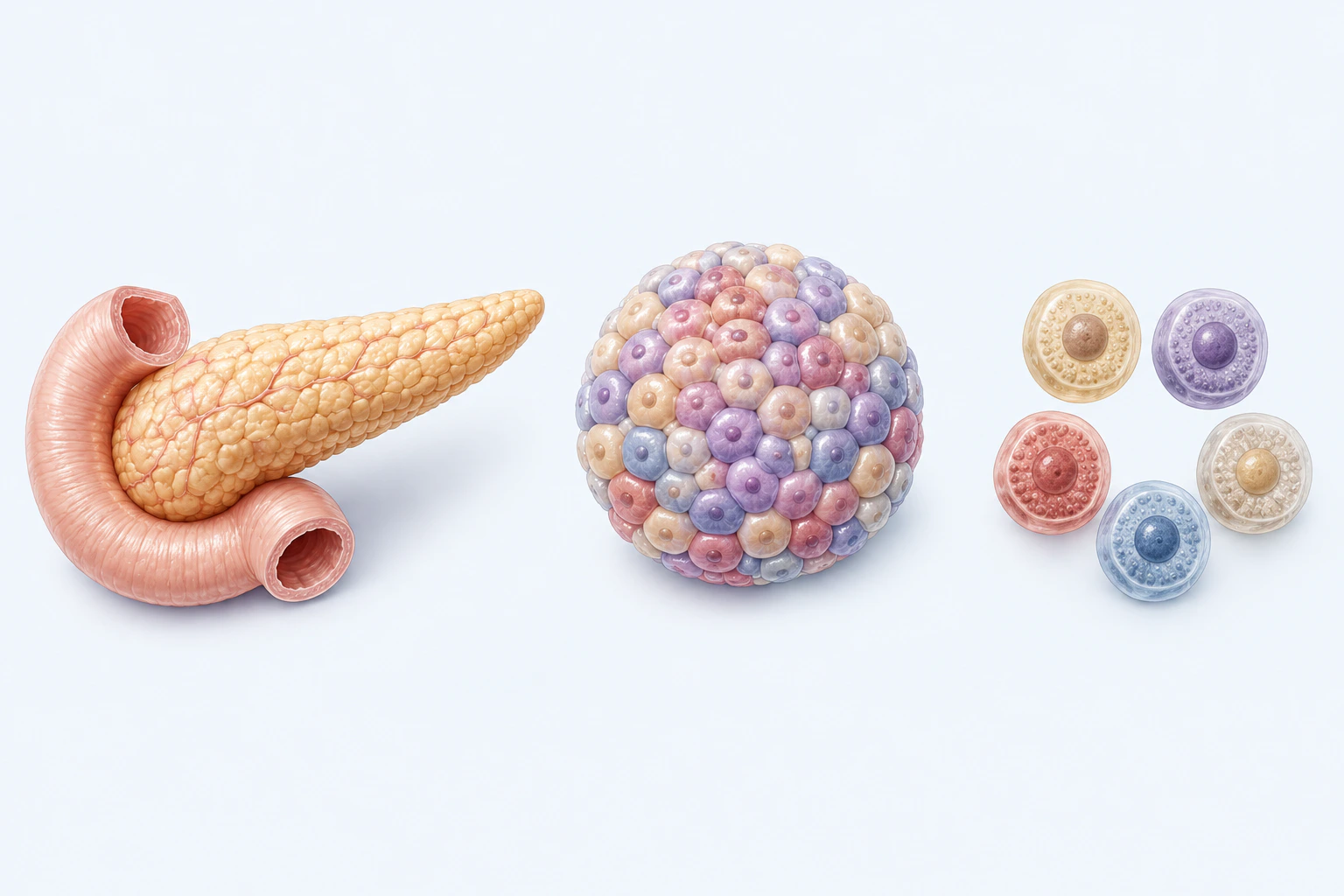
Neutral pancreatic and cellular orientation for type 1 diabetes. Cell colors and counts are illustrative and do not identify cell types, abundance, loss, immune attack, insulin state, glucose level, age, stage, treatment, or diagnosis.
Scientific media record1 verified identifier
- Subject
- Diabetes Mellitus, Type 1condition
- Identifiers
- MeSH:D003922
- Review
- Editorial review completeIdentifiers authority-verified · Accessibility validated · · type-1-diabetes|type-1-diabetes-pathology-v1.webp
- Digital source
- Trained-algorithmic mediaCreated with a trained generative algorithm and reviewed by WikiBiome for subject identity, scientific framing, identifiers, provenance, and accessibility.
- Scientific basis
- Diabetes Mellitus, Type 1 — MeSHWhat Is Diabetes?
- License
- CC BY-SA 4.0Created
Type 1 diabetes is an autoimmune disease in which immune-mediated destruction of insulin-producing beta cells in the pancreatic islets of Langerhans leads to lifelong insulin dependence. T1D is fundamentally distinct from type 2 diabetes, which is characterized by insulin resistance rather than autoimmune beta cell loss.
T1D accounts for approximately 5–10% of all diabetes cases, with incidence rising 3–4% annually in Europe.[1]Luo 2023 — Gut microbiota and type 1 diabetes: a two-sample bidirectional Mendelian randomization studyLuo M, Sun M, Wang T et al. · 2023Open reference 1 ↓ Global incidence is highest in Finland, Sardinia, and Scandinavia, reflecting the combined influence of HLA-DR genetic risk, environmental exposures, and early-life microbiome composition.
Three environmental factors now have strong mechanistic evidence: heavy metal status (particularly zinc and iron), enteroviral infection, and Gut Microbiome Dysbiosis—and all three converge during the developmentally critical first three years of life.
Evidence map66 cited passagesInspect provenance +
Type 1 diabetes is an autoimmune disease in which immune-mediated destruction of insulin-producing beta cells in the pancreatic islets of Langerhans leads to lifelong insulin dependence. T1D is fundamentally distinct from type 2 diabetes, which is characterized by insulin resistance rather than autoimmune beta cell loss. T1D accounts for approximately 5–10%
Copper exposure is relevant to T1D through its interaction with the gut microbiome. High blood copper levels—as seen in occupational exposure and dietary excess—shape gut microbial communities in ways that parallel T1D-associated dysbiosis. In a 12-week RCT in workers with elevated copper and nickel exposure, probiotic intervention reduced blood Cu by 34
| Taxon | Level | OR (95% CI) | p-value | Method | Source | |-------|-------|-------------|---------|--------|--------| | Bacteroidetes | Phylum | 1.24 (1.01–1.53) | 0.044 | IVW | | | Bacteroidia | Class | 1.28 (1.06–1.53) | 0.009 | IVW | | | Bacteroidales | Order | 1.28 (1.06–1.53) | 0.009 | IVW | |
| Taxon | Level | OR (95% CI) | p-value | PFDR | Source | |-------|-------|-------------|---------|-------|--------| | Eubacterium eligens group | Genus | 0.64 (0.50–0.81) | 2.84×10⁻⁴ | 0.031 | | | Family XI (Firmicutes) | Family | 0.87 (0.79–0.96) | 0.007 | 0.378 | | | Lachnospiraceae UCG008 | Genus | 0.86 (0.75–0.97) | 0.019 | 0.588 | | | Ruminococcaceae U
The inverse Bacteroidetes/Firmicutes causal pattern mirrors observational data: Bacteroidetes enrichment and Firmicutes depletion are both causally associated with increased T1D risk. The MR study leveraged FinnGen T1D GWAS (n = 264,137 Europeans), giving it substantially more power than prior analyses.
Enteroviral infection and microbiome disruption converge as complementary environmental T1D triggers. Coxsackievirus B4 (CVB4) infection in non-obese diabetic (NOD) mice restructures the gut microbiome before T1D onset, producing a community composition strikingly similar to spontaneously diabetic mice:
GPR43 (free fatty acid receptor 2, activated by SCFAs) expression was significantly reduced in dysbiotic microbiome recipients—reducing the capacity for SCFA-driven regulatory immune signaling
Bifidobacterium genus causally protects against diabetic kidney disease (DKD) in T1D: OR = 0.566 (95% CI 0.396–0.809, p = 0.0018, IVW); Bifidobacteriaceae family OR = 0.561 and Bifidobacteriales order OR = 0.561 (consistent across taxonomic levels)
Actinobacteria phylum (which includes Bifidobacterium as its dominant anaerobe) causally reduces DKD risk in T1D: OR = 0.445 (95% CI 0.269–0.738, p = 0.0017)
| Condition | Shared Metal Pattern | Shared Microbial Pattern | Clinical Relevance | |-----------|---------------------|--------------------------|-------------------| | celiac disease | Zinc and iron depletion | Bifidobacterium depleted, Bacteroides altered | Both autoimmune, often co-occurring; shared HLA-DQ2/DQ8 genetic risk | | type 2 diabetes | Zinc dep
The CVB4 virus experiments demonstrate that steps 1–4 can be triggered by viral infection through microbiome restructuring alone, independent of direct beta cell viral infection.
—Two-sample bidirectional MR, n=264,137; inverse association for the source-labeled Eubacterium eligens group (FDR-significant), not a species-resolved intervention result
—CVB4-induced dysbiosis FMT experiments; GPR43/Treg mechanism; gut barrier compromise
—MR evidence for Bifidobacterium protection against DKD in T1D (OR=0.566); complication-microbiota bidirectionality
—RCT evidence for Cu/Ni reduction via probiotic gut microbiome modulation; Blautia-antioxidant connection
| Metal | Status | Mechanism | |-------|--------|-----------| | Cadmium | Elevated | Aggravates diabetic nephropathy via TLR4/NF-kB; downregulates ZIP14 zinc transporter; linked to leaky gut via microbiome disruption | | Lead | Elevated | Prenatal exposure depletes B. bifidum/B. longum—the same species protective against T1D; disrupts early-life microbiome
The taxonomic signature of T1D is anchored by the TEDDY cohort (n=783, 10,913 stool samples) and validated by the INNODIA study (n=292).
bacteroides (B. dorei)—The paradoxical pathobiont. B. dorei dominates Finnish children who later develop T1D, peaking at 7 months of age. Its LPS is structurally distinct—acting as a TLR4 antagonist rather than agonist. This means B. dorei does not trigger inflammation; instead, it prevents immune education by blocking the normal LPS-driven immune traini
veillonella—Lactate consumer that diverts lactate away from butyrate-producing pathways toward propionate production. This is mechanistically significant because it contributes to the butyrate:other-SCFA ratio shift that is the ecological fulcrum of T1D.
escherichia coli—Enriched with iron acquisition genes (FecB, IsdDEF analogs), LPS biosynthesis genes, and type VI secretion systems. The iron acquisition enrichment supports Primitive 8 (Siderophore Competition).
sutterella KLE1602—Enriched in T1D patients with faster glycemic deterioration. A potential progressor biomarker for identifying patients at risk of aggressive disease course.
akkermansia muciniphila—Paradoxically enriched in the CVB4-induced diabetogenic microbiome configuration. Despite its generally protective reputation, Akkermansia's mucin-degrading activity may contribute to barrier thinning in the T1D context. This is a context-dependent pathogenicity—protective in metabolic syndrome but potentially harmful in autoimmun
faecalibacterium prausnitzii—The most clinically actionable depletion. Inversely correlated with HbA1c (P=0.0019) in established T1D. Depleted in TEDDY progressors. The primary butyrate producer whose loss drives the SCFA ratio shift.
bifidobacterium (B. longum, B. breve, B. bifidum)—Depleted across T1D cohorts. Particularly vulnerable to antibiotic exposure. Prenatal lead exposure depletes the same species, establishing a metal → microbiome → autoimmunity upstream pathway.
Showing 24 of 66 evidence-bearing passages. Every remaining citation is still indexed in the reference record below.
One disease. Five evidence layers.
A generated systems view of the metals, organisms, host sequestration signals, ecological conditions, and microbial functions indexed for Type 1 Diabetes.
Evidence layer
Taxonomic signature
Organisms reported as enriched or depleted, with their indexed functional context kept beside the name.Bacteroidetes causally increases T1D risk (MR OR=1.24-1.28); B. dorei and B. vulgatus elevated in children progressing to T1D; LPS activates innate immunity and triggers islet inflammation
Pro-inflammatory; mucin-degrading; enriched in autoimmune contexts
Enriched in T1D cases; LPS-producing
Lactate consumer — diverts lactate away from butyrate pathway toward propionate
Iron acquisition genes (FecB, IsdDEF); LPS biosynthesis; virulence factor enrichment
Gram-negative bloom; LPS biosynthesis genes enriched in T1D metagenomes
KLE1602 strain enriched in T1D progressors — potential biomarker for aggressive disease
Paradoxically enriched in CVB4-induced diabetogenic microbiome; context-dependent pathogenicity
Part of Bacteroidales order showing causal T1D risk (MR OR=1.28); contributes to LPS burden
Primary butyrate producer — inversely correlated with HbA1c (P=0.0019); depletion is the core SCFA deficit
Most replicated depletion finding in T1D observational studies; promotes Treg differentiation and barrier integrity; causally protects against diabetic kidney disease (MR OR=0.566)
Butyrate producer; depleted in TEDDY T1D progressors
SCFA producer; depleted in T1D cases
Butyrate producer; depleted alongside F. prausnitzii and Roseburia
Fiber-fermenting; depleted in Western-diet Bacteroides-dominant T1D microbiomes
L. rhamnosus GG protective when given before 27 days; depletion removes immune education
Multiple protective genera: UCG008 (MR OR=0.86), Dorea (MR OR=0.81); depletion in pre-T1D children consistent across cohorts
Butyrate-producing clostridia; depletion contributes to SCFA ratio shift
B. obeum inversely associated with C-peptide decline — protective for residual beta-cell function
Source-labeled group showed an inverse MR association with T1D (OR=0.64, FDR-adjusted p=0.031); the group label does not resolve Lachnospira eligens alone
Causally protective (MR OR=0.82); Firmicutes family contributing to SCFA production
Evidence layer
Nutritional immunity
Host metal-withholding, inflammatory, antioxidant, and microbial-metabolite signals indexed in the signature.Elevated host signals
9Depleted protective signals
6Evidence layer
Ecological state
The environmental conditions that connect the organism-level observations into a system.Evidence layer
Virulence functions
Microbial structures, enzymes, and acquisition systems implicated by the linked evidence.The disease record, in full.
The original WikiBiome disease narrative remains intact beneath the generated signature atlas.
Metal Associations#
Zinc: From Insulin Architecture to Autoantigen#
Zinc is indispensable for insulin production, storage, and secretion at multiple levels. Insulin is stored in beta cell granules as zinc-insulin hexamers—each hexamer coordinated by two zinc (Zn)²⁺ ions; zinc deficiency impairs crystallization, reducing insulin content per granule.
The ZnT8 transporter (SLC30A8) transports zinc²⁺ into insulin secretory granules and is itself a major autoantigen in T1D—anti-ZnT8 autoantibodies are present in 60–80% of newly diagnosed T1D patients, making it one of the most specific T1D biomarkers.
zinc²⁺ is co-released with insulin during exocytosis; extracellular zinc acts as a paracrine signal suppressing glucagon secretion from neighboring alpha cells—a loop disrupted when zinc is depleted.
SLC30A8 loss-of-function variants paradoxically protect against T2D (65% risk reduction), illustrating how the same zinc transporter plays opposing roles in T1D (autoantigen) and T2D (risk modifier).
Zinc deficiency reduces regulatory T cell (Treg) function and shifts the Th1/Th2 balance toward Th1-dominant autoimmunity—directly relevant to islet Metal-Driven Inflammation. Metallothioneins (zinc-binding proteins) in beta cells provide antioxidant defense; their depletion increases vulnerability to immune attack.
See Zinc for broader systemic zinc biology.
Iron: Beta Cell Toxicity and Inflammatory Amplification#
Iron accumulation in pancreatic islets is directly toxic to beta cells through multiple pathways. Hereditary hemochromatosis (HFE mutations) causes pancreatic iron overload and "bronze diabetes"—30–60% of hemochromatosis patients develop diabetes. iron (Fe)²⁺ generates hydroxyl radicals via Fenton chemistry, damaging beta cell membranes, DNA, and insulin-producing machinery.
Iron-loaded beta cells show reduced insulin secretion in response to glucose stimulation; islet iron content correlates inversely with insulin secretory capacity.
Iron-driven Oxidative Stress may generate neoantigens (oxidatively modified proteins) that trigger autoimmune recognition—potentially explaining how iron overload initiates rather than merely worsens T1D.
Ferroptosis-like beta cell death may release damage-associated molecular patterns (DAMPs) that activate dendritic cells and initiate the autoimmune cascade. Hepcidin, the master iron-regulatory hormone, is expressed in beta cells and modulates local iron homeostasis.
See Iron for systemic iron homeostasis.
Copper: Microbiome-Mediated Metal Dynamics#
Copper exposure is relevant to T1D through its interaction with the gut microbiome. High blood copper levels—as seen in occupational exposure and dietary excess—shape gut microbial communities in ways that parallel T1D-associated dysbiosis.
In a 12-week RCT in workers with elevated copper and nickel exposure, probiotic intervention reduced blood copper (Cu) by 34.45% and blood nickel (Ni) by 38.34% compared to conventional yogurt, while simultaneously enriching Blautia and depleting Bacteroides species through increased fecal metal excretion.[2]Feng 2022 — Human supplementation with Pediococcus acidilactici GR-1 decreases heavy metal levels through modifying the gut microbiota and metabolomeFeng P, Yang J, Zhao S et al. · 2022Open reference 2 ↓
The enriched Blautia species correlated positively with antioxidant metabolites and negatively with blood copper—suggesting a mechanistic link between metal detoxification and anti-inflammatory microbial metabolism. This metal-microbiome connection is mechanistically relevant to T1D because the same Bacteroides-enriched, Butyrate-depleted community structure that characterizes high-copper dysbiosis also characterizes pre-T1D gut ecology.
Microbial Associations#
Causal Evidence: Mendelian Randomization#
Observational associations between gut microbiota and T1D are abundant but vulnerable to confounding. Two-sample Mendelian randomization studies using large GWAS datasets now provide genetically-instrumented causal evidence for specific taxa.
Taxa with causal evidence for increasing T1D risk:
| Taxon | Level | OR (95% CI) | p-value | Method | Source |
|---|---|---|---|---|---|
| Bacteroidetes | Phylum | 1.24 (1.01–1.53) | 0.044 | IVW | [1]Luo 2023 — Gut microbiota and type 1 diabetes: a two-sample bidirectional Mendelian randomization studyLuo M, Sun M, Wang T et al. · 2023Open reference 1 ↓ |
| Bacteroidia | Class | 1.28 (1.06–1.53) | 0.009 | IVW | [1]Luo 2023 — Gut microbiota and type 1 diabetes: a two-sample bidirectional Mendelian randomization studyLuo M, Sun M, Wang T et al. · 2023Open reference 1 ↓ |
| Bacteroidales | Order | 1.28 (1.06–1.53) | 0.009 | IVW | [1]Luo 2023 — Gut microbiota and type 1 diabetes: a two-sample bidirectional Mendelian randomization studyLuo M, Sun M, Wang T et al. · 2023Open reference 1 ↓ |
Taxa with causal evidence for decreasing T1D risk (protective):
| Taxon | Level | OR (95% CI) | p-value | P_FDR | Source |
|---|---|---|---|---|---|
| Eubacterium eligens group | Genus | 0.64 (0.50–0.81) | 2.84×10⁻⁴ | 0.031 | [1]Luo 2023 — Gut microbiota and type 1 diabetes: a two-sample bidirectional Mendelian randomization studyLuo M, Sun M, Wang T et al. · 2023Open reference 1 ↓ |
| Family XI (Firmicutes) | Family | 0.87 (0.79–0.96) | 0.007 | 0.378 | [1]Luo 2023 — Gut microbiota and type 1 diabetes: a two-sample bidirectional Mendelian randomization studyLuo M, Sun M, Wang T et al. · 2023Open reference 1 ↓ |
| Lachnospiraceae UCG008 | Genus | 0.86 (0.75–0.97) | 0.019 | 0.588 | [1]Luo 2023 — Gut microbiota and type 1 diabetes: a two-sample bidirectional Mendelian randomization studyLuo M, Sun M, Wang T et al. · 2023Open reference 1 ↓ |
| Ruminococcaceae UCG010 | Genus | 0.81 (0.66–0.99) | 0.038 | 0.588 | [1]Luo 2023 — Gut microbiota and type 1 diabetes: a two-sample bidirectional Mendelian randomization studyLuo M, Sun M, Wang T et al. · 2023Open reference 1 ↓ |
| Dorea | Genus | 0.81 (0.66–1.00) | 0.048 | 0.540 | [1]Luo 2023 — Gut microbiota and type 1 diabetes: a two-sample bidirectional Mendelian randomization studyLuo M, Sun M, Wang T et al. · 2023Open reference 1 ↓ |
| Peptococcaceae | Family | 0.82 (0.68–0.98) | 0.034 | 0.588 | [1]Luo 2023 — Gut microbiota and type 1 diabetes: a two-sample bidirectional Mendelian randomization studyLuo M, Sun M, Wang T et al. · 2023Open reference 1 ↓ |
| Tenericutes | Phylum | 0.80 (0.64–0.99) | 0.037 | 0.138 | [1]Luo 2023 — Gut microbiota and type 1 diabetes: a two-sample bidirectional Mendelian randomization studyLuo M, Sun M, Wang T et al. · 2023Open reference 1 ↓ |
The source-labeled Eubacterium eligens group signal is the most robust inverse result in the dataset—FDR-significant (P_FDR = 0.031) with no heterogeneity or pleiotropy detected. The group label is broader than a verified species assignment and must not be treated as species-resolved evidence for Lachnospira eligens.
The inverse Bacteroidetes/Firmicutes causal pattern mirrors observational data: Bacteroidetes enrichment and Firmicutes depletion are both causally associated with increased T1D risk. The MR study leveraged FinnGen T1D GWAS (n = 264,137 Europeans), giving it substantially more power than prior analyses.[1]Luo 2023 — Gut microbiota and type 1 diabetes: a two-sample bidirectional Mendelian randomization studyLuo M, Sun M, Wang T et al. · 2023Open reference 1 ↓
Pre-Onset Dysbiosis Pattern#
Prospective studies (TEDDY, DIABIMMUNE, BABYDIET) show that gut microbiome composition diverges before seroconversion to islet autoantibodies—the microbiome shift precedes clinical onset by months to years.
Consistently depleted in pre-T1D and T1D. Bifidobacterium: Reduced across multiple cohorts from infancy onward; promotes Treg differentiation and intestinal barrier integrity; its depletion is the most replicated microbiome finding in T1D observational studies.
Faecalibacterium prausnitzii and other SCFA producers: Loss of butyrate production compromises gut barrier and epithelial fuel supply. Lachnospiraceae members: Multiple genera within this family show protective MR signals (UCG008 OR = 0.86) and are depleted in pre-T1D children.
Consistently enriched in pre-T1D and T1D. Bacteroides dorei and B. vulgatus: Elevated in children who progress to T1D; these species produce lipopolysaccharide (LPS) that activates innate immunity and may trigger islet inflammation.
Bacteroidetes-dominated community structure: Broader shift toward Bacteroidetes predominance, including increased Bacteroidota/Firmicutes ratio.
Viral Dysbiosis: The CVB4 Mechanism#
Enteroviral infection and microbiome disruption converge as complementary environmental T1D triggers. Coxsackievirus B4 (CVB4) infection in non-obese diabetic (NOD) mice restructures the gut microbiome before T1D onset, producing a community composition strikingly similar to spontaneously diabetic mice.[3]Morse 2023 — Virus induced dysbiosis promotes type 1 diabetes onsetMorse ZJ, Simister RL, Crowe SA et al. · 2023Open reference 3 ↓
CVB4 increases Actinobacteriota and Verrucomicrobiota phyla and contracts Firmicutes within days 7–21 post-infection.
Bifidobacteria and Akkermansia emerge as conspicuous members of the CVB4-induced diabetogenic microbiome—paradoxically elevated relative to healthy controls; the authors propose that strain-specific antibody responses to certain Bifidobacteria strains may contribute to autoimmunity.
Fecal microbiota transfer (FMT) of the CVB4-modified microbiome to microbiome-depleted naïve mice enhanced T1D susceptibility (61.2% hyperglycemic at 5 weeks vs 18.2% in control FMT, p < 0.05)—demonstrating that the dysbiotic microbiome alone, without virus, is sufficient to promote autoimmunity.
CVB4 infection caused ~2-fold reduction in gut barrier integrity (FITC-dextran assay), reduced tight-junction proteins (claudin-1, tjp1), elevated serum LPS, and enabled bacterial translocation to the pancreatic lymph nodes (PLN) by day 7 post-infection.
GPR43 (free fatty acid receptor 2, activated by SCFAs) expression was significantly reduced in dysbiotic microbiome recipients—reducing the capacity for SCFA-driven regulatory immune signaling.[3]Morse 2023 — Virus induced dysbiosis promotes type 1 diabetes onsetMorse ZJ, Simister RL, Crowe SA et al. · 2023Open reference 3 ↓
Foxp3+ CD4+ regulatory T cells were depleted in the intestinal lamina propria of CVB4 FMT recipients, with reduced IL-10 production in colon—directly implicating SCFA/Treg axis disruption as the mechanism connecting dysbiosis to autoimmunity.
Microbial Associations and Diabetic Complications#
The gut microbiome's relevance to T1D extends beyond disease onset to long-term complications. Mendelian randomization analysis of gut microbiota and diabetic complications in T1D patients finds.
Bifidobacterium genus causally protects against diabetic kidney disease (DKD) in T1D: OR = 0.566 (95% CI 0.396–0.809, p = 0.0018, IVW); Bifidobacteriaceae family OR = 0.561 and Bifidobacteriales order OR = 0.561 (consistent across taxonomic levels).[4]Liu 2024 — Causal relationship between gut microbiota and diabetic complications: a two-sample Mendelian randomization studyLiu J, Chen Y, Peng C · 2024Open reference 4 ↓
Actinobacteria phylum (which includes Bifidobacterium as its dominant anaerobe) causally reduces DKD risk in T1D: OR = 0.445 (95% CI 0.269–0.738, p = 0.0017).[4]Liu 2024 — Causal relationship between gut microbiota and diabetic complications: a two-sample Mendelian randomization studyLiu J, Chen Y, Peng C · 2024Open reference 4 ↓
At the stricter p < 1×10⁻⁶ threshold, Bifidobacteriaceae family protective effect against early DKD in T1D remains robust (OR = 0.423, 95% CI 0.275–0.65, p = 8.65×10⁻⁵).
Reverse MR: Diabetic retinopathy (DR) in T1D affects LachnospiraceaeUCG010 abundance—suggesting bidirectional relationships where complications worsen dysbiosis and dysbiosis accelerates complications.
This pattern implies that the Bifidobacterium depletion characteristic of T1D onset is not only contributing to autoimmune initiation but also to downstream nephropathy—a single microbial deficit with effects across the disease arc.
Developmental Vulnerability#
The convergence of metal status and microbiome composition during early life creates a period of heightened T1D risk. Infant zinc status affects thymic T cell development and immune tolerance establishment—zinc deficiency during this window may impair the Treg repertoire that would normally prevent islet autoimmunity.
Breastfeeding duration shapes both zinc/iron delivery and microbiome colonization (promoting Bifidobacterium); breastfeeding is inversely associated with T1D incidence. Antibiotic exposure in the first year of life disrupts Bifidobacterium colonization and is associated with increased T1D incidence in multiple cohort studies.
The "window of opportunity" for microbiome-immune programming (0–3 years) coincides with the critical period of immune tolerance and Treg establishment.
Early iron supplementation protocols must balance anemia prevention against potential islet iron loading—the optimal iron intake during the critical window remains undefined.
See Developmental Metal Vulnerability: Critical Windows of Susceptibility for broader developmental windows.
Associated Conditions#
T1D shares overlapping metallomic and taxonomic signatures with several conditions:
| Condition | Shared Metal Pattern | Shared Microbial Pattern | Clinical Relevance | |
|---|---|---|---|---|
| Celiac Disease | Zinc and iron depletion | Bifidobacterium depleted, Bacteroides altered | Both autoimmune, often co-occurring; shared HLA-DQ2/DQ8 genetic risk | |
| Type 2 Diabetes | Zinc depletion, iron dysregulation, copper elevation | Bifidobacterium depleted, Faecalibacterium depleted | Shared metabolic environment despite distinct pathogenesis | |
| Hashimoto's Thyroiditis | Zinc, iron, selenium depletion | Bifidobacterium and Lactobacillus depleted | Autoimmune clustering; T1D patients have 3× higher thyroid autoimmunity risk | |
| Multiple Sclerosis | Iron dysregulation | Bacteroidetes enriched, butyrate producers depleted | Shared Bacteroidetes enrichment pattern; shared Treg dysfunction mechanism | |
| [[chronic-kidney-disease | diabetic-kidney-disease]] | Zinc depletion | Bifidobacterium depleted (causally linked) | Direct complication of T1D; Bifidobacterium depletion contributes to both DKD onset and progression[4]Liu 2024 — Causal relationship between gut microbiota and diabetic complications: a two-sample Mendelian randomization studyLiu J, Chen Y, Peng C · 2024Open reference 4 ↓ |
Gut-Immune Axis: Mechanistic Summary#
The mechanistic pathway from gut dysbiosis to islet autoimmunity involves several converging processes:
- LPS barrier breach: Bacteroidetes-enriched community produces more LPS; compromised gut barrier allows LPS translocation to the portal system and PLN
- Bacterial antigen presentation: Translocated bacteria reach PLN and activate islet-reactive T cells through molecular mimicry with islet antigens (GAD65, IA-2, ZnT8)
- SCFA depletion: Loss of Firmicutes SCFA producers (Eubacterium, Lachnospiraceae, Peptococcaceae) reduces GPR43 signaling, impairing Treg differentiation and anti-inflammatory cytokine (IL-10, IL-4) production
- Treg depletion: Reduced intestinal Foxp3+ CD4+ Tregs allow autoreactive T cells to escape peripheral tolerance
- Zinc dysregulation: Islet zinc depletion exposes ZnT8 as an autoantigen; systemic zinc deficiency further impairs Treg function
The CVB4 virus experiments demonstrate that steps 1–4 can be triggered by viral infection through microbiome restructuring alone, independent of direct beta cell viral infection.[3]Morse 2023 — Virus induced dysbiosis promotes type 1 diabetes onsetMorse ZJ, Simister RL, Crowe SA et al. · 2023Open reference 3 ↓
Open Questions#
Unresolved questions identified by the current evidence record.
01Which organisms within the source-labeled Eubacterium eligens group drive the strongest inverse MR association, and can the result be reproduced at species or strain resolution?+
The current WikiBiome record identifies this as an unresolved evidence gap.
02Does the paradoxical Bifidobacteria elevation in CVB4-infected diabetogenic mice reflect specific diabetogenic strains vs. broadly protective strains—and can strain-level resolution resolve the Bifidobacterium paradox in T1D?+
The current WikiBiome record identifies this as an unresolved evidence gap.
03Can the Bifidobacterium-DKD causal protective signal (OR = 0.566) be translated into a complication-prevention intervention in established T1D patients?+
The current WikiBiome record identifies this as an unresolved evidence gap.
04What explains the Bacteroidota/Firmicutes ratio shift in pre-T1D—is it driven by elevated Bacteroidetes, depleted Firmicutes, or both independently?+
The current WikiBiome record identifies this as an unresolved evidence gap.
05Does ferroptosis contribute to beta cell death in T1D, and could ferroptosis inhibitors (which target iron-mediated oxidative death) preserve beta cell mass?+
The current WikiBiome record identifies this as an unresolved evidence gap.
06Is copper/nickel exposure a genuine T1D risk modifier, or only relevant at occupational exposure levels?+
The current WikiBiome record identifies this as an unresolved evidence gap.
Connections#
- Zinc—Essential for insulin crystallization/secretion; ZnT8 as major T1D autoantigen; zinc (Zn)-dependent Treg immune regulation
- Iron—Iron overload damages beta cells via Fenton chemistry; hemochromatosis-associated diabetes
- Copper—Gut microbiome-mediated metal dynamics; Blautia enrichment under high-copper (Cu) conditions
- Bifidobacterium—Consistently depleted in T1D; causally protective against DKD complication; disrupted by CVB4 viral infection
- Bacteroides—Bacteroidetes enriched in T1D; Bacteroidia and Bacteroidales causally increase T1D risk (MR evidence)
- Lachnospiraceae—Multiple source-defined genera or groups (UCG008, Eubacterium eligens group, Dorea) with inverse MR associations
- dysbiosis—Early-life microbiome disruption precedes autoimmune seroconversion; CVB4-driven dysbiosis is transferable via FMT
- gut-barrier—Compromised tight junctions enable LPS/bacterial translocation to PLN; CVB4 reduces barrier by 2-fold
- Immune Balance—Th1/Th2 shift, Treg dysfunction, GPR43/SCFA signaling in T1D pathogenesis
- Developmental Metal Vulnerability: Critical Windows of Susceptibility—Critical windows for metal-microbiome-immune programming in infancy
References 20
Numbered by first appearance in the article, then reconciled with its declared source list.
- 1
Luo M, Sun M, Wang T et al. (2023). Luo 2023 — Gut microbiota and type 1 diabetes: a two-sample bidirectional Mendelian randomization study. Frontiers in Cellular and Infection Microbiology.
- 2
Feng P, Yang J, Zhao S et al. (2022). Feng 2022 — Human supplementation with Pediococcus acidilactici GR-1 decreases heavy metal levels through modifying the gut microbiota and metabolome. npj Biofilms and Microbiomes.
- 3
Morse ZJ, Simister RL, Crowe SA et al. (2023). Morse 2023 — Virus induced dysbiosis promotes type 1 diabetes onset. Frontiers in Immunology.
- 4
Liu J, Chen Y, Peng C (2024). Liu 2024 — Causal relationship between gut microbiota and diabetic complications: a two-sample Mendelian randomization study. Diabetology & Metabolic Syndrome.
- 5
Various (2015). Microbiome and Autoimmunity 2015 — Dysbiosis and Autoantibodies in Type 1 Diabetes. Various.
- 6
Various (2017). Microbiome and Immune System 2017 — Modulation of Type 1 Diabetes Risk by the Intestinal Microbiome. Various.
- 7
Various (2020). Probiotics Treatment 2020 — Probiotics for the Treatment of Type 1 and Type 2 Diabetes. Various.
- 8
Various (2023). Metabolic Pathways 2023-2025 — Gut Microbiome Metabolic Activity in Type 1 Diabetes. Various.
- 9
Various (2021). 16S rRNA — Gut Microbiota in Adult Patients with Type 1 and 2 Diabetes Based on 16S rRNA Gene Fragment Analysis. Various.
- 10
Jumana Abuqwider, Alessandra Corrado, Giuseppe Scida et al. (2023). Abuqwider 2023 — Gut Microbiome and Blood Glucose Control in Type 1 Diabetes: A Systematic Review. Frontiers in Endocrinology.
- 11
Yujie Sun, Xiaoyu Zhang, Yingying Liu et al. (2024). Sun et al. 2024 — Zinc-Curcumin Complex Reverses Cadmium-Aggravated Diabetic Nephropathy via Microbiome Mediation. Frontiers in Pharmacology.
- 12
Fatemeh Rezazadegan, Maryam Mahmoudi, Seyed Mohammad Mousavi (2025). Rezazadegan et al. 2025 — Heavy Metals and Gut Microbiota: A Systematic Review. Journal of Health, Population and Nutrition.
- 13
Tommi Vatanen, Eric A. Franzosa, Randall Schwager et al. (2018). Vatanen et al. 2018 — The Human Gut Microbiome in Early-Onset Type 1 Diabetes from the TEDDY Study. Nature.
- 14
Tommi Vatanen, Jarno Honkanen, Leena Engman et al. (2024). Vatanen et al. 2024 — Gut Microbiome Associations with Glycemic Control in Type 1 Diabetes (INNODIA). Diabetologia.
- 15
Austin G. Davis-Richardson, Eric W. Triplett (2015). Davis-Richardson & Triplett 2015 — Bacteroides dorei as a Model for T1D Microbiome Pathogenesis. Diabetologia.
- 16
Christopher T. Brown, Austin G. Davis-Richardson, Adriana Giongo et al. (2011). Brown et al. 2011 — Gut Metagenomics and Functional Model of T1D Autoimmunity. PLoS ONE.
- 17
Zachary S. Morse, Rachel H. Bonami (2023). Morse et al. 2023 — Virus-Induced Dysbiosis Drives Type 1 Diabetes Susceptibility. Frontiers in Immunology.
- 18
Claudio M. Calabrese, Alice Valentini, Giorgio Calabrese (2021). Calabrese et al. 2021 — Mediterranean Diet and the Type 1 Diabetes Microbiome. Frontiers in Nutrition.
- 19
Mikael Knip, Jarno Honkanen (2017). Knip & Honkanen 2017 — Modulation of Type 1 Diabetes Risk by the Intestinal Microbiome. Current Diabetes Reports.
- 20
Nathalie Hoyer, Alexander Kurilshikov, Alexandra Zhernakova et al. (2025). Hoyer et al. 2025 — Repeated FMT for Type 1 Diabetes Gastroenteropathy. Diabetologia.
Article network
Mentioned here 18
Pages linking here 20
Connect the evidence
Publicly readable discussion by ORCID-authenticated researchers. Questions, interpretation, methods, corrections, and new evidence stay attached to this record.
No discussion yet. Start with a precise question or a source-backed observation.
Activity and accepted changes
Accepted researcher context, editorial status, public discussion, and upstream Git revisions are shown together. Pending, declined, and withdrawn proposals remain private.
- published revision
Backfill oxidative stress concept links
Karen Pendergrass · +1 −1
Inspect exact Git diff ↗ - published revision
Backfill gut microbiome concept links
Karen Pendergrass · +1 −1
Inspect exact Git diff ↗ - published revision
Backfill inflammation concept links
Karen Pendergrass · +1 −1
Inspect exact Git diff ↗ - published revision
Complete corpus-wide Dysbiosis linking
Karen Pendergrass · +2 −2
Inspect exact Git diff ↗ - published revision
Complete Tight junctions contextual coverage
Karen Pendergrass · +1 −1
Inspect exact Git diff ↗ - published revision
Reconcile Eubacterium species with current names
Karen Pendergrass · +5 −5
Inspect exact Git diff ↗ - published revision
massive wiki expansion: 149 stubs fixed, 100+ new pages, Rule 15 scan, keystone papers
WikiBiome Deploy Bot · +28 −28
Inspect exact Git diff ↗ - published revision
pre-overnight checkpoint 2026-04-18
WikiBiome Deploy Bot · +2 −2
Inspect exact Git diff ↗ - published revision
Batch: fix 1025 broken wikilinks, wire 13 STOP pages, deepen PPD/GERD/T1D/8 microbes
WikiBiome Deploy Bot · +155 −68
Inspect exact Git diff ↗ - published revision
Deepen metal/concept entities + 8 new sources for T1D/schizophrenia
WikiBiome Deploy Bot · +319 −0
Inspect exact Git diff ↗ - published revision
v2 migration Priority 2: All 29 disease entity pages upgraded with associated_conditions, seo_target, wikipedia_differentiation
WikiBiome Deploy Bot · +6 −0
Inspect exact Git diff ↗ - published revision
WikiBiome v2 migration: signature pages + safety fixes + gap analysis
WikiBiome Deploy Bot · +10 −1
Inspect exact Git diff ↗ - published revision
WikiBiome update — 2026-04-11 22:49
WikiBiome Deploy Bot · +99 −0
Inspect exact Git diff ↗