
 metals · microbes · host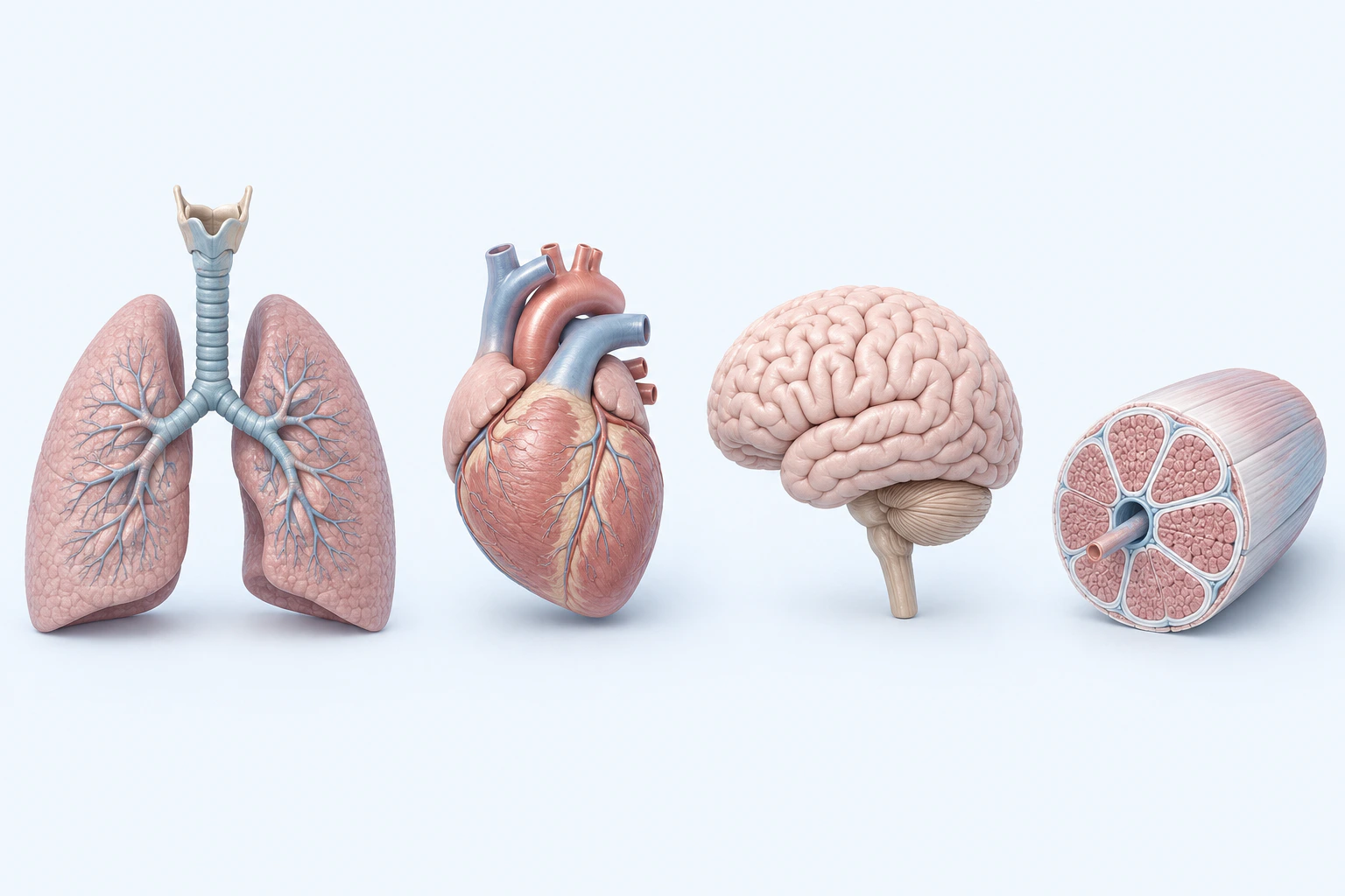
Heterogeneous multisystem orientation for Long COVID. These four selected undamaged models do not imply universal organ involvement or identify a lesion, biomarker, mechanism, symptom set, stage, severity, disability, prognosis, or diagnosis.
Scientific media record1 verified identifier
- Subject
- Post-Acute COVID-19 Syndromecondition
- Identifiers
- MeSH:D000094024
- Review
- Editorial review completeIdentifiers authority-verified · Accessibility validated · · long-covid|long-covid-pathology-v1.webp
- Digital source
- Trained-algorithmic mediaCreated with a trained generative algorithm and reviewed by WikiBiome for subject identity, scientific framing, identifiers, provenance, and accessibility.
- Scientific basis
- Post-Acute COVID-19 Syndrome — MeSHLong COVID Signs and Symptoms
- License
- CC BY-SA 4.0Created
Post-acute sequelae of SARS-CoV-2 infection (PASC), affecting an estimated 10-30% of COVID-19 survivors (WHO and CDC post-acute surveillance estimates) with symptoms persisting >12 weeks. Characterized by fatigue, cognitive dysfunction ("brain fog"), exercise intolerance, and multi-system involvement.
The emerging microbiome evidence reveals that Long COVID is not simply "slow recovery"—it is a self-perpetuating Dysbiosis-translocation-Metal-Driven Inflammation feedback loop where persistent Gut Microbiome disruption drives ongoing symptoms through the gut-lung and gut-brain axes.[1]Rego & Araújo-Filho 2024 — The Impact of Gut Microbiota on Long COVID: Insights and ChallengesAmália Cinthia Meneses do Rêgo, Irami Araújo-Filho · 2024Open reference 1 ↓[2]Ancona et al 2023 — Gut and Airway Microbiota Dysbiosis in COVID-19 and Long-COVIDGiuseppe Ancona, Laura Alagna, Claudia Alteri et al. · 2023Open reference 2 ↓
Evidence map62 cited passagesInspect provenance +
Post-acute sequelae of SARS-CoV-2 infection (PASC), affecting an estimated 10-30% of COVID-19 survivors (WHO and CDC post-acute surveillance estimates) with symptoms persisting 12 weeks. Characterized by fatigue, cognitive dysfunction ("brain fog"), exercise intolerance, and multi-system involvement. The emerging microbiome evidence reveals that Long COVID i
The microbiome disruption in Long COVID is distinguished from acute COVID and recovery by three features: (1) persistence—dysbiosis maintained months to years post-infection, (2) non-linear recovery—beneficial taxa peak at 3 months then regress as pathogens persist at 6 months, and (3) strain-level collapse—not just species depletion but loss of intra-
faecalibacterium prausnitzii—replicated across 6+ studies (,,,,, ); primary butyrate producer
roseburia—replicated across 5+ studies; SCFA producer,
bifidobacterium—immune education; SCFA production
lactobacillus—immune modulation; barrier function
akkermansia muciniphila—mucus layer maintenance
lachnospiraceae family—SCFA producers
Coordinated bacterial + fungal + viral community disruption. Bacteriophage populations shift in parallel; reduced phage diversity limits natural pathobiont predation.
Dysbiosis → SCFA depletion → barrier dysfunction → LPS translocation → systemic inflammation → further dysbiosis. This feedback loop explains why symptoms persist long after viral clearance. Approximately ~30% of hospitalized COVID patients had positive blood cultures matching gut organisms—direct evidence that barrier dysfunction drives bacteremia in the
Mendelian randomization analyses provide causal support: specific gut taxa (Lachnospiraceae, Ruminococcaceae) show causal relationships with Long COVID risk, and broader COVID-19 susceptibility shows causal microbiome contributions.
iron sequestered: Hepcidin elevation drives iron sequestration as host antiviral defense—functional anemia, not true deficiency. Iron was 10-fold decreased in COVID+ human milk.
selenium depleted: 2-fold decrease in COVID+ mothers; lower Se associated with COVID mortality.
zinc elevated: 1.7-fold increase in human milk as antiviral defense.
glutathione depleted: Reduced antioxidant metabolites.
LPS translocation crosses the BBB; reduced butyrate impairs BBB tight junctions; altered tryptophan metabolism (reduced kynurenine/AhR signaling) drives neuroinflammation and microglial activation, explaining "brain fog" and cognitive symptoms,.
SCFA depletion impairs respiratory mucosal immunity; reduced sIgA production; impaired Treg migration to respiratory tract.
Post-acute sequelae of SARS-CoV-2 (PASC) affects 10-30% of COVID survivors with symptoms persisting 12 weeks. The signature is distinctive for its self-perpetuating feedback loop: persistent gut dysbiosis → SCFA depletion → barrier dysfunction → bacterial translocation → systemic inflammation → further dysbiosis. This loop explains why symptoms persist long
iron sequestered: 10-fold decrease in COVID+ human milk. Hepcidin elevation drives iron sequestration as antiviral host defense—functional anemia, not true deficiency. This is a Primitive 2 insight: iron supplementation would feed siderophore-producing Proteobacteria pathobionts.
Glutathione depleted: Reduced antioxidant metabolites including glutathione and cysteine.
| Marker | Direction | Evidence | |--------|-----------|---------| | IL-6 | Persistently elevated |, | | TNF-alpha, IL-1beta | Elevated | Persistent Th1 activation | | PD-1/TIM-3 on CD8+ T cells | Elevated | T-cell exhaustion despite activation—hallmark of chronic antigen exposure | | LPS in circulation | Elevated | From translocation; ~30% of hospitalized
| Taxon | Role | Key Evidence | |-------|------|-------------| | Proteobacteria / enterobacteriaceae | LPS production; siderophore iron acquisition; blood translocation | (n=96), (n=112) | | streptococcus (S. equinus) | Facultative aerobe; persists 6 months post-recovery | (prospective, n=53) | | enterococcus | Translocation marker | | | candida albicans | M
| Taxon | Lost Function | Key Evidence | |-------|--------------|-------------| | faecalibacterium prausnitzii | Primary butyrate producer; anti-inflammatory | 6+ studies: Ancona, Didenko, Rego, Ghannoum, Ke, Mazzarelli | | roseburia | SCFA producer; barrier support | 5+ studies: Ancona, Didenko, Rego, Ghannoum, Ke | | bifidobacterium | Immune education; SCF
Persistence: Long COVID patients maintain Grade II dysbiosis while recovered patients show partial resolution
Showing 24 of 62 evidence-bearing passages. Every remaining citation is still indexed in the reference record below.
One disease. Five evidence layers.
A generated systems view of the metals, organisms, host sequestration signals, ecological conditions, and microbial functions indexed for Long COVID.
Evidence layer
Taxonomic signature
Organisms reported as enriched or depleted, with their indexed functional context kept beside the name.LPS producers; translocate to blood (~30% of hospitalized patients); siderophore iron acquisition
S. equinus persists at 6 months post-recovery; facultative aerobe enriched in dysbiotic niche
Facultative aerobe; translocation marker; dysbiosis indicator persisting post-recovery
Enriched in acute COVID dysbiosis; persists in some Long COVID
Multi-kingdom co-expansion with bacterial pathobionts; fungal dysbiosis component
Enhanced LPS synthesis genes in dysbiotic strains; pro-inflammatory
Fungal co-infection in severe/Long COVID
LPS-producing facultative anaerobe; translocation to bloodstream documented; siderophore-mediated iron piracy
Proteobacteria pathobiont; blood culture-confirmed translocation in ~30% hospitalized patients
Primary butyrate producer — replicated across 6+ studies; depletion drives SCFA insufficiency
Major SCFA producer — replicated across 5+ studies; loss impairs barrier function
Immune education; SCFA production; depletion impairs Treg development
Barrier function; immune modulation; loss weakens gut-lung axis immunity
Mucus layer maintenance; oxygen scavenging; loss exposes epithelium
SCFA-producing family; Mendelian randomization shows causal relationship with Long COVID risk
Evidence layer
Nutritional immunity
Host metal-withholding, inflammatory, antioxidant, and microbial-metabolite signals indexed in the signature.Elevated host signals
11Depleted protective signals
8Evidence layer
Ecological state
The environmental conditions that connect the organism-level observations into a system.Evidence layer
Virulence functions
Microbial structures, enzymes, and acquisition systems implicated by the linked evidence.The disease record, in full.
The original WikiBiome disease narrative remains intact beneath the generated signature atlas.
Microbiome Signature#
The microbiome disruption in Long COVID is distinguished from acute COVID and recovery by three features: (1) persistence—dysbiosis maintained months to years post-infection, (2) non-linear recovery—beneficial taxa peak at 3 months then regress as pathogens persist at 6 months,[3]Li et al. 2025 — Long-term Alterations in Gut Microbiota Following Mild COVID-19 RecoveryDa Li, Da-Ya Zhang, Shi-Ju Chen et al. · 2025Open reference 3 ↓ and (3) strain-level collapse—not just species depletion but loss of intra-species genetic diversity in beneficial taxa.[4]Ke, Weiss, Liu 2022 — Dissecting the role of the human microbiome in COVID-19 via metagenome-assembled genomesShanlin Ke, Scott T. Weiss, Yang-Yu Liu · 2022Open reference 4 ↓
Depleted Taxa#
The hallmark is systematic loss of obligate anaerobic SCFA producers.
Faecalibacterium prausnitzii—replicated across 6+ studies ([2]Ancona et al 2023 — Gut and Airway Microbiota Dysbiosis in COVID-19 and Long-COVIDGiuseppe Ancona, Laura Alagna, Claudia Alteri et al. · 2023Open reference 2 ↓[5]Didenko et al 2025 — Intestinal Microbiota and Short-Chain Fatty Acids in Patients with Post-COVID Immune ResponseV.I. Didenko, I.A. Klenina, O.M. Tatarchuk et al. · 2025Open reference 5 ↓[1]Rego & Araújo-Filho 2024 — The Impact of Gut Microbiota on Long COVID: Insights and ChallengesAmália Cinthia Meneses do Rêgo, Irami Araújo-Filho · 2024Open reference 1 ↓[6]Ghannoum et al. 2021 — Microbiome-Driven Approach to Combating Depression During COVID-19Mahmoud A. Ghannoum, Mary Kate Ford, Robert A. Bonomo et al. · 2021Open reference 6 ↓[4]Ke, Weiss, Liu 2022 — Dissecting the role of the human microbiome in COVID-19 via metagenome-assembled genomesShanlin Ke, Scott T. Weiss, Yang-Yu Liu · 2022Open reference 4 ↓[7]Mazzarelli et al. 2021 — 16S rRNA Gene Sequencing of Rectal Swab in COVID-19 PatientsAntonio Mazzarelli, Maria Letizia Giancola, Anna Farina et al. · 2021Open reference 7 ↓); primary Butyrate producer.
Roseburia—replicated across 5+ studies; SCFA producer.[5]Didenko et al 2025 — Intestinal Microbiota and Short-Chain Fatty Acids in Patients with Post-COVID Immune ResponseV.I. Didenko, I.A. Klenina, O.M. Tatarchuk et al. · 2025Open reference 5 ↓[1]Rego & Araújo-Filho 2024 — The Impact of Gut Microbiota on Long COVID: Insights and ChallengesAmália Cinthia Meneses do Rêgo, Irami Araújo-Filho · 2024Open reference 1 ↓
Bifidobacterium—immune education; SCFA production.[2]Ancona et al 2023 — Gut and Airway Microbiota Dysbiosis in COVID-19 and Long-COVIDGiuseppe Ancona, Laura Alagna, Claudia Alteri et al. · 2023Open reference 2 ↓ Lactobacillus—immune modulation; barrier function.[8]Xu et al. 2022 — Effective Regulation of Gut Microbiota With Probiotics and Prebiotics to Prevent/Alleviate COVID-19 via Gut-Lung AxisLei Xu, Chung S. Yang, Yanan Liu et al. · 2022Open reference 8 ↓ Akkermansia muciniphila—mucus layer maintenance.[1]Rego & Araújo-Filho 2024 — The Impact of Gut Microbiota on Long COVID: Insights and ChallengesAmália Cinthia Meneses do Rêgo, Irami Araújo-Filho · 2024Open reference 1 ↓
Lachnospiraceae family—SCFA producers.[5]Didenko et al 2025 — Intestinal Microbiota and Short-Chain Fatty Acids in Patients with Post-COVID Immune ResponseV.I. Didenko, I.A. Klenina, O.M. Tatarchuk et al. · 2025Open reference 5 ↓
Enriched Taxa#
Facultative aerobes and pathobionts bloom. Proteobacteria (phylum-level, including Enterobacteriaceae, Klebsiella pneumoniae, Escherichia coli)—LPS production; translocation to blood. Streptococcus (S. equinus persists at 6 months). Enterococcus—facultative aerobe; translocation marker.
Candida albicans—multi-kingdom co-expansion with bacterial pathobionts. Fusobacterium nucleatum—enhanced LPS synthesis genes in dysbiotic strains.
Multi-Kingdom Dysbiosis#
Coordinated bacterial + fungal + viral community disruption. Bacteriophage populations shift in parallel; reduced phage diversity limits natural pathobiont predation.[9]Lu et al. 2021 — Alterations in the Composition of Intestinal DNA Virome in Patients With COVID-19Zhen-Hua Lu, Hao-Wei Zhou, Wei-Kang Wu et al. · 2021Open reference 9 ↓
The Self-Perpetuating Loop#
Dysbiosis → SCFA depletion → barrier dysfunction → LPS translocation → systemic inflammation → further dysbiosis. This feedback loop explains why symptoms persist long after viral clearance.
Approximately ~30% of hospitalized COVID patients had positive blood cultures matching gut organisms—direct evidence that barrier dysfunction drives bacteremia in the acute phase.[10]Bernard-Raichon et al. 2022 — Gut microbiome dysbiosis in antibiotic-treated COVID-19 patients is associated with microbial translocation and bacteremiaLucie Bernard-Raichon, Mericien Venzon, Jon Klein et al. · 2022Open reference 10 ↓
Mendelian randomization analyses provide causal support: specific gut taxa (Lachnospiraceae, Ruminococcaceae) show causal relationships with Long COVID risk,[11]Li et al 2024 — The Causal Role of Gut Microbiota in Susceptibility of Long COVID: A Mendelian Randomization StudyZuming Li, Qinghua Xia, Jieni Feng et al. · 2024Open reference 11 ↓ and broader COVID-19 susceptibility shows causal microbiome contributions.[12]Zhong et al 2023 — Causal Effects of Gut Microbiome on COVID-19 Susceptibility and Severity: A Mendelian Randomization StudyMeng-Mei Zhong, Jia-Hao Xie, Yao Feng et al. · 2023Open reference 12 ↓
Metal Associations#
Iron sequestered: Hepcidin elevation drives iron sequestration as host antiviral defense—functional anemia, not true deficiency. Iron was 10-fold decreased in COVID+ human milk.[13]Metallomic and Untargeted Metabolomic Signatures of Human Milk from SARS-CoV-2 Positive MothersArias-Borrego A, Soto Cruz FJ, Selma-Royo M et al. · 2022Open reference 13 ↓
Selenium depleted: 2-fold decrease in COVID+ mothers; lower selenium (Se) associated with COVID mortality.[13]Metallomic and Untargeted Metabolomic Signatures of Human Milk from SARS-CoV-2 Positive MothersArias-Borrego A, Soto Cruz FJ, Selma-Royo M et al. · 2022Open reference 13 ↓ Zinc elevated: 1.7-fold increase in human milk as antiviral defense.[13]Metallomic and Untargeted Metabolomic Signatures of Human Milk from SARS-CoV-2 Positive MothersArias-Borrego A, Soto Cruz FJ, Selma-Royo M et al. · 2022Open reference 13 ↓ Glutathione (GSH) depleted: Reduced antioxidant metabolites.[14]Saito et al 2024 — Metabolomic and Immune Alterations in Long COVID Patients with Chronic Fatigue SyndromeSuguru Saito, Shima Shahbaz, Xian Luo et al. · 2024Open reference 14 ↓
Gut-Brain Axis#
LPS translocation crosses the BBB; reduced butyrate impairs BBB tight junctions; altered tryptophan metabolism (reduced kynurenine/AhR signaling) drives neuroinflammation and microglial activation, explaining "brain fog" and cognitive symptoms.[15]Plummer et al 2023 — Gut-Brain Pathogenesis of Post-Acute COVID-19 Neurocognitive SymptomsAllison M. Plummer, Yvette L. Matos, Henry C. Lin et al. · 2023Open reference 15 ↓[16]Brown et al. 2024 — Pathophysiology, Diagnosis, and Management of Neuroinflammation in COVID-19Rachel L Brown, Laura Benjamin, Michael P Lunn et al. · 2024Open reference 16 ↓
Gut-Lung Axis#
SCFA depletion impairs respiratory mucosal immunity; reduced sIgA production; impaired Treg migration to respiratory tract.[8]Xu et al. 2022 — Effective Regulation of Gut Microbiota With Probiotics and Prebiotics to Prevent/Alleviate COVID-19 via Gut-Lung AxisLei Xu, Chung S. Yang, Yanan Liu et al. · 2022Open reference 8 ↓
Open Questions#
Unresolved questions identified by the current evidence record.
01Can targeted microbiome restoration (FMT, specific probiotics, fiber) resolve Long COVID symptoms?+
The current WikiBiome record identifies this as an unresolved evidence gap.
02Does the strain-level diversity collapse require FMT or can dietary intervention restore it?+
The current WikiBiome record identifies this as an unresolved evidence gap.
03Why does recovery stall at 3-6 months in some patients but not others?+
The current WikiBiome record identifies this as an unresolved evidence gap.
04Is the estrobolome disruption driving the female predominance of Long COVID?+
The current WikiBiome record identifies this as an unresolved evidence gap.
Connections#
- dysbiosis—self-perpetuating loop unique to Long COVID
- Gut-Brain Axis—LPS translocation, tryptophan dysmetabolism, neuroinflammation
- Iron—hepcidin-mediated sequestration (Primitive 2)
- Selenium—depleted; associated with mortality
- Intestinal Permeability—Paneth cell and goblet cell loss documented
- Depression—shared SCFA depletion and tryptophan dysmetabolism patterns
References 18
Numbered by first appearance in the article, then reconciled with its declared source list.
- 1
Amália Cinthia Meneses do Rêgo, Irami Araújo-Filho (2024). Rego & Araújo-Filho 2024 — The Impact of Gut Microbiota on Long COVID: Insights and Challenges. Journal of Scientific Case Reports.
- 2
Giuseppe Ancona, Laura Alagna, Claudia Alteri et al. (2023). Ancona et al 2023 — Gut and Airway Microbiota Dysbiosis in COVID-19 and Long-COVID. Frontiers in Immunology.
- 3
Da Li, Da-Ya Zhang, Shi-Ju Chen et al. (2025). Li et al. 2025 — Long-term Alterations in Gut Microbiota Following Mild COVID-19 Recovery. Frontiers in Cellular and Infection Microbiology.
- 4
Shanlin Ke, Scott T. Weiss, Yang-Yu Liu (2022). Ke, Weiss, Liu 2022 — Dissecting the role of the human microbiome in COVID-19 via metagenome-assembled genomes. Nature Communications.
- 5
V.I. Didenko, I.A. Klenina, O.M. Tatarchuk et al. (2025). Didenko et al 2025 — Intestinal Microbiota and Short-Chain Fatty Acids in Patients with Post-COVID Immune Response. Gastroenterology.
- 6
Mahmoud A. Ghannoum, Mary Kate Ford, Robert A. Bonomo et al. (2021). Ghannoum et al. 2021 — Microbiome-Driven Approach to Combating Depression During COVID-19. Frontiers in Nutrition.
- 7
Antonio Mazzarelli, Maria Letizia Giancola, Anna Farina et al. (2021). Mazzarelli et al. 2021 — 16S rRNA Gene Sequencing of Rectal Swab in COVID-19 Patients. PLOS ONE.
- 8
Lei Xu, Chung S. Yang, Yanan Liu et al. (2022). Xu et al. 2022 — Effective Regulation of Gut Microbiota With Probiotics and Prebiotics to Prevent/Alleviate COVID-19 via Gut-Lung Axis. Frontiers in Pharmacology.
- 9
Zhen-Hua Lu, Hao-Wei Zhou, Wei-Kang Wu et al. (2021). Lu et al. 2021 — Alterations in the Composition of Intestinal DNA Virome in Patients With COVID-19. Frontiers in Cellular and Infection Microbiology.
- 10
Lucie Bernard-Raichon, Mericien Venzon, Jon Klein et al. (2022). Bernard-Raichon et al. 2022 — Gut microbiome dysbiosis in antibiotic-treated COVID-19 patients is associated with microbial translocation and bacteremia. Nature Communications.
- 11
Zuming Li, Qinghua Xia, Jieni Feng et al. (2024). Li et al 2024 — The Causal Role of Gut Microbiota in Susceptibility of Long COVID: A Mendelian Randomization Study. Frontiers in Microbiology.
- 12
Meng-Mei Zhong, Jia-Hao Xie, Yao Feng et al. (2023). Zhong et al 2023 — Causal Effects of Gut Microbiome on COVID-19 Susceptibility and Severity: A Mendelian Randomization Study. Frontiers in Immunology.
- 13
Arias-Borrego A, Soto Cruz FJ, Selma-Royo M et al. (2022). Metallomic and Untargeted Metabolomic Signatures of Human Milk from SARS-CoV-2 Positive Mothers. Molecular Nutrition and Food Research.
- 14
Suguru Saito, Shima Shahbaz, Xian Luo et al. (2024). Saito et al 2024 — Metabolomic and Immune Alterations in Long COVID Patients with Chronic Fatigue Syndrome. Frontiers in Immunology.
- 15
Allison M. Plummer, Yvette L. Matos, Henry C. Lin et al. (2023). Plummer et al 2023 — Gut-Brain Pathogenesis of Post-Acute COVID-19 Neurocognitive Symptoms. Frontiers in Neuroscience.
- 16
Rachel L Brown, Laura Benjamin, Michael P Lunn et al. (2024). Brown et al. 2024 — Pathophysiology, Diagnosis, and Management of Neuroinflammation in COVID-19. BMJ (British Medical Journal).
- 17
Natascha Brigo, Wolfram Mayr, Maja Taenzer et al. (2025). Brigo et al. 2025 — Uremic Bacterial Metabolites in Post-COVID-19 Syndrome. Frontiers in Cellular and Infection Microbiology.
- 18
Khrystyna Duve, Pavlo Petakh, Oleksandr Kamyshnyi (2024). Duve et al. 2024 — COVID-19-Associated Encephalopathy: Neuroinflammation and Microbiota-Gut-Brain Axis. Frontiers in Microbiology.
Article network
Mentioned here 18
Pages linking here 6
Connect the evidence
Publicly readable discussion by ORCID-authenticated researchers. Questions, interpretation, methods, corrections, and new evidence stay attached to this record.
No discussion yet. Start with a precise question or a source-backed observation.
Activity and accepted changes
Accepted researcher context, editorial status, public discussion, and upstream Git revisions are shown together. Pending, declined, and withdrawn proposals remain private.
- published revision
Backfill gut microbiome concept links
Karen Pendergrass · +1 −1
Inspect exact Git diff ↗ - published revision
Backfill inflammation concept links
Karen Pendergrass · +1 −1
Inspect exact Git diff ↗ - published revision
Complete corpus-wide Dysbiosis linking
Karen Pendergrass · +2 −2
Inspect exact Git diff ↗ - published revision
Complete Tight junctions contextual coverage
Karen Pendergrass · +1 −1
Inspect exact Git diff ↗ - published revision
massive wiki expansion: 149 stubs fixed, 100+ new pages, Rule 15 scan, keystone papers
WikiBiome Deploy Bot · +18 −18
Inspect exact Git diff ↗ - published revision
ingest: 87 papers (17 endometriosis, 67 erectile dysfunction, 3 cross-condition)
WikiBiome Deploy Bot · +3 −3
Inspect exact Git diff ↗ - published revision
cycle 1: health check + lint fixes + 8 ingests + 2 stubs + gestational-diabetes signature
WikiBiome Deploy Bot · +1 −0
Inspect exact Git diff ↗ - published revision
pre-overnight checkpoint 2026-04-18
WikiBiome Deploy Bot · +2 −2
Inspect exact Git diff ↗ - published revision
Batch: fix 1025 broken wikilinks, wire 13 STOP pages, deepen PPD/GERD/T1D/8 microbes
WikiBiome Deploy Bot · +18 −15
Inspect exact Git diff ↗ - published revision
Batch: fix 1025 broken wikilinks, wire 13 STOP pages, deepen PPD/GERD/T1D/8 microbes
WikiBiome Deploy Bot · +14 −14
Inspect exact Git diff ↗ - published revision
Priority 5 batch migration: 100 source pages + source_count fixes + broken-links + orphans + index recount
WikiBiome Deploy Bot · +1 −1
Inspect exact Git diff ↗ - published revision
Deepen metal/concept entities + 8 new sources for T1D/schizophrenia
WikiBiome Deploy Bot · +217 −0
Inspect exact Git diff ↗ - published revision
Deepen metal/concept entities + 8 new sources for T1D/schizophrenia
WikiBiome Deploy Bot · +15 −13
Inspect exact Git diff ↗ - published revision
Citation integrity pass: 38 citations added, 24 parenthesized citations fixed
WikiBiome Deploy Bot · +7 −7
Inspect exact Git diff ↗ - published revision
Long COVID + Female Infertility signatures, entities, and ASD cleanup
WikiBiome Deploy Bot · +78 −0
Inspect exact Git diff ↗