
 metals · microbes · host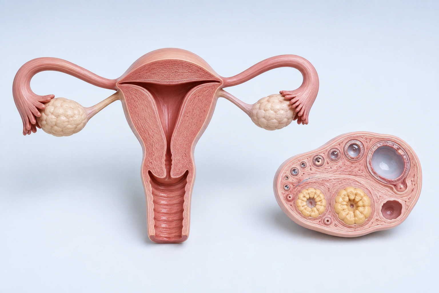
Neutral reproductive anatomy and ovarian-cycle orientation for female infertility. The ovarian cutaway depicts representative maturational follicles, not cysts, an ovarian-reserve measurement, an asserted cause, treatment, prognosis, or diagnosis.
Scientific media record1 verified identifier
- Subject
- Infertility, Femalecondition
- Identifiers
- MeSH:D007247
- Review
- Editorial review completeIdentifiers authority-verified · Accessibility validated · · female-infertility|female-infertility-pathology-v1.webp
- Digital source
- Trained-algorithmic mediaCreated with a trained generative algorithm and reviewed by WikiBiome for subject identity, scientific framing, identifiers, provenance, and accessibility.
- Scientific basis
- Infertility, Female — MeSHPossible causes of female infertility
- License
- CC BY-SA 4.0Created
Affects approximately 48 million women worldwide (WHO global infertility prevalence estimates).
From a microbiome-metallomics perspective, female infertility sits at the intersection of the estrobolome (microbial estrogen metabolism), heavy metal reproductive toxicity (cadmium (Cd), lead (Pb) as endocrine disruptors),[1]Female Infertility Associated with Blood Lead and Cadmium LevelsLee S, Min JY, Min KB · 2020Open reference 1 ↓[2]Association between heavy metals exposure and infertility among American women aged 20-44 years: A cross-sectional analysis from 2013 to 2018 NHANES dataLin J, Lin X, Qiu J et al. · 2023Open reference 2 ↓ and an emerging concept: the gut-ovarian axis—direct causal links between Gut Microbiome composition and ovarian function.[3]Fecal microbiota transplantation from patients with polycystic ovary syndrome induces metabolic disorders and ovarian dysfunction in germ-free miceHuang F, Deng Y, Zhou M et al. · 2024Open reference 3 ↓[4]Estropausal gut microbiota transplant improves measures of ovarian function in adult miceKim M, Wang J, Pilley SE et al. · 2026Open reference 4 ↓
Evidence map48 cited passagesInspect provenance +
Affects approximately 48 million women worldwide (WHO global infertility prevalence estimates). From a microbiome-metallomics perspective, female infertility sits at the intersection of the estrobolome (microbial estrogen metabolism), heavy metal reproductive toxicity (Cd, Pb as endocrine disruptors),, and an emerging concept: the gut-ovarian axis—direct c
FMT transfers PCOS phenotype: Fecal transplant from PCOS patients into germ-free mice transferred the full metabolic + ovarian dysfunction phenotype—insulin resistance, obesity, and disrupted ovarian function. The microbiome can "set the hormonal phenotype."
FMT reverses ovarian aging: Heterochronic FMT from young mice reversed age-related ovarian transcriptome changes, reduced inflammation, and increased fertility, Nature Aging. A young microbiome can rejuvenate aged ovaries.
Bifidobacterium longum improves IVF response: B. longum abundance correlated with good ovarian stimulation response; gavage in mice improved outcomes.
Enriched in premature ovarian insufficiency (POI),
Eggerthella is a known beta-glucuronidase producer—deconjugates estrogen metabolites, driving estrogen recirculation
Eggerthella lenta caused ovarian fibrosis in mouse models; estrogen treatment ameliorated it
HRT reversed both the dysbiosis and the ovarian fibrosis
Enriched in POI: Eggerthella, Butyricimonas, Dorea, Sutterella,
5-bile-acid panel achieves AUC = 0.964 for distinguishing DOR from normal reserve
cadmium binds ERalpha at picomolar concentrations; accumulates in ovarian tissue over decades (half-life 12-30 years in the human body),,
lead disrupts hypothalamic-pituitary-ovarian axis; associated with reduced fertility in epidemiological studies,
nickel binds ERalpha noncompetitively; epigenetic carcinogenesis
NHANES analyses link heavy metal mixtures (Cd, Pb, As, Hg) to elevated female infertility risk in US population-level data
Beyond the gut, the reproductive tract itself harbors microbial communities that influence fertility. Lactobacillus-dominant vaginal communities (particularly L. crispatus) correlate with successful conception and implantation, while dysbiotic communities (bacterial vaginosis-associated taxa such as Gardnerella, Atopobium, Prevotella) are associated with red
Dietary cadmium (leafy greens, shellfish, offal, cocoa grown on contaminated soil) and occupational/ambient lead exposure (water infrastructure, dust, tobacco smoke) are the dominant non-occupational routes in women of reproductive age,. Because cadmium has a 12-30 year biological half-life, fertility risk reflects cumulative exposure across the premenarchal
FMT from PCOS patients into germ-free mice transferred insulin resistance + obesity + disrupted ovarian function. The microbiome can "set the hormonal phenotype."
Heterochronic FMT from young mice reversed age-related ovarian transcriptome changes, reduced inflammation, and increased fertility (, Nature Aging 2026).
Bifidobacterium longum abundance correlated with good ovarian stimulation response (follicle-to-oocyte index = 0.5); gavage in mice validated the effect.
Enriched in POI patients
5-bile-acid panel achieves AUC = 0.964 for DOR diagnosis (, n=182)
Female infertility affects approximately 48 million women worldwide (WHO). This signature reveals three converging pathways: (1) the estrobolome—microbial estrogen metabolism driven by beta-glucuronidase-producing taxa like eggerthella lenta that disrupt ovarian function, (2) metalloestrogen accumulation—cadmium and nickel binding estrogen receptors at p
| Metal | Direction | Key Evidence | |-------|-----------|-------------| | cadmium | Elevated | Binds ERalpha with affinity near estradiol (Kd 4.5x10^-10 M); accumulates in ovarian tissue over decades (half-life 12-30 years); 2-fold increase in blood Cd = 1.84x infertility odds; NHANES urinary Cd significantly higher in infertile women | | lead | Elevated |
Cadmium is the paradigmatic metalloestrogen. It binds ERalpha with a dissociation constant (Kd 4.5x10^-10 M) nearly equivalent to estradiol, activating estrogen target genes (CycD1, c-myc) at concentrations as low as 1 uM. It also activates the membrane-bound estrogen receptor GPR30/GPER, inducing proliferative responses at 50-500 nM in ER-negative cells. Be
Showing 24 of 48 evidence-bearing passages. Every remaining citation is still indexed in the reference record below.
One disease. Five evidence layers.
A generated systems view of the metals, organisms, host sequestration signals, ecological conditions, and microbial functions indexed for Female Infertility.
Evidence layer
Taxonomic signature
Organisms reported as enriched or depleted, with their indexed functional context kept beside the name.Beta-glucuronidase producer; enriched in POI; caused ovarian fibrosis in mouse models; estrogen deconjugation drives estrogen recirculation
Enriched in POI; gram-negative anaerobe associated with gut inflammation
Enriched in premature ovarian insufficiency; associated with altered sex hormone profiles
Enriched in POI; correlates with FSH and LH levels
Reclassified Bacteroides; enriched in PCOS (transferable via FMT)
Major butyrate producer depleted in POI; loss reduces anti-inflammatory SCFA buffer
B. longum abundance correlates with good IVF ovarian stimulation response; gavage in mice improved outcomes
L. crispatus dominance in vaginal microbiome correlates with successful conception; depleted in PCOS vaginal communities
SCFA producers depleted across infertility subtypes; loss impairs barrier function and bile acid metabolism
Evidence layer
Nutritional immunity
Host metal-withholding, inflammatory, antioxidant, and microbial-metabolite signals indexed in the signature.Elevated host signals
3Depleted protective signals
4Evidence layer
Ecological state
The environmental conditions that connect the organism-level observations into a system.Evidence layer
Virulence functions
Microbial structures, enzymes, and acquisition systems implicated by the linked evidence.The disease record, in full.
The original WikiBiome disease narrative remains intact beneath the generated signature atlas.
The Gut-Ovarian Axis#
Three independent lines of evidence establish causal microbiome-to-ovarian links:
- FMT transfers PCOS phenotype: Fecal transplant from PCOS patients into germ-free mice transferred the full metabolic + ovarian dysfunction phenotype—insulin resistance, obesity, and disrupted ovarian function.[3]Fecal microbiota transplantation from patients with polycystic ovary syndrome induces metabolic disorders and ovarian dysfunction in germ-free miceHuang F, Deng Y, Zhou M et al. · 2024Open reference 3 ↓ The microbiome can "set the hormonal phenotype."
- FMT reverses ovarian aging: Heterochronic FMT from young mice reversed age-related ovarian transcriptome changes, reduced Metal-Driven Inflammation, and increased fertility,[4]Estropausal gut microbiota transplant improves measures of ovarian function in adult miceKim M, Wang J, Pilley SE et al. · 2026Open reference 4 ↓ Nature Aging. A young microbiome can rejuvenate aged ovaries.
- Bifidobacterium longum improves IVF response: B. longum abundance correlated with good ovarian stimulation response; gavage in mice improved outcomes.[5]Metagenomic analysis revealed the association between gut microbiota and different ovary responses to controlled ovarian stimulationFo X, Pei M, Liu P et al. · 2024Open reference 5 ↓
Estrobolome Connection#
Eggerthella lenta is the critical organism linking gut Dysbiosis to ovarian disease.
Enriched in premature ovarian insufficiency (POI).[6]Hormone Replacement Therapy Reverses Gut Microbiome and Serum Metabolome Alterations in Premature Ovarian InsufficiencyJiang L, Fei H, Tong J et al. · 2021Open reference 6 ↓[7]Association between premature ovarian insufficiency and gut microbiotaWu J, Zhuo Y, Liu Y et al. · 2021Open reference 7 ↓ Eggerthella is a known beta-glucuronidase producer—deconjugates estrogen metabolites, driving estrogen recirculation.[6]Hormone Replacement Therapy Reverses Gut Microbiome and Serum Metabolome Alterations in Premature Ovarian InsufficiencyJiang L, Fei H, Tong J et al. · 2021Open reference 6 ↓
Eggerthella lenta caused ovarian fibrosis in mouse models; estrogen treatment ameliorated it.[6]Hormone Replacement Therapy Reverses Gut Microbiome and Serum Metabolome Alterations in Premature Ovarian InsufficiencyJiang L, Fei H, Tong J et al. · 2021Open reference 6 ↓ HRT reversed both the dysbiosis and the ovarian fibrosis.[6]Hormone Replacement Therapy Reverses Gut Microbiome and Serum Metabolome Alterations in Premature Ovarian InsufficiencyJiang L, Fei H, Tong J et al. · 2021Open reference 6 ↓
Premature Ovarian Insufficiency (POI)#
Two independent microbiome studies show. Enriched in POI: Eggerthella, Butyricimonas, Dorea, Sutterella.[7]Association between premature ovarian insufficiency and gut microbiotaWu J, Zhuo Y, Liu Y et al. · 2021Open reference 7 ↓[6]Hormone Replacement Therapy Reverses Gut Microbiome and Serum Metabolome Alterations in Premature Ovarian InsufficiencyJiang L, Fei H, Tong J et al. · 2021Open reference 6 ↓ Depleted in POI: Faecalibacterium prausnitzii (major Butyrate producer), Bulleidia.
Microbial alterations correlated with FSH, LH, E2, AMH, and FSH/LH ratio.
Diminished Ovarian Reserve (DOR)#
Bile acids produced by gut bacteria are measurable in follicular fluid and depleted in DOR. Lithocholic acid, chenodeoxycholic acid, ursodeoxycholic acid, deoxycholic acid, cholic acid all significantly lower. 5-bile-acid panel achieves AUC = 0.964 for distinguishing DOR from normal reserve.[8]Ding 2024 — Bile Acids in Follicular Fluid as Therapeutic Targets for Diminished Ovarian ReserveDing S, Li W, Xiong X et al. · 2024Open reference 8 ↓
Metal Connections#
Female reproductive tissues are uniquely vulnerable to metalloestrogens. Cadmium binds ERalpha at picomolar concentrations; accumulates in ovarian tissue over decades (half-life 12-30 years in the human body).[9]Role of Cadmium and Nickel in Estrogen Receptor Signaling and Breast Cancer: Metalloestrogens or Not?Aquino NB, Sevigny MB, Sabangan J et al. · 2012Open reference 9 ↓[10]The Effects of Cadmium ToxicityGiuseppe Genchi, Maria Stefania Sinicropi, Graziantonio Lauria et al. · 2020Open reference 10 ↓[11]Exposure to Cadmium and Its Impacts on Human Health: A Short ReviewPuthiyavalappil Rasin, Ashwathi A V, Sabeel M Basheer et al. · 2025Open reference 11 ↓
Lead disrupts hypothalamic-pituitary-ovarian axis; associated with reduced fertility in epidemiological studies.[1]Female Infertility Associated with Blood Lead and Cadmium LevelsLee S, Min JY, Min KB · 2020Open reference 1 ↓[12]Relationship between risk factors for infertility in women and lead, cadmium, and arsenic blood levels: a cross-sectional study from TaiwanLei HL, Wei HJ, Ho HY et al. · 2015Open reference 12 ↓
Nickel binds ERalpha noncompetitively; epigenetic carcinogenesis.[9]Role of Cadmium and Nickel in Estrogen Receptor Signaling and Breast Cancer: Metalloestrogens or Not?Aquino NB, Sevigny MB, Sabangan J et al. · 2012Open reference 9 ↓
NHANES analyses link heavy metal mixtures (cadmium (Cd), lead (Pb), arsenic (As), mercury (Hg)) to elevated female infertility risk in US population-level data.[2]Association between heavy metals exposure and infertility among American women aged 20-44 years: A cross-sectional analysis from 2013 to 2018 NHANES dataLin J, Lin X, Qiu J et al. · 2023Open reference 2 ↓
Vaginal and Endometrial Microbiome#
Beyond the gut, the reproductive tract itself harbors microbial communities that influence fertility. Lactobacillus-dominant vaginal communities (particularly L. crispatus) correlate with successful conception and implantation, while dysbiotic communities (bacterial vaginosis-associated taxa such as Gardnerella, Atopobium, Prevotella) are associated with reduced IVF success, recurrent implantation failure, and early pregnancy loss.
Shotgun metagenomics in PCOS patients shows parallel vaginal dysbiosis—reduced Lactobacillus with enriched anaerobes and altered phageome composition.[13]Differential enrichment of bacteria and phages in the vaginal microbiomes in PCOS and obesity: shotgun sequencing analysisZheng S, Chen H, Yang H et al. · 2024Open reference 13 ↓
The endometrial microbiome, historically assumed sterile, hosts a low-biomass community whose composition at embryo transfer may predict live birth—a rapidly evolving field where WikiBiome will track emerging signatures.
Environmental Exposures#
Dietary cadmium (leafy greens, shellfish, offal, cocoa grown on contaminated soil) and occupational/ambient lead exposure (water infrastructure, dust, tobacco smoke) are the dominant non-occupational routes in women of reproductive age.[10]The Effects of Cadmium ToxicityGiuseppe Genchi, Maria Stefania Sinicropi, Graziantonio Lauria et al. · 2020Open reference 10 ↓[11]Exposure to Cadmium and Its Impacts on Human Health: A Short ReviewPuthiyavalappil Rasin, Ashwathi A V, Sabeel M Basheer et al. · 2025Open reference 11 ↓
Because cadmium has a 12-30 year biological half-life, fertility risk reflects cumulative exposure across the premenarchal and reproductive years, not point-in-time serum levels—a critical distinction for interpreting biomarker studies.
Smoking doubles body cadmium burden and is among the strongest modifiable fertility risk factors in epidemiological analyses.[1]Female Infertility Associated with Blood Lead and Cadmium LevelsLee S, Min JY, Min KB · 2020Open reference 1 ↓
Open Questions#
Unresolved questions identified by the current evidence record.
01Can targeted probiotic (B. longum) supplementation improve IVF outcomes in poor responders?+
The current WikiBiome record identifies this as an unresolved evidence gap.
02Does Eggerthella-targeting (antibiotic or probiotic competition) prevent or reverse POI?+
The current WikiBiome record identifies this as an unresolved evidence gap.
03Can microbiome-based prediction (bile acid panel, taxa profiles) identify infertility risk before clinical manifestation?+
The current WikiBiome record identifies this as an unresolved evidence gap.
04What is the relative contribution of gut vs. vaginal/endometrial microbiome to infertility?+
The current WikiBiome record identifies this as an unresolved evidence gap.
05Does cadmium-driven estrobolome dysfunction explain rising infertility rates in contaminated regions?+
The current WikiBiome record identifies this as an unresolved evidence gap.
Connections#
- Estrogen Recirculation—Eggerthella-mediated beta-glucuronidase activity
- Beta-Glucuronidase—key enzyme linking gut microbiome to estrogen metabolism
- Polycystic Ovary Syndrome—PCOS phenotype transferable via FMT; shared signature exists
- Endometriosis—shared metalloestrogen biology (cadmium (Cd), nickel (Ni))
- Cadmium—metalloestrogen; ovarian tissue accumulation
- Bile Acid Metabolism—follicular fluid bile acids as ovarian reserve biomarkers
- Metalloestrogens—cadmium, nickel, arsenic (As) binding ERalpha
References 13
Numbered by first appearance in the article, then reconciled with its declared source list.
- 1
Lee S, Min JY, Min KB (2020). Female Infertility Associated with Blood Lead and Cadmium Levels. International Journal of Environmental Research and Public Health.
- 2
Lin J, Lin X, Qiu J et al. (2023). Association between heavy metals exposure and infertility among American women aged 20-44 years: A cross-sectional analysis from 2013 to 2018 NHANES data. Frontiers in Public Health.
- 3
Huang F, Deng Y, Zhou M et al. (2024). Fecal microbiota transplantation from patients with polycystic ovary syndrome induces metabolic disorders and ovarian dysfunction in germ-free mice. BMC Microbiology.
- 4
Kim M, Wang J, Pilley SE et al. (2026). Estropausal gut microbiota transplant improves measures of ovarian function in adult mice. Nature Aging.
- 5
Fo X, Pei M, Liu P et al. (2024). Metagenomic analysis revealed the association between gut microbiota and different ovary responses to controlled ovarian stimulation. Scientific Reports.
- 6
Jiang L, Fei H, Tong J et al. (2021). Hormone Replacement Therapy Reverses Gut Microbiome and Serum Metabolome Alterations in Premature Ovarian Insufficiency. Frontiers in Endocrinology.
- 7
Wu J, Zhuo Y, Liu Y et al. (2021). Association between premature ovarian insufficiency and gut microbiota. BMC Pregnancy and Childbirth.
- 8
Ding S, Li W, Xiong X et al. (2024). Ding 2024 — Bile Acids in Follicular Fluid as Therapeutic Targets for Diminished Ovarian Reserve. Journal of Ovarian Research.
- 9
★Aquino NB, Sevigny MB, Sabangan J et al. (2012). Role of Cadmium and Nickel in Estrogen Receptor Signaling and Breast Cancer: Metalloestrogens or Not?. Journal of Environmental Science and Health Part C - Environmental Carcinogenesis and Ecotoxicology Reviews.
- 10
Giuseppe Genchi, Maria Stefania Sinicropi, Graziantonio Lauria et al. (2020). The Effects of Cadmium Toxicity. International Journal of Environmental Research and Public Health.
- 11
★Puthiyavalappil Rasin, Ashwathi A V, Sabeel M Basheer et al. (2025). Exposure to Cadmium and Its Impacts on Human Health: A Short Review. Journal of Hazardous Materials Advances.
- 12
Lei HL, Wei HJ, Ho HY et al. (2015). Relationship between risk factors for infertility in women and lead, cadmium, and arsenic blood levels: a cross-sectional study from Taiwan. BMC Public Health.
- 13
Zheng S, Chen H, Yang H et al. (2024). Differential enrichment of bacteria and phages in the vaginal microbiomes in PCOS and obesity: shotgun sequencing analysis. Frontiers in Microbiomes.
Article network
Mentioned here 16
Pages linking here 8
Connect the evidence
Publicly readable discussion by ORCID-authenticated researchers. Questions, interpretation, methods, corrections, and new evidence stay attached to this record.
No discussion yet. Start with a precise question or a source-backed observation.
Activity and accepted changes
Accepted researcher context, editorial status, public discussion, and upstream Git revisions are shown together. Pending, declined, and withdrawn proposals remain private.
- published revision
Backfill gut microbiome concept links
Karen Pendergrass · +1 −1
Inspect exact Git diff ↗ - published revision
Backfill inflammation concept links
Karen Pendergrass · +1 −1
Inspect exact Git diff ↗ - published revision
Complete corpus-wide Dysbiosis linking
Karen Pendergrass · +1 −1
Inspect exact Git diff ↗ - published revision
massive wiki expansion: 149 stubs fixed, 100+ new pages, Rule 15 scan, keystone papers
WikiBiome Deploy Bot · +14 −14
Inspect exact Git diff ↗ - published revision
cycle 1: health check + lint fixes + 8 ingests + 2 stubs + gestational-diabetes signature
WikiBiome Deploy Bot · +1 −0
Inspect exact Git diff ↗ - published revision
pre-overnight checkpoint 2026-04-18
WikiBiome Deploy Bot · +2 −2
Inspect exact Git diff ↗ - published revision
Batch: fix 1025 broken wikilinks, wire 13 STOP pages, deepen PPD/GERD/T1D/8 microbes
WikiBiome Deploy Bot · +6 −6
Inspect exact Git diff ↗ - published revision
Batch: fix 1025 broken wikilinks, wire 13 STOP pages, deepen PPD/GERD/T1D/8 microbes
WikiBiome Deploy Bot · +4 −4
Inspect exact Git diff ↗ - published revision
Deepen metal/concept entities + 8 new sources for T1D/schizophrenia
WikiBiome Deploy Bot · +150 −0
Inspect exact Git diff ↗ - published revision
Deepen metal/concept entities + 8 new sources for T1D/schizophrenia
WikiBiome Deploy Bot · +19 −11
Inspect exact Git diff ↗ - published revision
Citation integrity pass: 38 citations added, 24 parenthesized citations fixed
WikiBiome Deploy Bot · +6 −6
Inspect exact Git diff ↗ - published revision
Long COVID + Female Infertility signatures, entities, and ASD cleanup
WikiBiome Deploy Bot · +78 −0
Inspect exact Git diff ↗