

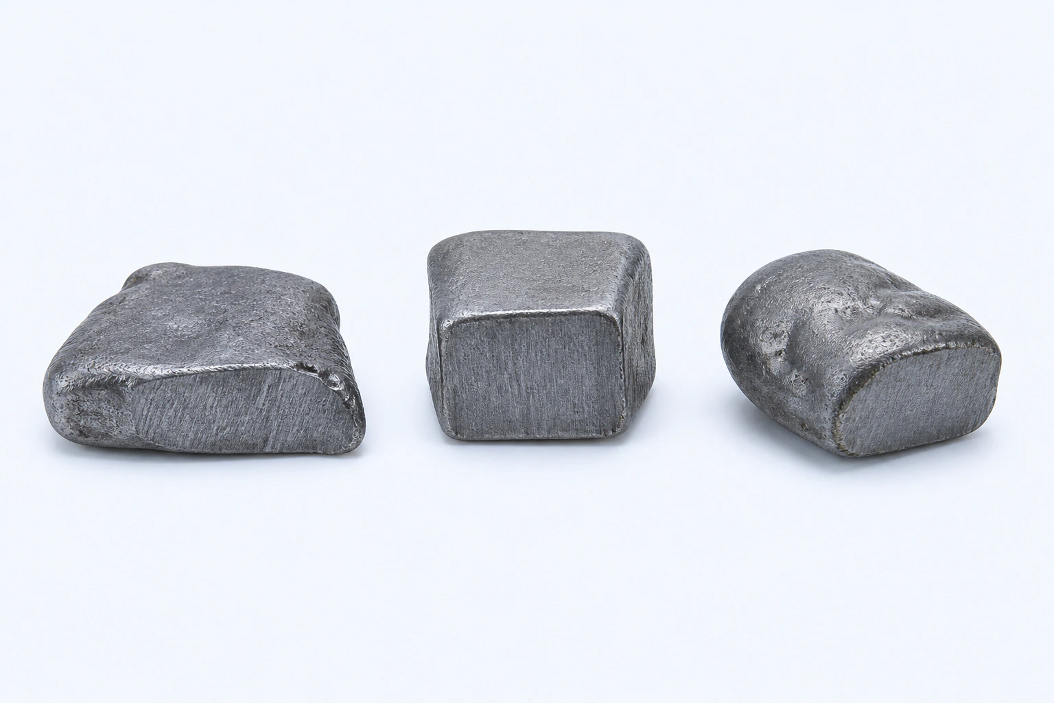
Elemental lead (Pb), shown as three representative dense-looking gray solid specimens. Form and surface vary with purity, processing, and tarnish; this is not a product, analytical reference material, or a photograph.
Scientific media record2 verified identifiers
- Subject
- Leadelement
- Identifiers
- Atomic number 82PubChem CID:5352425
- Review
- Editorial review completeIdentifiers authority-verified · Accessibility validated · · lead|lead-technical-specimen-v1.webp
- Digital source
- Trained-algorithmic mediaCreated with a trained generative algorithm and reviewed by WikiBiome for subject identity, scientific framing, identifiers, provenance, and accessibility.
- Scientific basis
- Lead — PubChem ElementLead, PubChem CID 5352425
- License
- CC BY-SA 4.0Created
Lead is a purely toxic heavy metal with no known biological function. It is the most extensively studied metal in relation to neurodevelopmental harm and is increasingly recognized as a contributor to chronic disease across virtually every organ system, even at levels once considered safe.
Its toxicity operates primarily through calcium mimicry, heme biosynthesis disruption, and Oxidative Stress.[1]Toxic Mechanisms of Five Heavy Metals: Mercury, Lead, Chromium, Cadmium, and ArsenicBalali-Mood M, Naseri K, Tahergorabi Z et al. · 2021Open reference 1 ↓[2]Toxicity, Mechanism and Health Effects of Some Heavy MetalsMonisha Jaishankar, Tenzin Tseten, Naresh Anbalagan et al. · 2014Open reference 2 ↓ No level of lead exposure can be considered safe.[3]Metal toxicity exposure in Alzheimer's disease - literature reviewJakubowska E, Hoppe-Mitera E, Sionek I et al. · 2024Open reference 3 ↓[4]Renal health and the environment: heavy metal nephrotoxicitySabath E, Robles-Osorio ML · 2012Open reference 4 ↓
What sets lead apart from other toxic metals is its ability to infiltrate calcium-dependent signaling pathways throughout the body.
Because lead (Pb)(II) mimics calcium(II) (Ca2+), it enters cells through calcium channels, accumulates in bone as a long-term reservoir, crosses the blood-brain barrier, and disrupts neurotransmission, enzyme function, and gene expression at concentrations far below those that produce overt symptoms.[5]Interaction of Heavy Metal Lead with Gut Microbiota: Implications for Autism Spectrum DisorderTizabi Y, Bennani S, El Kouhen N et al. · 2023Open reference 5 ↓[6]Heavy metals in the diet: unraveling the molecular pathways linked to neurodegenerative disease riskGuevara-Ramirez P, Tamayo-Trujillo R, Cadena-Ullauri S et al. · 2024Open reference 6 ↓
Approximately 99% of blood lead is protein-bound, and bone serves as the primary long-term reservoir with a half-life of decades.[5]Interaction of Heavy Metal Lead with Gut Microbiota: Implications for Autism Spectrum DisorderTizabi Y, Bennani S, El Kouhen N et al. · 2023Open reference 5 ↓
What Wikipedia does not cover is lead's profound disruption of the Gut Microbiome—its role as a selective pressure favoring pathogenic taxa, its destruction of the intestinal barrier, the bidirectional relationship in which Dysbiosis impairs the microbiome's own capacity for lead detoxification, and the co-selection of antibiotic resistance genes under lead pressure.
Evidence map57 cited passagesInspect provenance +
Lead is a purely toxic heavy metal with no known biological function. It is the most extensively studied metal in relation to neurodevelopmental harm and is increasingly recognized as a contributor to chronic disease across virtually every organ system, even at levels once considered safe. Its toxicity operates primarily through calcium mimicry, heme biosynt
What sets lead apart from other toxic metals is its ability to infiltrate calcium-dependent signaling pathways throughout the body. Because Pb(II) mimics Ca2+, it enters cells through calcium channels, accumulates in bone as a long-term reservoir, crosses the blood-brain barrier, and disrupts neurotransmission, enzyme function, and gene expression at concent
Lead has no biological role. It is purely toxic. Unlike essential metals such as iron, zinc, and copper, lead is not a required cofactor for any known enzyme or biological process. Its toxicity arises entirely from its ability to mimic and displace essential divalent cations.
Lead competes with Ca2+ for binding sites on ion channels, transporters, and intracellular proteins. This disrupts neurotransmitter release at GABA and glutamate receptors, cell adhesion, signal transduction, protein folding, and apoptosis,. Lead binds to erythrocytes and readily crosses both the blood-brain barrier and the placental barrier, disrupting neur
Pb uses the divalent metal transporter DMT1 (SLC11A2), which is also the primary iron importer. Iron deficiency upregulates DMT1 and dramatically increases Pb absorption, explaining why iron-deficient children have disproportionately higher blood lead levels. In the brain, lead competes with iron and manganese for DMT1 transport in dopaminergic neurons, dire
Pb inhibits two critical enzymes in the heme synthesis pathway: aminolevulinic acid dehydratase (ALAD) and ferrochelatase. This blocks heme synthesis, causes anemia, and accumulates the neurotoxic precursor aminolevulinic acid (ALA).
Lead depletes GSH, SOD, CAT, and GPx while increasing lipid peroxidation (MDA) and H2O2. At 500 mg/L PbA, these changes are measurable in liver and kidney tissue. In PCOS patients, serum Pb was significantly elevated alongside depleted glutathione and SOD (both P < 0.001), with strong negative correlations between Pb and GSH levels.
Lead competes with zinc for protein binding sites, effectively creating functional zinc deficiency. This is proposed as a unifying mechanism in autism spectrum disorder, where toxic metals reduce zinc bioavailability by competing for protein binding sites, producing overlapping gut pathologies including barrier dysfunction, increased permeability, inflammati
Early-life Pb exposure produces latent effects on gene expression through DNA methylation changes. Pb promotes amyloid-beta accumulation through APP gene demethylation: early-life exposure leads to hypomethylation of the APP gene, causing overexpression of amyloid precursor protein that manifests decades later,,.
Contaminated soil (legacy leaded gasoline, paint), drinking water (lead pipes and solder), and ambient air near industrial sites remain major exposure pathways. Occupational sources include battery manufacturing, smelting, mining, and construction or demolition of older buildings. Korean adults carry substantially higher blood lead levels than US, Canadian,
Diet is the main exposure source for non-professionally exposed populations. Baby foods and infant formulas contain detectable lead at low but measurable levels. All 10 commercial baby food products tested from Houston, TX contained lead (0.0-0.008 ug/g), with contamination originating from food type and soil rather than packaging. Baby food jars from Teneri
Tampons contain detectable Pb. Some traditional remedies and cosmetics are additional sources. Elevated levels are more common in populations with lower education, lower income, and smoking.
Lead directly damages the intestinal barrier by reducing colonic MUC2, ZO-1, claudin-1, and occludin—the core tight junction proteins that maintain gut integrity,. This barrier breach creates a vicious cycle: lead damages the gut, allowing more lead and other metals to enter systemic circulation, amplifying the original insult.
Enriched taxa: Firmicutes, Bacteroidetes (phylum level); Enterobacteriaceae (family level),
Depleted taxa: Lactobacillaceae, Lachnospiraceae, Ruminococcaceae, Oscillibacter, Ruminococcus, Coprococcus, Blautia,,
Cross-metal pathobiont: Collinsella is enriched across multiple metal exposures including lead, and Desulfovibrio is enriched across metal exposures, contributing to hydrogen sulfide production and further barrier damage
Akkermansia muciniphila is decreased under lead exposure, removing a critical mucus layer protector
In an 8-week exposure study in Balb/C mice (100 or 500 ppm Pb), decreased Lachnospiraceae and Ruminococcaceae coincided with increased oxidative stress and defense/detoxification metabolic pathways.
Lead exposure reduces vitamin E, primary bile acids, cholesterol, and coprostanol in the gut metabolome. Depletion of butyrate-producing bacteria (Coprococcus, Roseburia) under lead exposure reduces SCFA availability for colonocyte energy and barrier maintenance,. The metabolic disruption extends beyond the gut: heavy metal load in children correlated with e
The relationship between lead and the gut microbiome is bidirectional: lead drives dysbiosis, and dysbiosis impairs the microbiome's capacity for lead detoxification—a positive feedback loop,. The gut microbiota is the first line of defense against heavy metal toxicity, bioaccumulating, binding, and transforming metals via enzymatic reactions to facilitat
When this detoxification capacity is compromised—for example, by a high-fat diet that depletes Lactobacillus—less metal is excreted via feces and more enters systemic circulation. High-fat diet mice accumulated significantly more lead in kidney tissue with more severe renal damage, and excreted less metal via feces, compared to normal-diet controls rec
Prenatal Pb exposure negatively affects child gut microbiome composition years later, particularly Bacteroides caccae. A systematic review of over 3,000 subjects confirmed that prenatal lead specifically depleted Bifidobacterium bifidum and B. longum—the same species depleted in infants who go on to develop type 1 diabetes. In infants, lead is among the m
A particularly concerning dimension of Pb-microbiome interaction is the co-selection of antibiotic resistance. In CKD patients from mining regions in Chile, lead-resistant gut bacteria simultaneously carried resistance genes to gentamicin, cefazolin, ceftazidime, and ciprofloxacin—a co-resistance pattern driven by shared resistance mechanisms on mobile ge
Iron sequestration amplifies lead toxicity: When the host upregulates hepcidin and sequesters iron (as in infection), DMT1 expression increases to compensate, inadvertently increasing lead absorption.
Showing 24 of 57 evidence-bearing passages. Every remaining citation is still indexed in the reference record below.
Contents
1. Biological Roles2. Dietary and Environmental Sources3. Microbiome Interactions4. Nutritional Immunity5. Conditions Associated6. Interactions with Other Metals7. Key Studies8. Biomarkers9. Open Questions10. Cross-ReferencesBiological Roles#
Lead has no biological role. It is purely toxic. Unlike essential metals such as iron, zinc, and copper, lead is not a required cofactor for any known enzyme or biological process.
Its toxicity arises entirely from its ability to mimic and displace essential divalent cations.[1]Toxic Mechanisms of Five Heavy Metals: Mercury, Lead, Chromium, Cadmium, and ArsenicBalali-Mood M, Naseri K, Tahergorabi Z et al. · 2021Open reference 1 ↓
Calcium Mimicry and Mis-metallation#
Lead competes with calcium(II) (Ca2+) for binding sites on ion channels, transporters, and intracellular proteins. This disrupts neurotransmitter release at GABA and glutamate receptors, cell adhesion, signal transduction, protein folding, and apoptosis.[2]Toxicity, Mechanism and Health Effects of Some Heavy MetalsMonisha Jaishankar, Tenzin Tseten, Naresh Anbalagan et al. · 2014Open reference 2 ↓[5]Interaction of Heavy Metal Lead with Gut Microbiota: Implications for Autism Spectrum DisorderTizabi Y, Bennani S, El Kouhen N et al. · 2023Open reference 5 ↓
Lead binds to erythrocytes and readily crosses both the blood-brain barrier and the placental barrier, disrupting neurotransmission and calcium-dependent processes in the developing brain.[6]Heavy metals in the diet: unraveling the molecular pathways linked to neurodegenerative disease riskGuevara-Ramirez P, Tamayo-Trujillo R, Cadena-Ullauri S et al. · 2024Open reference 6 ↓
DMT1-Mediated Transport and Iron Competition#
lead (Pb) uses the divalent metal transporter DMT1 (SLC11A2), which is also the primary iron importer. Iron deficiency upregulates DMT1 and dramatically increases lead absorption, explaining why iron-deficient children have disproportionately higher blood lead levels.[4]Renal health and the environment: heavy metal nephrotoxicitySabath E, Robles-Osorio ML · 2012Open reference 4 ↓
In the brain, lead competes with iron and manganese for DMT1 transport in dopaminergic neurons, directly linking lead exposure to iron dysregulation and potential ferroptosis.[7]Finkelstein 2022 -- Lead Exposure, Gut Microbiome, and Parkinson's Disease RiskYoram Finkelstein, Seth Bhatt, Danielle Bhatt · 2022Open reference 7 ↓
Heme Biosynthesis Disruption#
lead (Pb) inhibits two critical enzymes in the heme synthesis pathway: aminolevulinic acid dehydratase (ALAD) and ferrochelatase. This blocks heme synthesis, causes anemia, and accumulates the neurotoxic precursor aminolevulinic acid (ALA).[1]Toxic Mechanisms of Five Heavy Metals: Mercury, Lead, Chromium, Cadmium, and ArsenicBalali-Mood M, Naseri K, Tahergorabi Z et al. · 2021Open reference 1 ↓
Oxidative Stress Cascade#
Lead depletes GSH, SOD, CAT, and GPx while increasing lipid peroxidation (MDA) and H2O2. At 500 mg/L PbA, these changes are measurable in liver and kidney tissue.[1]Toxic Mechanisms of Five Heavy Metals: Mercury, Lead, Chromium, Cadmium, and ArsenicBalali-Mood M, Naseri K, Tahergorabi Z et al. · 2021Open reference 1 ↓
In PCOS patients, serum lead (Pb) was significantly elevated alongside depleted glutathione and SOD (both P < 0.001), with strong negative correlations between lead and GSH levels.[8]Antioxidant Status in Relation to Heavy Metals Induced Oxidative Stress in Patients with Polycystic Ovarian Syndrome (PCOS)Manal Abudawood, Hajera Tabassum, Atheer H. Alanazi et al. · 2021Open reference 8 ↓
Zinc Displacement#
Lead competes with zinc for protein binding sites, effectively creating functional zinc deficiency.
This is proposed as a unifying mechanism in autism spectrum disorder, where toxic metals reduce zinc bioavailability by competing for protein binding sites, producing overlapping gut pathologies including barrier dysfunction, increased permeability, Metal-Driven Inflammation, and dysbiosis.[9]Metal Dyshomeostasis as a Driver of Gut Pathology in Autism Spectrum DisordersO'Grady K, Grabrucker AM · 2025Open reference 9 ↓[10]Metal Profiles in Autism Spectrum Disorders: A Crosstalk between Toxic and Essential MetalsBlazewicz A, Grabrucker AM · 2023Open reference 10 ↓[11]The Metallome as a Link Between the 'Omes' in Autism Spectrum DisordersJanelle E. Stanton, Sigita Malijauskaite, Kieran McGourty et al. · 2021Open reference 11 ↓
Epigenetic Modification#
Early-life lead (Pb) exposure produces latent effects on gene expression through DNA methylation changes.
lead promotes amyloid-beta accumulation through APP gene demethylation: early-life exposure leads to hypomethylation of the APP gene, causing overexpression of amyloid precursor protein that manifests decades later.[12]Exposure of metal toxicity in Alzheimer's disease: An extensive reviewIslam F, Shohag S, Akhter S et al. · 2022Open reference 12 ↓[13]Heavy Metals Exposure and Alzheimer's Disease and Related DementiasBakulski KM, Seo YA, Hickman RC et al. · 2020Open reference 13 ↓[3]Metal toxicity exposure in Alzheimer's disease - literature reviewJakubowska E, Hoppe-Mitera E, Sionek I et al. · 2024Open reference 3 ↓
Dietary and Environmental Sources#
Environmental Sources#
Contaminated soil (legacy leaded gasoline, paint), drinking water (lead pipes and solder), and ambient air near industrial sites remain major exposure pathways.[14]Heavy Metal Pollution in the Environment and Their Toxicological Effects on HumansJessica Briffa, Emmanuel Sinagra, Renald Blundell · 2020Open reference 14 ↓
Occupational sources include battery manufacturing, smelting, mining, and construction or demolition of older buildings.[15]Heavy Metal Pollution in the Environment and Their Toxicological Effects on HumansBriffa J, Sinagra E, Blundell R · 2020Open reference 15 ↓
Korean adults carry substantially higher blood lead levels than US, Canadian, or European populations (1.58 vs 0.86 ug/dL), reflecting regional variation in exposure history.[16]A Comparative Study on the Paradoxical Relationship Between Heavy Metal Exposure and Kidney FunctionJee Hyun Rho, Seungho Lee, Jung-Yeon Kwon et al. · 2025Open reference 16 ↓
Dietary Sources#
Diet is the main exposure source for non-professionally exposed populations.[6]Heavy metals in the diet: unraveling the molecular pathways linked to neurodegenerative disease riskGuevara-Ramirez P, Tamayo-Trujillo R, Cadena-Ullauri S et al. · 2024Open reference 6 ↓ Baby foods and infant formulas contain detectable lead at low but measurable levels.
All 10 commercial baby food products tested from Houston, TX contained lead (0.0-0.008 ug/g), with contamination originating from food type and soil rather than packaging.[17]Evaluation of Heavy Metals in Commercial Baby FoodsGaruba OD, Anglin JC, Good S et al. · 2024Open reference 17 ↓
Baby food jars from Tenerife, Spain showed lead levels producing margin of exposure (MOE) values of 112.5-450, far below the safe threshold.[18]Baby Food Jars as a Dietary Source of Essential (K, Na, Ca, Mg, Fe, Zn, Cu, Co, Mo, Mn) and Toxic Elements (Al, Cd, Pb, B, Ba, V, Sr, Li, Ni)Gonzalez-Suarez S, Paz-Montelongo S, Niebla-Canelo D et al. · 2022Open reference 18 ↓ Approximately 60% of ingested Heavy Metals are absorbed in the intestine.[19]Heavy Metal-Gut Microbiota Interactions: Probiotics Modulation and Biosensors DetectionLiliana Anchidin-Norocel, Oana C. Iatcu, Andrei Lobiuc et al. · 2025Open reference 19 ↓
Consumer Products and Socioeconomic Gradient#
Tampons contain detectable lead (Pb).[20]Tampons as a Source of Exposure to Metal(loid)sJenni A. Shearston, Kristen Upson, Milo Gordon et al. · 2024Open reference 20 ↓ Some traditional remedies and cosmetics are additional sources.[14]Heavy Metal Pollution in the Environment and Their Toxicological Effects on HumansJessica Briffa, Emmanuel Sinagra, Renald Blundell · 2020Open reference 14 ↓ Elevated levels are more common in populations with lower education, lower income, and smoking.[21]Association of low-level heavy metal exposure with risk of chronic kidney disease and long-term mortalityKuo PF, Huang YT, Chuang MH et al. · 2024Open reference 21 ↓
Microbiome Interactions#
This section covers what is arguably the least appreciated dimension of lead toxicity: its profound and bidirectional interaction with the gut microbiome. This relationship is central to understanding why lead produces different disease outcomes in different individuals, and it is content that cannot be found on Wikipedia.
Gut Barrier Destruction#
Lead directly damages the intestinal barrier by reducing colonic MUC2, ZO-1, claudin-1, and occludin—the core tight junction proteins that maintain gut integrity.[22]Effects of Heavy Metals on Gut Barrier Integrity and Gut MicrobiotaSweta Ghosh, Syam P. Nukavarpu, Venkatakrishna Rao Jala · 2024Open reference 22 ↓[19]Heavy Metal-Gut Microbiota Interactions: Probiotics Modulation and Biosensors DetectionLiliana Anchidin-Norocel, Oana C. Iatcu, Andrei Lobiuc et al. · 2025Open reference 19 ↓
This barrier breach creates a vicious cycle: lead damages the gut, allowing more lead and other metals to enter systemic circulation, amplifying the original insult.[23]Zhang 2021 -- Lead Exposure and Gut Microbiome Alterations in NeurodegenerationPengya Zhang, Huizhen Zheng, Guangbo Qu · 2021Open reference 23 ↓
Dysbiosis Pattern#
Lead exposure consistently alters gut microbiome composition in a dose-dependent and time-dependent manner. Across animal and human studies. Enriched taxa: Firmicutes, Bacteroidetes (phylum level); Enterobacteriaceae (family level).[23]Zhang 2021 -- Lead Exposure and Gut Microbiome Alterations in NeurodegenerationPengya Zhang, Huizhen Zheng, Guangbo Qu · 2021Open reference 23 ↓[5]Interaction of Heavy Metal Lead with Gut Microbiota: Implications for Autism Spectrum DisorderTizabi Y, Bennani S, El Kouhen N et al. · 2023Open reference 5 ↓
Depleted taxa: Lactobacillaceae, Lachnospiraceae, Ruminococcaceae, Oscillibacter, Ruminococcus, Coprococcus, Blautia.[23]Zhang 2021 -- Lead Exposure and Gut Microbiome Alterations in NeurodegenerationPengya Zhang, Huizhen Zheng, Guangbo Qu · 2021Open reference 23 ↓[24]Toxic and Essential Metals: Metabolic Interactions with the Gut Microbiota and Health ImplicationsQinheng Zhu, Boyan Chen, Fu Zhang et al. · 2024Open reference 24 ↓[25]Gut dysbiosis in animals due to environmental chemical exposuresRosenfeld CS · 2017Open reference 25 ↓
Cross-metal pathobiont: Collinsella is enriched across multiple metal exposures including lead, and Desulfovibrio is enriched across metal exposures, contributing to hydrogen sulfide production and further barrier damage.[26]Rezazadegan et al. 2025 — Heavy Metals and Gut Microbiota: A Systematic ReviewFatemeh Rezazadegan, Maryam Mahmoudi, Seyed Mohammad Mousavi · 2025Open reference 26 ↓
Akkermansia muciniphila is decreased under lead exposure, removing a critical mucus layer protector.[27]Gut Microbiota: A Target for Heavy Metal Toxicity and a Probiotic Protective StrategyHui Duan, Leilei Yu, Fengwei Tian et al. · 2020Open reference 27 ↓
In an 8-week exposure study in Balb/C mice (100 or 500 ppm lead (Pb)), decreased Lachnospiraceae and Ruminococcaceae coincided with increased oxidative stress and defense/detoxification metabolic pathways.[25]Gut dysbiosis in animals due to environmental chemical exposuresRosenfeld CS · 2017Open reference 25 ↓
Metabolic Disruption#
Lead exposure reduces vitamin E, primary bile acids, cholesterol, and coprostanol in the gut metabolome.[28]Multi-Omics Reveals that Lead Exposure Disturbs Gut Microbiome Development, Key Metabolites, and Metabolic PathwaysBei Gao, Liang Chi, Ridwan Mahbub et al. · 2017Open reference 28 ↓
Depletion of Butyrate-producing bacteria (Coprococcus, Roseburia) under lead exposure reduces SCFA availability for colonocyte energy and barrier maintenance.[29]Liu 2020 — High-Fat Diet Affects Heavy Metal Accumulation and Kidney Toxicity via Gut MicrobiotaLiu, Liu, Liu et al. · 2020Open reference 29 ↓[23]Zhang 2021 -- Lead Exposure and Gut Microbiome Alterations in NeurodegenerationPengya Zhang, Huizhen Zheng, Guangbo Qu · 2021Open reference 23 ↓
The metabolic disruption extends beyond the gut: heavy metal load in children correlated with elevated microbiome-associated catecholamine precursor metabolites (phenylalanine, tyrosine, L-dopa derivatives), accounting for 32% of variance in social behaviors.[30]Krajewski 2025 -- Heavy metals, noradrenaline/adrenaline ratio, and microbiome-associated hormone precursor metabolites: biomarkers for social behaviour, ADHD symptoms, and executive function in childrenKristin Krajewski · 2025Open reference 30 ↓
The Bidirectional Relationship#
The relationship between lead and the gut microbiome is bidirectional: lead drives dysbiosis, and dysbiosis impairs the microbiome's capacity for lead detoxification—a positive feedback loop.[27]Gut Microbiota: A Target for Heavy Metal Toxicity and a Probiotic Protective StrategyHui Duan, Leilei Yu, Fengwei Tian et al. · 2020Open reference 27 ↓[24]Toxic and Essential Metals: Metabolic Interactions with the Gut Microbiota and Health ImplicationsQinheng Zhu, Boyan Chen, Fu Zhang et al. · 2024Open reference 24 ↓
The gut microbiota is the first line of defense against heavy metal toxicity, bioaccumulating, binding, and transforming metals via enzymatic reactions to facilitate fecal excretion.[27]Gut Microbiota: A Target for Heavy Metal Toxicity and a Probiotic Protective StrategyHui Duan, Leilei Yu, Fengwei Tian et al. · 2020Open reference 27 ↓
Specific mechanisms include siderophore production by Pseudomonas aeruginosa, sulfide production by sulfate-reducing bacteria, and metal transport proteins.[27]Gut Microbiota: A Target for Heavy Metal Toxicity and a Probiotic Protective StrategyHui Duan, Leilei Yu, Fengwei Tian et al. · 2020Open reference 27 ↓
When this detoxification capacity is compromised—for example, by a high-fat diet that depletes Lactobacillus—less metal is excreted via feces and more enters systemic circulation.
High-fat diet mice accumulated significantly more lead in kidney tissue with more severe renal damage, and excreted less metal via feces, compared to normal-diet controls receiving the same lead dose.[29]Liu 2020 — High-Fat Diet Affects Heavy Metal Accumulation and Kidney Toxicity via Gut MicrobiotaLiu, Liu, Liu et al. · 2020Open reference 29 ↓
Prenatal Exposure and the Developing Microbiome#
Prenatal lead (Pb) exposure negatively affects child gut microbiome composition years later, particularly Bacteroides caccae.[31]Prenatal Lead Exposure is Negatively Associated with the Gut Microbiome in ChildhoodEggers S, Midya V, Bixby M et al. · 2023Open reference 31 ↓
A systematic review of over 3,000 subjects confirmed that prenatal lead specifically depleted Bifidobacterium bifidum and B. longum—the same species depleted in infants who go on to develop type 1 diabetes.[26]Rezazadegan et al. 2025 — Heavy Metals and Gut Microbiota: A Systematic ReviewFatemeh Rezazadegan, Maryam Mahmoudi, Seyed Mohammad Mousavi · 2025Open reference 26 ↓
In infants, lead is among the metals that shape gut microbial community structure, with different metals selecting for different organisms—a direct demonstration that metal exposure patterns predict microbial community composition.[32]Yan 2025 — Association Between Infants' Serum Levels of 26 Metals and Gut Microbiota: A Hospital-Based Cross-Sectional Study in ChinaXing Yan, Jun Qiu, Ruiwen Huang et al. · 2025Open reference 32 ↓
Antibiotic Resistance Gene Enrichment#
A particularly concerning dimension of lead (Pb)-microbiome interaction is the co-selection of antibiotic resistance.
In CKD patients from mining regions in Chile, lead-resistant gut bacteria simultaneously carried resistance genes to gentamicin, cefazolin, ceftazidime, and ciprofloxacin—a co-resistance pattern driven by shared resistance mechanisms on mobile genetic elements.[33]Miranda 2022 — Characterization of Metal(loid)s and Antibiotic Resistance in Bacteria of Human Gut Microbiota from CKD SubjectsMaría V. Miranda, Fernanda C. González, Osvaldo S. Paredes-Godoy et al. · 2022Open reference 33 ↓
More broadly, lead accumulating in the environment triggers co-selection of antibiotic resistance through shared efflux pumps, biofilm formation, and intracellular sequestration mechanisms. Providencia vermicola sequesters lead via the plasmid-borne bmtA gene, and Vibrio harveyi bioprecipitates lead(II) as Pb9(PO4)6.[34]Co-selection of multi-antibiotic resistance in bacterial pathogens in metal and microplastic contaminated environments: an emerging health threatImran M, Das KR, Naik MM · 2019Open reference 34 ↓
Nutritional Immunity#
Lead has no essential biological function, so the host does not sequester it through classical nutritional immunity pathways the way it sequesters iron or zinc. However, lead intersects with nutritional immunity indirectly.
Iron sequestration amplifies lead toxicity: When the host upregulates hepcidin and sequesters iron (as in infection), DMT1 expression increases to compensate, inadvertently increasing lead absorption.[4]Renal health and the environment: heavy metal nephrotoxicitySabath E, Robles-Osorio ML · 2012Open reference 4 ↓
Glutathione depletion: Lead depletes the host's primary metal-detoxification molecule, glutathione. PCOS patients with elevated lead (Pb) showed significantly decreased GSH (6.24 vs 8.09 mg/ml, P < 0.001) with strong negative correlations between lead and GSH.[8]Antioxidant Status in Relation to Heavy Metals Induced Oxidative Stress in Patients with Polycystic Ovarian Syndrome (PCOS)Manal Abudawood, Hajera Tabassum, Atheer H. Alanazi et al. · 2021Open reference 8 ↓
Alpha-klotho mediation: Alpha-klotho, an anti-aging renoprotective protein, mediates the relationship between low-dose metal exposure and chronic kidney disease risk.
Lead had a posterior inclusion probability of 0.608 in a Bayesian kernel machine regression model of CKD risk, and alpha-klotho mediates the metal-CKD association through antioxidant enzyme regulation, NF-kappaB inhibition, and autophagy promotion.[35]The association between low-concentration heavy metal exposure and chronic kidney disease risk through alpha-klothoLiu S, Wang H, Cao Y et al. · 2025Open reference 35 ↓
Conditions Associated#
Nervous System#
Blood lead levels >10 ug/dL affect IQ in children.[2]Toxicity, Mechanism and Health Effects of Some Heavy MetalsMonisha Jaishankar, Tenzin Tseten, Naresh Anbalagan et al. · 2014Open reference 2 ↓ Even low blood lead (Pb) measured at ages 7-8 is associated with more autistic behaviors at ages 11-12.[5]Interaction of Heavy Metal Lead with Gut Microbiota: Implications for Autism Spectrum DisorderTizabi Y, Bennani S, El Kouhen N et al. · 2023Open reference 5 ↓
Higher tibia lead (cumulative lifetime exposure) is associated with cognitive decline in older adults.[13]Heavy Metals Exposure and Alzheimer's Disease and Related DementiasBakulski KM, Seo YA, Hickman RC et al. · 2020Open reference 13 ↓ Lead workers show respiratory symptoms, reduced PFT values, elevated BLL, and serum IgE.[1]Toxic Mechanisms of Five Heavy Metals: Mercury, Lead, Chromium, Cadmium, and ArsenicBalali-Mood M, Naseri K, Tahergorabi Z et al. · 2021Open reference 1 ↓
See Developmental Metal Vulnerability: Critical Windows of Susceptibility for critical windows of neurodevelopmental susceptibility.
Alzheimer's Disease and Dementia#
Lead is the most extensively studied metal for AD risk, with 21 mechanistic studies in a recent review alone—more than any other metal.[36]Associations of Environmental Exposure to Arsenic, Manganese, Lead, and Cadmium with Alzheimer's Disease: A Review of Recent Evidence from Mechanistic StudiesGiasuddin Ahmed, Md. Shiblur Rahaman, Enrique Perez et al. · 2025Open reference 36 ↓
Cumulative bone lead (tibia/patella) provides better exposure estimates than blood lead for late-life risk.[13]Heavy Metals Exposure and Alzheimer's Disease and Related DementiasBakulski KM, Seo YA, Hickman RC et al. · 2020Open reference 13 ↓
Early-life lead (Pb) exposure produces latent AD-related gene expression changes via epigenetic mechanisms including APP gene hypomethylation and BACE1 activity upregulation.[12]Exposure of metal toxicity in Alzheimer's disease: An extensive reviewIslam F, Shohag S, Akhter S et al. · 2022Open reference 12 ↓[3]Metal toxicity exposure in Alzheimer's disease - literature reviewJakubowska E, Hoppe-Mitera E, Sionek I et al. · 2024Open reference 3 ↓
Approximately 95% of AD cases are sporadic with no observable family history, suggesting environmental factors like metal exposure play significant roles.[3]Metal toxicity exposure in Alzheimer's disease - literature reviewJakubowska E, Hoppe-Mitera E, Sionek I et al. · 2024Open reference 3 ↓
In brain metallomic profiling, lead is among the metals measured that distinguish neurodegenerative disease subtypes. PCA/PLS-DA of multi-element brain profiles achieves clear separation between Alzheimer's disease, dementia with Lewy bodies, and Parkinson's disease dementia, demonstrating that diseases have diagnostically distinct metallomic signatures.[37]Scholefield et al. 2024 — Brain Metallomic Signatures Distinguish DLB from AD and PDDMelissa Scholefield, Stephanie J. Church, Jingshu Xu et al. · 2024Open reference 37 ↓
Autism Spectrum Disorder#
Lead is consistently elevated in hair, blood, teeth, and nails of ASD children. Hair lead was dramatically elevated in severe ASD (1.778 vs 0.881 ug/g in controls, P < 0.001), with a dose-dependent relationship to severity.[38]Evaluation of Heavy Metals and Essential Minerals in the Hair of Children with Autism Spectrum Disorder and Their Association with Symptom SeverityXulan Zhou, Xiaochun Xia, Liming Li et al. · 2025Open reference 38 ↓[10]Metal Profiles in Autism Spectrum Disorders: A Crosstalk between Toxic and Essential MetalsBlazewicz A, Grabrucker AM · 2023Open reference 10 ↓
The gut-brain axis disruption model posits that lead (Pb) alters gut microbiota, increases Neuroinflammation via microglial activation, and disrupts GABA/glutamate balance through calcium mimicry.[5]Interaction of Heavy Metal Lead with Gut Microbiota: Implications for Autism Spectrum DisorderTizabi Y, Bennani S, El Kouhen N et al. · 2023Open reference 5 ↓
Children with the lowest social behaviors had a sixfold increase in odds of high heavy metal loads.[30]Krajewski 2025 -- Heavy metals, noradrenaline/adrenaline ratio, and microbiome-associated hormone precursor metabolites: biomarkers for social behaviour, ADHD symptoms, and executive function in childrenKristin Krajewski · 2025Open reference 30 ↓
A PRISMA systematic review of 37 controlled studies confirmed that lead, mercury, cadmium, and zinc deficiency produce overlapping gut pathologies—barrier dysfunction, permeability, inflammation, and dysbiosis.
The unifying mechanism: toxic metals reduce zinc bioavailability through protein-binding competition, mimicking zinc deficiency, with 30-70% of children with ASD suffering some form of GI disturbance.[9]Metal Dyshomeostasis as a Driver of Gut Pathology in Autism Spectrum DisordersO'Grady K, Grabrucker AM · 2025Open reference 9 ↓
Parkinson's Disease#
Chronic lead exposure is associated with elevated PD risk in epidemiological analyses.
Lead-induced gut microbiome alterations mirror PD-characteristic patterns (increased Enterobacteriaceae, decreased Lactobacillaceae), and lead's competition with iron and manganese for DMT1 in both the gut and the substantia nigra creates a "double hit" scenario where lead both directly damages neurons and indirectly promotes neurodegeneration through gut dysbiosis.[7]Finkelstein 2022 -- Lead Exposure, Gut Microbiome, and Parkinson's Disease RiskYoram Finkelstein, Seth Bhatt, Danielle Bhatt · 2022Open reference 7 ↓[23]Zhang 2021 -- Lead Exposure and Gut Microbiome Alterations in NeurodegenerationPengya Zhang, Huizhen Zheng, Guangbo Qu · 2021Open reference 23 ↓
Chronic Kidney Disease#
Elevated blood lead (Pb) (>=1.5 ug/dL) is independently associated with increased CKD risk (OR 1.41, 95% CI 1.15-1.74).[21]Association of low-level heavy metal exposure with risk of chronic kidney disease and long-term mortalityKuo PF, Huang YT, Chuang MH et al. · 2024Open reference 21 ↓ CKD patients have higher blood lead but lower urinary lead excretion, suggesting reduced elimination creates a vicious cycle.[39]Susceptibility to Environmental Heavy Metal Toxicity among Americans with Kidney DiseaseDanziger J, Dodge LE, Hu H et al. · 2022Open reference 39 ↓
Black race significantly modifies the association: 0.13 ug/dL more lead per 10 mL/min lower eGFR in Black vs 0.03 in White participants.[39]Susceptibility to Environmental Heavy Metal Toxicity among Americans with Kidney DiseaseDanziger J, Dodge LE, Hu H et al. · 2022Open reference 39 ↓
lead causes mitochondrial damage, GSH depletion, NF-kappaB activation, and renin-angiotensin system activation in the proximal tubule.[4]Renal health and the environment: heavy metal nephrotoxicitySabath E, Robles-Osorio ML · 2012Open reference 4 ↓
A longitudinal study with 4 repeated measurements (n=384, 2016-2021) confirmed synergistic effects between lead, cadmium (Cd), and chromium (Cr) on renal biomarkers: the triple lead-cadmium-chromium interaction was significant for urinary albumin-to-creatinine ratio (UACR), and females and smokers showed higher kidney damage at the same exposure levels.[40]Complex interplay of heavy metals and renal injury: New perspectives from longitudinal epidemiological evidenceYin G, Zhao S, Zhao M et al. · 2024Open reference 40 ↓
A paradoxical finding in environmentally vulnerable areas of Korea: eGFR appeared to increase with higher heavy metal levels, likely reflecting the reverse causality of impaired kidneys reducing metal excretion rather than metals improving kidney function.[16]A Comparative Study on the Paradoxical Relationship Between Heavy Metal Exposure and Kidney FunctionJee Hyun Rho, Seungho Lee, Jung-Yeon Kwon et al. · 2025Open reference 16 ↓
Cardiovascular Disease#
An overview of 8 systematic reviews (153 studies, ~160,000+ participants) confirmed that lead (Pb) exposure above 10 ug/dL drives oxidative stress via ROS, inhibits NO bioavailability, reduces Na+/K+ ATPase and myofibril phosphorylation, disrupts elastin synthesis, and disturbs copper (Cu)/zinc (Zn) homeostasis in the cardiovascular system.
Mortality risk rises 5.9-fold at lead >10 ug/dL in CKD patients.[41]Nucera 2024 — Non-essential heavy metal effects in cardiovascular diseases: overview of systematic reviewsNucera S, Serra M, Caminiti R et al. · 2024Open reference 41 ↓ Hypertension, CAD, PAD, heart failure, and stroke are all associated with elevated lead levels.[41]Nucera 2024 — Non-essential heavy metal effects in cardiovascular diseases: overview of systematic reviewsNucera S, Serra M, Caminiti R et al. · 2024Open reference 41 ↓
Reproductive System#
Blood lead (Pb) is associated with female infertility, particularly in ages 35-44 and BMI >= 25 (OR 2.62, 95% CI 1.19-5.77).[42]Association between heavy metals exposure and infertility among American women aged 20-44 years: A cross-sectional analysis from 2013 to 2018 NHANES dataLin J, Lin X, Qiu J et al. · 2023Open reference 42 ↓
A two-fold increase in blood lead is associated with a 2.60-fold increased odds of infertility (95% CI 1.05-6.41), and even at very low geometric mean levels (lead = 0.50 ug/dL), associations remain significant.[43]Female Infertility Associated with Blood Lead and Cadmium LevelsLee S, Min JY, Min KB · 2020Open reference 43 ↓
Lead is elevated in PCOS patients (23.1 vs 15.5 ppb, P < 0.001) with positive correlation to TNF-alpha and HsCRP.[44]Are Heavy Metal Exposure and Trace Element Levels Related to Metabolic and Endocrine Problems in Polycystic Ovary Syndrome?Kirmizi DA, Baser E, Turksoy VA et al. · 2020Open reference 44 ↓ Lead also shows a weak but consistent association with higher odds of elevated depressive symptoms during pregnancy.[45]Early pregnancy essential and non-essential metal mixtures and maternal antepartum and postpartum depressive symptomsRokoff LB, Cardenas A, Lin PI et al. · 2023Open reference 45 ↓
Serum lead is significantly associated with increased bacterial vaginosis risk (OR = 1.35, 95% CI: 1.06-1.72, P = 0.016 for highest tertile) in a dose-response relationship across 2,493 women, likely through endocrine disruption of the hormonal milieu that maintains vaginal Lactobacillus dominance.[46]Feng 2025 — Heavy Metal Exposure and Bacterial VaginosisYu-Xue Feng, Ming-Zhi Tan, Hui-Han Qiu et al. · 2025Open reference 46 ↓
Breast Cancer#
copper (Cu), cadmium (Cd), and lead (Pb) concentrations are higher in breast cancer patients in all biological specimens. lead activates ERa and the Ras/Raf/MEK/ERK pathway, functioning as a metalloestrogen.[47]Relationships Between Biological Heavy Metals and Breast Cancer: A Systematic Review and Meta-AnalysisLiu L, Chen J, Liu C et al. · 2022Open reference 47 ↓
Rheumatoid Arthritis and Fibromyalgia#
lead (Pb), cadmium (Cd), and chromium (Cr) are significantly elevated in both RA and fibromyalgia patients. lead inversely correlates with vitamin D and directly correlates with DAS28 disease activity score.[48]Impact of heavy metals on serum vitamin D3 and PTH in fibromyalgia and rheumatoid arthritis and their correlation to disease activityHaddad R, Elbeialy A, El Sawy S et al. · 2024Open reference 48 ↓
In a large NHANES analysis (n=14,319), lead showed a negative SHAP value for arthritis overall, though arsenic metabolites and tungsten were the strongest positive predictors for RA specifically.[49]Analyzing the impact of heavy metal exposure on osteoarthritis and rheumatoid arthritis: an approach based on interpretable machine learningFan W, Pi Z, Kong K et al. · 2024Open reference 49 ↓
Depression#
Lead and cadmium had stronger impact on depressive symptoms in women than mercury in BKMR single-variable analysis. Higher quantile levels of combined metal and behavioral exposures were associated with increased depression risk in a mixture-effects model.[50]Ogundare 2024 — Association of Combined Effect of Metals Exposure and Behavioral Factors on Depressive Symptoms in WomenOlamide Ogundare, Emmanuel Obeng-Gyasi · 2024Open reference 50 ↓
Thyroid Disease#
Lead may interfere with the thyroid gland directly or indirectly by influencing iodine intake. A link between thyroid volume and lead (Pb) concentrations has been proposed, and exposure to combinations of heavy metals rather than single metals may account for thyroid toxicity.
Cadmium, arsenic, nickel, and lead are classified as endocrine-disrupting chemicals (EDCs) with thyroid-disrupting potential.[51]Street et al. 2024 — The Impact of Environmental Factors and Contaminants on Thyroid Function and Disease from Fetal to Adult LifeStreet ME, Shulhai A, Petraroli M et al. · 2024Open reference 51 ↓[52]Effects of Trace Elements on Endocrine Function and Pathogenesis of Thyroid Diseases — A Literature ReviewBrylinski L, Kostelecka K, Wolinski F et al. · 2025Open reference 52 ↓
Gastrointestinal Conditions#
Lead exposure paradoxically can mitigate chemically induced colitis in mice at subchronic (6-week) doses, suggesting hormesis-like immunosuppressive effects.[53]Does oral exposure to cadmium and lead mediate susceptibility to colitis? The dark-and-bright sides of heavy metals in gut ecologyBreton J, Daniel C, Vignal C et al. · 2016Open reference 53 ↓
Infant reflux/dysphagia cohorts showed detectable urinary lead, though levels remained below ATSDR toxic thresholds in the study population.[54]Heavy metal exposures in aerodigestive clinic cohort of infants with reflux or dysphagiaDu N, Du M, Punshon T et al. · 2025Open reference 54 ↓
Interactions with Other Metals#
Calcium: Primary interaction—lead (Pb) competes with calcium(II) (Ca2+) for channels, binding sites, and signaling molecules. Lead enters cells through calcium channels and disrupts all calcium-dependent processes.[2]Toxicity, Mechanism and Health Effects of Some Heavy MetalsMonisha Jaishankar, Tenzin Tseten, Naresh Anbalagan et al. · 2014Open reference 2 ↓[41]Nucera 2024 — Non-essential heavy metal effects in cardiovascular diseases: overview of systematic reviewsNucera S, Serra M, Caminiti R et al. · 2024Open reference 41 ↓
Cadmium: Synergistic nephrotoxicity; combined elevated lead + cadmium (Cd) mortality risk HR 1.32 (P for interaction < 0.01).[21]Association of low-level heavy metal exposure with risk of chronic kidney disease and long-term mortalityKuo PF, Huang YT, Chuang MH et al. · 2024Open reference 21 ↓ The triple lead-cadmium-chromium (Cr) interaction is synergistic for UACR.[40]Complex interplay of heavy metals and renal injury: New perspectives from longitudinal epidemiological evidenceYin G, Zhao S, Zhao M et al. · 2024Open reference 40 ↓
cobalt (Co)-exposure to lead and cadmium at the vaginal level both independently increase bacterial vaginosis risk.[46]Feng 2025 — Heavy Metal Exposure and Bacterial VaginosisYu-Xue Feng, Ming-Zhi Tan, Hui-Han Qiu et al. · 2025Open reference 46 ↓
Zinc: lead competes with zinc (Zn) for protein binding sites, effectively creating functional zinc deficiency—proposed as a unifying mechanism in ASD.[10]Metal Profiles in Autism Spectrum Disorders: A Crosstalk between Toxic and Essential MetalsBlazewicz A, Grabrucker AM · 2023Open reference 10 ↓[9]Metal Dyshomeostasis as a Driver of Gut Pathology in Autism Spectrum DisordersO'Grady K, Grabrucker AM · 2025Open reference 9 ↓
In the metallome framework, lead displaces zinc from metalloprotein binding sites across the proteome, transcriptome, and epigenome.[11]The Metallome as a Link Between the 'Omes' in Autism Spectrum DisordersJanelle E. Stanton, Sigita Malijauskaite, Kieran McGourty et al. · 2021Open reference 11 ↓
Iron: lead shares the DMT1 divalent metal transporter with iron; iron deficiency increases lead absorption.[4]Renal health and the environment: heavy metal nephrotoxicitySabath E, Robles-Osorio ML · 2012Open reference 4 ↓ In the brain, lead competes with iron for DMT1 in dopaminergic neurons, linking lead exposure to ferroptosis risk.[7]Finkelstein 2022 -- Lead Exposure, Gut Microbiome, and Parkinson's Disease RiskYoram Finkelstein, Seth Bhatt, Danielle Bhatt · 2022Open reference 7 ↓
Mercury, Arsenic: cobalt-exposure to lead + cadmium + As + mercury (Hg) is the real-world scenario.
All four metals consistently disrupt gut microbiota composition, share overlapping dysbiosis phenotypes including Collinsella enrichment, and converge on cardiovascular damage through ROS, endothelial dysfunction, and inflammation.[26]Rezazadegan et al. 2025 — Heavy Metals and Gut Microbiota: A Systematic ReviewFatemeh Rezazadegan, Maryam Mahmoudi, Seyed Mohammad Mousavi · 2025Open reference 26 ↓[41]Nucera 2024 — Non-essential heavy metal effects in cardiovascular diseases: overview of systematic reviewsNucera S, Serra M, Caminiti R et al. · 2024Open reference 41 ↓
Significant positive intercorrelations among all four metals suggest common co-exposure patterns.[8]Antioxidant Status in Relation to Heavy Metals Induced Oxidative Stress in Patients with Polycystic Ovarian Syndrome (PCOS)Manal Abudawood, Hajera Tabassum, Atheer H. Alanazi et al. · 2021Open reference 8 ↓
Key Studies#
| Source | Evidence Level | Key Contribution |
|---|---|---|
| [9]Metal Dyshomeostasis as a Driver of Gut Pathology in Autism Spectrum DisordersO'Grady K, Grabrucker AM · 2025Open reference 9 ↓ | Systematic review | lead (Pb), mercury (Hg), cadmium (Cd) + zinc (Zn) deficiency produce overlapping gut pathologies; unifying mechanism via zinc displacement |
| [41]Nucera 2024 — Non-essential heavy metal effects in cardiovascular diseases: overview of systematic reviewsNucera S, Serra M, Caminiti R et al. · 2024Open reference 41 ↓ | Overview of systematic reviews | 5.9x mortality at lead >10 ug/dL in CKD; convergent CVD mechanisms across 4 metals |
| [26]Rezazadegan et al. 2025 — Heavy Metals and Gut Microbiota: A Systematic ReviewFatemeh Rezazadegan, Maryam Mahmoudi, Seyed Mohammad Mousavi · 2025Open reference 26 ↓ | Systematic review | Prenatal lead depletes Bifidobacterium; Collinsella enriched across metals; n=3,000+ |
| [27]Gut Microbiota: A Target for Heavy Metal Toxicity and a Probiotic Protective StrategyHui Duan, Leilei Yu, Fengwei Tian et al. · 2020Open reference 27 ↓ | Review (keystone) | Bidirectional metal-microbiome framework; probiotic detoxification mechanisms |
| [21]Association of low-level heavy metal exposure with risk of chronic kidney disease and long-term mortalityKuo PF, Huang YT, Chuang MH et al. · 2024Open reference 21 ↓ | Prospective cohort | Combined lead+cadmium shows highest CKD risk (OR 1.65); synergistic mortality |
| [37]Scholefield et al. 2024 — Brain Metallomic Signatures Distinguish DLB from AD and PDDMelissa Scholefield, Stephanie J. Church, Jingshu Xu et al. · 2024Open reference 37 ↓ | Case-control | Brain metallomic signatures distinguish AD from DLB from PDD |
| [36]Associations of Environmental Exposure to Arsenic, Manganese, Lead, and Cadmium with Alzheimer's Disease: A Review of Recent Evidence from Mechanistic StudiesGiasuddin Ahmed, Md. Shiblur Rahaman, Enrique Perez et al. · 2025Open reference 36 ↓ | Review | 21 mechanistic studies on lead-AD: BBB disruption, neuroinflammation |
| [40]Complex interplay of heavy metals and renal injury: New perspectives from longitudinal epidemiological evidenceYin G, Zhao S, Zhao M et al. · 2024Open reference 40 ↓ | Prospective cohort | Triple lead-cadmium-chromium (Cr) synergism on UACR; sex and smoking modify effect |
| [33]Miranda 2022 — Characterization of Metal(loid)s and Antibiotic Resistance in Bacteria of Human Gut Microbiota from CKD SubjectsMaría V. Miranda, Fernanda C. González, Osvaldo S. Paredes-Godoy et al. · 2022Open reference 33 ↓ | Cross-sectional | lead-resistant CKD gut bacteria carry antibiotic resistance genes (co-selection) |
Biomarkers#
| Matrix | What It Reflects | Notes |
|---|---|---|
| Blood lead (BLL) | Recent/ongoing exposure | Mean ~2 ug/dL in US adults; >=1.5 ug/dL associated with CKD risk;[21]Association of low-level heavy metal exposure with risk of chronic kidney disease and long-term mortalityKuo PF, Huang YT, Chuang MH et al. · 2024Open reference 21 ↓ Korean mean 1.58 ug/dL[16]A Comparative Study on the Paradoxical Relationship Between Heavy Metal Exposure and Kidney FunctionJee Hyun Rho, Seungho Lee, Jung-Yeon Kwon et al. · 2025Open reference 16 ↓ |
| Bone lead (tibia) | Cumulative lifetime exposure | Best biomarker for AD risk; half-life of decades[13]Heavy Metals Exposure and Alzheimer's Disease and Related DementiasBakulski KM, Seo YA, Hickman RC et al. · 2020Open reference 13 ↓ |
| Urinary lead | Recent excretion | Lower in CKD patients despite higher blood levels, reflecting impaired elimination[39]Susceptibility to Environmental Heavy Metal Toxicity among Americans with Kidney DiseaseDanziger J, Dodge LE, Hu H et al. · 2022Open reference 39 ↓ |
| Hair/nails | Medium-term exposure | Used in ASD studies; severity-dependent: 1.778 vs 0.881 ug/g in severe ASD vs controls[38]Evaluation of Heavy Metals and Essential Minerals in the Hair of Children with Autism Spectrum Disorder and Their Association with Symptom SeverityXulan Zhou, Xiaochun Xia, Liming Li et al. · 2025Open reference 38 ↓ |
| Deciduous teeth | Early-life exposure | Used in birth cohort studies; accumulate metals during prenatal and early postnatal development[55]Akdag 2023 — Heavy Metal Toxicity: A Potential Risk Factor for AutismZehra Akdag, Oytun Erbas · 2023Open reference 55 ↓ |
Open Questions#
Unresolved questions identified by the current evidence record.
01Is there a safe threshold for lead (Pb)?+
Evidence increasingly suggests no—effects are detectable at levels once considered safe, including infertility associations at geometric mean lead (Pb) = 0.50 ug/dL.[43]Female Infertility Associated with Blood Lead and Cadmium LevelsLee S, Min JY, Min KB · 2020Open reference 43 ↓
02Mechanism of latent neurotoxicity: How does early-life lead (Pb) exposure produce AD-related gene expression changes that manifest decades later?+
The APP hypomethylation pathway is established, but the full epigenetic cascade remains incomplete.[3]Metal toxicity exposure in Alzheimer's disease - literature reviewJakubowska E, Hoppe-Mitera E, Sionek I et al. · 2024Open reference 3 ↓
03Gut microbiome as detoxification buffer: Can restoration of Lactobacillus and other metal-binding commensals reduce systemic lead (Pb) body burden?+
High-fat diet data show that diet-driven microbiome changes alter metal excretion capacity,[29]Liu 2020 — High-Fat Diet Affects Heavy Metal Accumulation and Kidney Toxicity via Gut MicrobiotaLiu, Liu, Liu et al. · 2020Open reference 29 ↓ but human intervention trials are lacking.
04Racial disparities: Why do Black individuals show greater susceptibility to lead (Pb)-CKD associations—is it iron deficiency, vitamin D status, or proximal tubular handling differences?[39]Susceptibility to Environmental Heavy Metal Toxicity among Americans with Kidney DiseaseDanziger J, Dodge LE, Hu H et al. · 2022Open reference 39 ↓+
The current WikiBiome record identifies this as an unresolved evidence gap.
05Metal mixture interactions: How does co-exposure to lead (Pb) + cadmium (Cd) + As (the real-world scenario) modify disease risk compared to single-metal exposures?+
The triple lead (Pb)-cadmium (Cd)-chromium (Cr) synergism on kidney biomarkers suggests non-additive effects.[40]Complex interplay of heavy metals and renal injury: New perspectives from longitudinal epidemiological evidenceYin G, Zhao S, Zhao M et al. · 2024Open reference 40 ↓
06cobalt (Co)-selection of resistance: Does lead (Pb)-driven enrichment of antibiotic-resistant gut bacteria contribute to treatment failure in CKD patients with urinary tract infections?[33]Miranda 2022 — Characterization of Metal(loid)s and Antibiotic Resistance in Bacteria of Human Gut Microbiota from CKD SubjectsMaría V. Miranda, Fernanda C. González, Osvaldo S. Paredes-Godoy et al. · 2022Open reference 33 ↓+
The current WikiBiome record identifies this as an unresolved evidence gap.
07Prenatal exposure windows: At what gestational stage does lead (Pb) exposure most strongly affect the child's developing gut microbiome, and is the effect reversible with postnatal intervention?[26]Rezazadegan et al. 2025 — Heavy Metals and Gut Microbiota: A Systematic ReviewFatemeh Rezazadegan, Maryam Mahmoudi, Seyed Mohammad Mousavi · 2025Open reference 26 ↓+
The current WikiBiome record identifies this as an unresolved evidence gap.
Cross-References#
Cadmium—synergistic nephrotoxicity, co-exposure in many settings, shared cardiovascular mechanisms. Arsenic—co-reviewed toxic metal; shared kidney, neurological, and gut microbiome targets. Zinc—lead (Pb) competes with zinc (Zn) for binding sites; functional zinc deficiency as unifying ASD mechanism.
Copper—co-measured in many disease studies; shared DMT1 transport; lead disrupts copper (Cu)/zinc homeostasis in cardiovascular system. Mercury—co-reviewed neurotoxin; shared ASD, AD, and PCOS associations; shared gut dysbiosis patterns. Iron—shared DMT1 transport; iron deficiency amplifies lead absorption; competition in dopaminergic neurons.
Chromium—co-elevated in RA patients; triple lead-cadmium (Cd)-chromium (Cr) synergism in nephrotoxicity. Nickel—co-measured in RA and cancer studies; co-exposure effects on gut microbiome. oxidative stress—central mechanism across all organ systems; GSH/SOD depletion.
gut-microbiota—lead-induced dysbiosis, barrier disruption, bidirectional detoxification failure. Metal Carcinogenesis—metalloestrogen activity in breast cancer. Glutathione (GSH)—lead depletes GSH and inhibits GSH-dependent antioxidant enzymes.
Mis-Metallation—lead entering via calcium (Ca) channels and displacing zinc from metalloprotein binding sites. Neurodegeneration and Metals—cumulative lead exposure associates with cognitive decline and AD risk. Environmental Metal Exposure—legacy paint, contaminated soil, water pipes, baby foods, consumer products.
Heavy Metals—lead is the most extensively studied purely toxic heavy metal. Biomarkers—blood lead level (BLL) is the standard exposure biomarker; bone lead reflects cumulative dose. Co-Selection—lead drives co-selection of antibiotic resistance genes in gut bacteria.
Developmental Metal Vulnerability: Critical Windows of Susceptibility—prenatal and early-life exposure windows. Ferroptosis—lead competition with iron (Fe) for DMT1 in dopaminergic neurons links to iron-dependent cell death.
References 59
Numbered by first appearance in the article, then reconciled with its declared source list.
- 1
★Balali-Mood M, Naseri K, Tahergorabi Z et al. (2021). Toxic Mechanisms of Five Heavy Metals: Mercury, Lead, Chromium, Cadmium, and Arsenic. Frontiers in Pharmacology.
- 2
★Monisha Jaishankar, Tenzin Tseten, Naresh Anbalagan et al. (2014). Toxicity, Mechanism and Health Effects of Some Heavy Metals. Interdisciplinary Toxicology.
- 3
Jakubowska E, Hoppe-Mitera E, Sionek I et al. (2024). Metal toxicity exposure in Alzheimer's disease - literature review. Journal of Education, Health and Sport.
- 4
Sabath E, Robles-Osorio ML (2012). Renal health and the environment: heavy metal nephrotoxicity. Nefrologia.
- 5
★Tizabi Y, Bennani S, El Kouhen N et al. (2023). Interaction of Heavy Metal Lead with Gut Microbiota: Implications for Autism Spectrum Disorder. Biomolecules.
- 6
★Guevara-Ramirez P, Tamayo-Trujillo R, Cadena-Ullauri S et al. (2024). Heavy metals in the diet: unraveling the molecular pathways linked to neurodegenerative disease risk. Food and Agricultural Immunology.
- 7
Yoram Finkelstein, Seth Bhatt, Danielle Bhatt (2022). Finkelstein 2022 -- Lead Exposure, Gut Microbiome, and Parkinson's Disease Risk. npj Parkinson's Disease.
- 8
Manal Abudawood, Hajera Tabassum, Atheer H. Alanazi et al. (2021). Antioxidant Status in Relation to Heavy Metals Induced Oxidative Stress in Patients with Polycystic Ovarian Syndrome (PCOS). Scientific Reports.
- 9
★O'Grady K, Grabrucker AM (2025). Metal Dyshomeostasis as a Driver of Gut Pathology in Autism Spectrum Disorders. Journal of Neurochemistry.
- 10
Blazewicz A, Grabrucker AM (2023). Metal Profiles in Autism Spectrum Disorders: A Crosstalk between Toxic and Essential Metals. International Journal of Molecular Sciences.
- 11
Janelle E. Stanton, Sigita Malijauskaite, Kieran McGourty et al. (2021). The Metallome as a Link Between the 'Omes' in Autism Spectrum Disorders. Frontiers in Molecular Neuroscience.
- 12
Islam F, Shohag S, Akhter S et al. (2022). Exposure of metal toxicity in Alzheimer's disease: An extensive review. Frontiers in Pharmacology.
- 13
★Bakulski KM, Seo YA, Hickman RC et al. (2020). Heavy Metals Exposure and Alzheimer's Disease and Related Dementias. Journal of Alzheimer's Disease.
- 14
Jessica Briffa, Emmanuel Sinagra, Renald Blundell (2020). Heavy Metal Pollution in the Environment and Their Toxicological Effects on Humans. Heliyon.
- 15
Briffa J, Sinagra E, Blundell R (2020). Heavy Metal Pollution in the Environment and Their Toxicological Effects on Humans. Heliyon.
- 16
Jee Hyun Rho, Seungho Lee, Jung-Yeon Kwon et al. (2025). A Comparative Study on the Paradoxical Relationship Between Heavy Metal Exposure and Kidney Function. Diagnostics.
- 17
Garuba OD, Anglin JC, Good S et al. (2024). Evaluation of Heavy Metals in Commercial Baby Foods. Archive of Food and Nutritional Science.
- 18
Gonzalez-Suarez S, Paz-Montelongo S, Niebla-Canelo D et al. (2022). Baby Food Jars as a Dietary Source of Essential (K, Na, Ca, Mg, Fe, Zn, Cu, Co, Mo, Mn) and Toxic Elements (Al, Cd, Pb, B, Ba, V, Sr, Li, Ni). Applied Sciences.
- 19
★Liliana Anchidin-Norocel, Oana C. Iatcu, Andrei Lobiuc et al. (2025). Heavy Metal-Gut Microbiota Interactions: Probiotics Modulation and Biosensors Detection. Biosensors.
- 20
Jenni A. Shearston, Kristen Upson, Milo Gordon et al. (2024). Tampons as a Source of Exposure to Metal(loid)s. Environment International.
- 21
Kuo PF, Huang YT, Chuang MH et al. (2024). Association of low-level heavy metal exposure with risk of chronic kidney disease and long-term mortality. PLOS ONE.
- 22
★Sweta Ghosh, Syam P. Nukavarpu, Venkatakrishna Rao Jala (2024). Effects of Heavy Metals on Gut Barrier Integrity and Gut Microbiota. Microbiota and Host.
- 23
Pengya Zhang, Huizhen Zheng, Guangbo Qu (2021). Zhang 2021 -- Lead Exposure and Gut Microbiome Alterations in Neurodegeneration. Microbiome.
- 24
★Qinheng Zhu, Boyan Chen, Fu Zhang et al. (2024). Toxic and Essential Metals: Metabolic Interactions with the Gut Microbiota and Health Implications. Frontiers in Nutrition.
- 25
Rosenfeld CS (2017). Gut dysbiosis in animals due to environmental chemical exposures. Frontiers in Cellular and Infection Microbiology.
- 26
Fatemeh Rezazadegan, Maryam Mahmoudi, Seyed Mohammad Mousavi (2025). Rezazadegan et al. 2025 — Heavy Metals and Gut Microbiota: A Systematic Review. Journal of Health, Population and Nutrition.
- 27
★Hui Duan, Leilei Yu, Fengwei Tian et al. (2020). Gut Microbiota: A Target for Heavy Metal Toxicity and a Probiotic Protective Strategy. Science of the Total Environment.
- 28
Bei Gao, Liang Chi, Ridwan Mahbub et al. (2017). Multi-Omics Reveals that Lead Exposure Disturbs Gut Microbiome Development, Key Metabolites, and Metabolic Pathways. Chemical Research in Toxicology.
- 29
Liu, Liu, Liu et al. (2020). Liu 2020 — High-Fat Diet Affects Heavy Metal Accumulation and Kidney Toxicity via Gut Microbiota. Frontiers in Microbiology.
- 30
Kristin Krajewski (2025). Krajewski 2025 -- Heavy metals, noradrenaline/adrenaline ratio, and microbiome-associated hormone precursor metabolites: biomarkers for social behaviour, ADHD symptoms, and executive function in children. Scientific Reports.
- 31
Eggers S, Midya V, Bixby M et al. (2023). Prenatal Lead Exposure is Negatively Associated with the Gut Microbiome in Childhood. Frontiers in Microbiology.
- 32
Xing Yan, Jun Qiu, Ruiwen Huang et al. (2025). Yan 2025 — Association Between Infants' Serum Levels of 26 Metals and Gut Microbiota: A Hospital-Based Cross-Sectional Study in China. Frontiers in Microbiology.
- 33
María V. Miranda, Fernanda C. González, Osvaldo S. Paredes-Godoy et al. (2022). Miranda 2022 — Characterization of Metal(loid)s and Antibiotic Resistance in Bacteria of Human Gut Microbiota from CKD Subjects. Biological Research.
- 34
Imran M, Das KR, Naik MM (2019). Co-selection of multi-antibiotic resistance in bacterial pathogens in metal and microplastic contaminated environments: an emerging health threat. Chemosphere.
- 35
Liu S, Wang H, Cao Y et al. (2025). The association between low-concentration heavy metal exposure and chronic kidney disease risk through alpha-klotho. Scientific Reports.
- 36
★Giasuddin Ahmed, Md. Shiblur Rahaman, Enrique Perez et al. (2025). Associations of Environmental Exposure to Arsenic, Manganese, Lead, and Cadmium with Alzheimer's Disease: A Review of Recent Evidence from Mechanistic Studies. Journal of Xenobiotics.
- 37
★Melissa Scholefield, Stephanie J. Church, Jingshu Xu et al. (2024). Scholefield et al. 2024 — Brain Metallomic Signatures Distinguish DLB from AD and PDD. Frontiers in Neuroscience.
- 38
Xulan Zhou, Xiaochun Xia, Liming Li et al. (2025). Evaluation of Heavy Metals and Essential Minerals in the Hair of Children with Autism Spectrum Disorder and Their Association with Symptom Severity. Biological Trace Element Research.
- 39
Danziger J, Dodge LE, Hu H et al. (2022). Susceptibility to Environmental Heavy Metal Toxicity among Americans with Kidney Disease. Kidney360.
- 40
Yin G, Zhao S, Zhao M et al. (2024). Complex interplay of heavy metals and renal injury: New perspectives from longitudinal epidemiological evidence. Ecotoxicology and Environmental Safety.
- 41
Nucera S, Serra M, Caminiti R et al. (2024). Nucera 2024 — Non-essential heavy metal effects in cardiovascular diseases: overview of systematic reviews. Frontiers in Cardiovascular Medicine.
- 42
Lin J, Lin X, Qiu J et al. (2023). Association between heavy metals exposure and infertility among American women aged 20-44 years: A cross-sectional analysis from 2013 to 2018 NHANES data. Frontiers in Public Health.
- 43
Lee S, Min JY, Min KB (2020). Female Infertility Associated with Blood Lead and Cadmium Levels. International Journal of Environmental Research and Public Health.
- 44
★Kirmizi DA, Baser E, Turksoy VA et al. (2020). Are Heavy Metal Exposure and Trace Element Levels Related to Metabolic and Endocrine Problems in Polycystic Ovary Syndrome?. Biological Trace Element Research.
- 45
Rokoff LB, Cardenas A, Lin PI et al. (2023). Early pregnancy essential and non-essential metal mixtures and maternal antepartum and postpartum depressive symptoms. Neurotoxicology.
- 46
Yu-Xue Feng, Ming-Zhi Tan, Hui-Han Qiu et al. (2025). Feng 2025 — Heavy Metal Exposure and Bacterial Vaginosis. PLOS ONE.
- 47
★Liu L, Chen J, Liu C et al. (2022). Relationships Between Biological Heavy Metals and Breast Cancer: A Systematic Review and Meta-Analysis. Frontiers in Nutrition.
- 48
Haddad R, Elbeialy A, El Sawy S et al. (2024). Impact of heavy metals on serum vitamin D3 and PTH in fibromyalgia and rheumatoid arthritis and their correlation to disease activity. Research Square (Preprint).
- 49
Fan W, Pi Z, Kong K et al. (2024). Analyzing the impact of heavy metal exposure on osteoarthritis and rheumatoid arthritis: an approach based on interpretable machine learning. Frontiers in Nutrition.
- 50
Olamide Ogundare, Emmanuel Obeng-Gyasi (2024). Ogundare 2024 — Association of Combined Effect of Metals Exposure and Behavioral Factors on Depressive Symptoms in Women. Toxics.
- 51
★Street ME, Shulhai A, Petraroli M et al. (2024). Street et al. 2024 — The Impact of Environmental Factors and Contaminants on Thyroid Function and Disease from Fetal to Adult Life. Frontiers in Endocrinology.
- 52
★Brylinski L, Kostelecka K, Wolinski F et al. (2025). Effects of Trace Elements on Endocrine Function and Pathogenesis of Thyroid Diseases — A Literature Review. Nutrients.
- 53
Breton J, Daniel C, Vignal C et al. (2016). Does oral exposure to cadmium and lead mediate susceptibility to colitis? The dark-and-bright sides of heavy metals in gut ecology. Scientific Reports.
- 54
Du N, Du M, Punshon T et al. (2025). Heavy metal exposures in aerodigestive clinic cohort of infants with reflux or dysphagia. Scientific Reports.
- 55
Zehra Akdag, Oytun Erbas (2023). Akdag 2023 — Heavy Metal Toxicity: A Potential Risk Factor for Autism. Journal of Experimental and Basic Medical Sciences.
- 56
Moody EC, Coca SG, Sanders AP (2018). Toxic metals and chronic kidney disease: A systematic review of recent literature. Current Environmental Health Reports.
- 57
Bakulski KM, et al. (2025). Contributions of heavy metal exposure to late-onset Alzheimer's disease. (Behavioural Brain Research / related journal).
- 58
★Federica Giambo, Sebastiano Italia, Michele Teodoro et al. (2021). Influence of Toxic Metal Exposure on the Gut Microbiota (Review). World Academy of Sciences Journal.
- 59
Aguilera M, Lamas B, Van Pamel E et al. (2021). Editorial: Risk of dietary hazardous substances and impact on human microbiota: possible role in several dysbiosis phenotypes. Frontiers in Microbiology.
Article network
Mentioned here 18
Pages linking here 123
Connect the evidence
Publicly readable discussion by ORCID-authenticated researchers. Questions, interpretation, methods, corrections, and new evidence stay attached to this record.
No discussion yet. Start with a precise question or a source-backed observation.
Activity and accepted changes
Accepted researcher context, editorial status, public discussion, and upstream Git revisions are shown together. Pending, declined, and withdrawn proposals remain private.
- published revision
Add reviewed neuroinflammation coverage batch
Karen Pendergrass · +1 −1
Inspect exact Git diff ↗ - published revision
Backfill heavy metals concept links
Karen Pendergrass · +2 −2
Inspect exact Git diff ↗ - published revision
Backfill oxidative stress concept links
Karen Pendergrass · +2 −2
Inspect exact Git diff ↗ - published revision
Backfill gut microbiome concept links
Karen Pendergrass · +2 −2
Inspect exact Git diff ↗ - published revision
Backfill inflammation concept links
Karen Pendergrass · +1 −1
Inspect exact Git diff ↗ - published revision
Complete corpus-wide Dysbiosis linking
Karen Pendergrass · +1 −1
Inspect exact Git diff ↗ - published revision
Complete Tight junctions contextual coverage
Karen Pendergrass · +1 −1
Inspect exact Git diff ↗ - published revision
massive wiki expansion: 149 stubs fixed, 100+ new pages, Rule 15 scan, keystone papers
WikiBiome Deploy Bot · +30 −30
Inspect exact Git diff ↗ - published revision
deepen 7 metal entity pages: iron, zinc, cadmium, lead, mercury, nickel, arsenic
WikiBiome Deploy Bot · +181 −90
Inspect exact Git diff ↗ - published revision
pre-overnight checkpoint 2026-04-18
WikiBiome Deploy Bot · +2 −2
Inspect exact Git diff ↗ - published revision
Deepen metal/concept entities + 8 new sources for T1D/schizophrenia
WikiBiome Deploy Bot · +4 −1
Inspect exact Git diff ↗ - published revision
wiki: bulk entity upgrades, new article pages, and site regeneration
WikiBiome Deploy Bot · +6 −0
Inspect exact Git diff ↗ - published revision
WikiBiome v2 migration: signature pages + safety fixes + gap analysis
WikiBiome Deploy Bot · +7 −0
Inspect exact Git diff ↗ - published revision
WikiBiome update — integrity fixes, metallomic diet pages, cross-condition analyses
WikiBiome Deploy Bot · +3 −1
Inspect exact Git diff ↗ - published revision
WikiBiome v7 — interactive microbiome metallomics encyclopedia
Karen Pendergrass · +130 −0
Inspect exact Git diff ↗