
 metals · microbes · host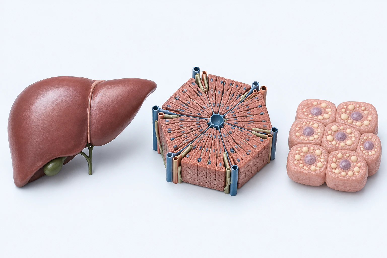
Representative NAFLD/MASLD liver, lobule, and intracellular-lipid orientation. The plate does not depict alcohol use, inflammation, fibrosis, cirrhosis, NASH/MASH classification, or stage.
Scientific media record1 verified identifier
- Subject
- Non-alcoholic Fatty Liver Diseasecondition
- Identifiers
- MeSH:D065626
- Review
- Editorial review completeIdentifiers authority-verified · Accessibility validated · · non-alcoholic-fatty-liver-disease|non-alcoholic-fatty-liver-disease-pathology-v1.webp
- Digital source
- Trained-algorithmic mediaCreated with a trained generative algorithm and reviewed by WikiBiome for subject identity, scientific framing, identifiers, provenance, and accessibility.
- License
- CC BY-SA 4.0Created
Non-alcoholic fatty liver disease (NAFLD), recently reclassified as metabolic dysfunction-associated steatotic liver disease (MASLD), encompasses a spectrum from simple hepatic steatosis to metabolic dysfunction-associated steatohepatitis (MASH, formerly NASH), fibrosis, and cirrhosis. Affecting roughly one in four adults globally, NAFLD is the hepatic manifestation of metabolic syndrome.
The gut-liver axis—the bidirectional communication between intestinal microbiota and the liver via the portal vein—is increasingly recognized as central to NAFLD pathogenesis.
One disease. Five evidence layers.
A generated systems view of the metals, organisms, host sequestration signals, ecological conditions, and microbial functions indexed for Non-Alcoholic Fatty Liver Disease.
Evidence layer
Taxonomic signature
Organisms reported as enriched or depleted, with their indexed functional context kept beside the name.No structured taxa indexed yet.
No structured taxa indexed yet.
Evidence layer
Nutritional immunity
Host metal-withholding, inflammatory, antioxidant, and microbial-metabolite signals indexed in the signature.Elevated host signals
0No structured signals indexed yet.
Depleted protective signals
0No structured signals indexed yet.
Evidence layer
Ecological state
The environmental conditions that connect the organism-level observations into a system.No structured ecological features indexed yet.
Evidence layer
Virulence functions
Microbial structures, enzymes, and acquisition systems implicated by the linked evidence.No structured virulence functions indexed yet.
The disease record, in full.
The original WikiBiome disease narrative remains intact beneath the generated signature atlas.
Microbiome Associations#
The gut-liver axis ensures that the liver is the first organ exposed to microbial products translocating from the gut. In NAFLD, several microbiome patterns emerge.
Proteobacteria (Pseudomonadota) enrichment—Increased Gram-negative bacteria elevate portal LPS levels, activating Kupffer cells via TLR4 and driving hepatic Metal-Driven Inflammation. Akkermansia muciniphila depletion—Loss of this barrier-protective organism increases intestinal permeability and metabolic endotoxemia.
Blautia depletion—Reduced bile salt hydrolase (BSH) activity alters the bile acid pool, disrupting Farnesoid X Receptor (FXR) signaling and impairing hepatic lipid metabolism. Collinsella enrichment—Alters bile acid profiles, reducing hepatic bile acid synthesis via disrupted FXR signaling and promoting lipid accumulation.
Ethanol-producing bacteria—Certain gut bacteria (e.g., Klebsiella pneumoniae) produce endogenous ethanol, contributing to hepatic injury even in the absence of alcohol consumption.
Metal Associations#
Cadmium—Disrupts the gut-liver axis in animal models, altering microbiome composition and accelerating hepatic steatosis. cadmium (Cd)-induced gut barrier damage increases portal LPS translocation.
Nickel—Increases hepatic glycogenolysis and elevates inducible nitric oxide synthase; in overweight women, nickel allergy prevalence reaches 59.7%, with NAFLD as a potential mediator.
Arsenic—Environmental arsenic exposure correlates with NAFLD prevalence in epidemiological studies; arsenic disrupts hepatic lipid metabolism and promotes insulin resistance. Iron—Hepatic iron overload is common in MASH; iron catalyzes lipid peroxidation and drives progression from steatosis to steatohepatitis.
Associated Conditions#
NAFLD shares significant microbiome and metabolic overlap with other conditions. Obesity—Shared metabolic endotoxemia, Akkermansia depletion, and adipose tissue inflammation. Type 2 Diabetes—Bidirectional relationship; insulin resistance drives hepatic lipogenesis while NAFLD worsens glycemic control. Atherosclerosis—NAFLD is an independent CVD risk factor; shared LPS-driven vascular inflammation.
Colorectal Cancer—Altered bile acid metabolism (shifted secondary bile acid pool) connects hepatic and colonic pathology.
Environmental Factors#
Dietary patterns strongly influence NAFLD through the gut-liver axis. High-fat, high-fructose Western diets reduce microbial diversity, deplete SCFA-producing commensals, and increase intestinal permeability. Dietary cadmium exposure from contaminated grains, leafy vegetables, and shellfish provides a chronic metal burden that compounds the metabolic insult.
Open Questions#
Unresolved questions identified by the current evidence record.
01Does microbial endogenous ethanol production drive NAFLD independently of diet, or is it a marker of broader Dysbiosis?+
The current WikiBiome record identifies this as an unresolved evidence gap.
02Can targeted restoration of BSH-expressing bacteria (Blautia, Lactobacillus) reverse hepatic steatosis through FXR reactivation?+
The current WikiBiome record identifies this as an unresolved evidence gap.
03What is the relative contribution of portal LPS versus bile acid dysregulation to NAFLD progression?+
The current WikiBiome record identifies this as an unresolved evidence gap.
Cross-References#
- Farnesoid X Receptor (FXR)—bile acid receptor whose disruption promotes hepatic steatosis
- Lipopolysaccharide—metabolic endotoxemia driver via portal vein
- Bile Acid Metabolism—microbial transformation controls hepatic signaling
- Obesity—shared metabolic and microbiome features
- Collinsella—enriched in NAFLD; disrupts FXR signaling
- Cadmium—gut-liver axis disruptor in animal models
References 8
Numbered by first appearance in the article, then reconciled with its declared source list.
- 1
Kosuke Fujimoto, Daichi Miyaoka, Satoshi Uematsu (2022). Characterization of the human gut virome in metabolic and autoimmune diseases. Inflammation and Regeneration.
- 2
Patrick A. de Jonge, Koen Wortelboer, Torsten P. M. Scheithauer et al. (2022). Gut Virome Profiling Identifies a Widespread Bacteriophage Family Associated with Metabolic Syndrome. Nature Communications.
- 3
Kosuke Fujimoto, Daichi Miyaoka, Satoshi Uematsu (2022). Characterization of the Human Gut Virome in Metabolic and Autoimmune Diseases. Inflammation and Regeneration.
- 4
Shukla S, Srivastava A, Verma D et al. (2023). Shukla et al. 2023 — Analysis of Gut Bacteriome of In Utero Arsenic-Exposed Mice. Frontiers in Microbiology.
- 5
Yijia Song, Sutong Liu, Lihui Zhang et al. (2025). The effect of gut microbiome-targeted therapies in nonalcoholic fatty liver disease: a systematic review and network meta-analysis. Frontiers in Nutrition.
- 6
Elizabeth Half, Nirit Keren, Leah Reshef et al. (2019). Fecal microbiome signatures of pancreatic cancer patients. Scientific Reports.
- 7
Quanxin Su, Kenan Wang, Yayin Luo et al. (2025). Su 2025 — Altered Gut Microbiota in Erectile Dysfunction Patients: A Pilot Study. Frontiers in Microbiology.
- 8
Kieran M. Tuohy, Francesca Fava, Roberto Viola (2014). The way to a man's heart is through his gut microbiota - dietary pro- and prebiotics for the management of cardiovascular risk. Proceedings of the Nutrition Society.
Article network
Mentioned here 14
Pages linking here 2
Connect the evidence
Publicly readable discussion by ORCID-authenticated researchers. Questions, interpretation, methods, corrections, and new evidence stay attached to this record.
No discussion yet. Start with a precise question or a source-backed observation.
Activity and accepted changes
Accepted researcher context, editorial status, public discussion, and upstream Git revisions are shown together. Pending, declined, and withdrawn proposals remain private.
- published revision
Backfill inflammation concept links
Karen Pendergrass · +1 −1
Inspect exact Git diff ↗ - published revision
Complete corpus-wide Dysbiosis linking
Karen Pendergrass · +1 −1
Inspect exact Git diff ↗ - published revision
massive wiki expansion: 149 stubs fixed, 100+ new pages, Rule 15 scan, keystone papers
WikiBiome Deploy Bot · +25 −19
Inspect exact Git diff ↗ - published revision
maintenance: 409 source fixes, 34 entity updates, 17 concept updates, 30 analysis outputs
WikiBiome Deploy Bot · +98 −0
Inspect exact Git diff ↗