

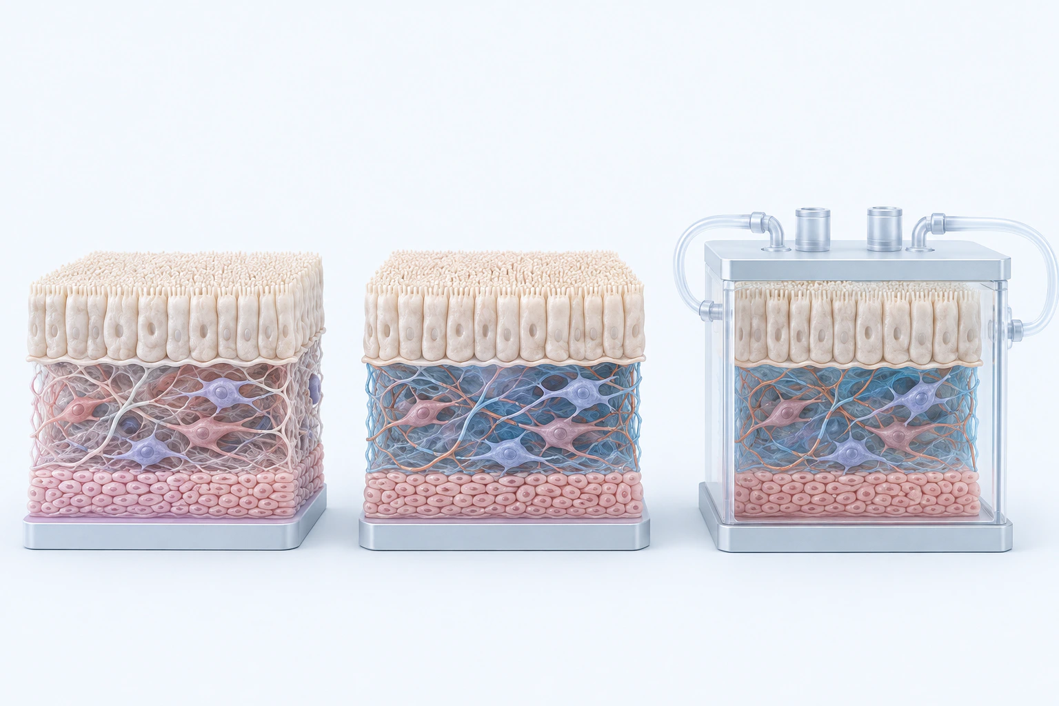
Qualitative local chemical-environment orientation. Color does not report a pH value, systemic acid-base state, disease, tissue damage, mechanism, severity, or diagnosis.
Scientific media record1 verified identifier
- Subject
- Acidic Microenvironmentbiological-process
- Identifiers
- WikiBiome:acidic-microenvironment
- Review
- Editorial review completeIdentifiers authority-verified · Accessibility validated · · acidic-microenvironment|acidic-microenvironment-mechanism-v1.webp
- Digital source
- Trained-algorithmic mediaCreated with a trained generative algorithm and reviewed by WikiBiome for subject identity, scientific framing, identifiers, provenance, and accessibility.
- Scientific basis
- Acidic MicroenvironmentChEBI Ontology
- License
- CC BY-SA 4.0Created
An acidic microenvironment is a local drop in pH—typically into the 4.5–6.5 range—inside a tissue, organ, or ecological niche.
Acidification arises from three recurring sources: bacterial Fermentative Metabolism that releases short-chain fatty acids and lactate ([1]Louis et al. 2022 — Microbial Lactate Utilisation and the Stability of the Gut MicrobiomeLouis P, et al. · 2022Open reference 1 ↓[2]Role of Gut Microbiota-Generated Short-Chain Fatty Acids in Metabolic and Cardiovascular HealthEdward S. Chambers, Tom Preston, Gary Frost et al. · 2018Open reference 2 ↓), tumor glycolysis (the Warburg effect) that accumulates lactate in cancerous tissue ([3]Microbiota disbiosis is associated with colorectal cancerZhiguang Gao, Bomin Guo, Renyuan Gao et al. · 2015Open reference 3 ↓), and inflammatory infiltration where activated immune cells release lactic acid during respiratory burst.
The consequences are ecological: acid-tolerant taxa thrive, acid-sensitive commensals lose ground, and the solubility of divalent metals rises sharply, amplifying metal-driven Dysbiosis ([4]Mechanism of Iron Ion Homeostasis in Intestinal Immunity and Gut Microbiota RemodelingHonghong Bao, Yi Wang, Hanlin Xiong et al. · 2024Open reference 4 ↓[5]Metal Homeostasis in Pathogenic StreptococciAkbari MS, Doran KS, Burcham LR · 2022Open reference 5 ↓).
This extends primitive-9-oxygen-state to pH: niche chemistry—both oxygenation and acidity—selects which organisms can persist.
Evidence map11 cited passagesInspect provenance +
An acidic microenvironment is a local drop in pH—typically into the 4.5–6.5 range—inside a tissue, organ, or ecological niche. Acidification arises from three recurring sources: bacterial fermentative metabolism that releases short-chain fatty acids and lactate (, ), tumor glycolysis (the Warburg effect) that accumulates lactate in cancerous tissue (), a
Sources of acidification. Colonic pH is set largely by microbial fermentation end products. butyrate, acetate, and propionate produced by SCFA-fermenting commensals maintain a mildly acidic lumen that is part of normal gut function (, ). Lactate is normally cross-fed between producing and utilizing taxa; failure of lactate utilization leads to lactate accumu
pH-dependent metal bioavailability. Lower pH raises proton competition at metal-binding sites and increases the free, soluble fraction of divalent cations. Iron homeostasis in the intestine is pH-sensitive, with ferrous iron (Fe²⁺) uptake via DMT1 favored under acidic conditions (). The same transporter family moves other divalent metals—zinc, manganese, n
Inflammatory bowel disease. Active IBD is associated with altered colonic metal handling and SCFA profiles consistent with a shifted pH environment; circulating trace-metal biomarkers track disease activity (). High-fiber, low-fat dietary patterns that favor SCFA producers show signal in Crohn's disease, consistent with restoring protective short-chain-fatty
Colorectal cancer. The tumor microenvironment is characteristically acidic from Warburg-style glycolysis, and the associated microbiota is enriched for acid-tolerant, pro-inflammatory taxa while depleted of butyrate producers (, ). Bacterial communities in the upper GI tract similarly track with local pH and oxygenation across the carcinogenic trajectory ().
Endometriosis. Lesional tissue shows elevated nickel, and dysbiosis patterns across vaginal, cervical, and gut compartments (,,, ). The pH-metal interaction is central: acidified lesions hyperabsorb divalent cations, feeding metal-dependent pathogens implicated in lesion biology.
Candidiasis. Lactobacillus-produced lactic acid is the primary maintainer of the vaginal acidic environment, and the balance between Lactobacillus and Candida shifts with pH and with the wider microbial community (, ).
Iron. Acidic tissue favors Fe²⁺ solubility and DMT1-mediated uptake; inflamed niches are iron-replete even while circulating iron falls (). Siderophore-producing pathogens exploit this local iron pool even when systemic nutritional immunity is withholding.
Zinc, manganese, nickel. Metal homeostasis in streptococci and other pathogens is tuned to the elevated divalent-cation availability of acidified tissue; transporters for Zn, Mn, and Ni become more productive at lower pH ().
Nickel in E. coli. Nickel, iron, and copper act synergistically in E. coli iron-sulfur metabolism—the pH-dependent rise in metal availability is a lever on virulence, not just a passive consequence ().
Cadmium displacement. Cadmium exposure restructures microbial communities and metabolite profiles in the gut, consistent with divalent-metal competition at acidic sites ().
Mechanism#
Sources of acidification. Colonic pH is set largely by microbial fermentation end products. Butyrate, acetate, and propionate produced by SCFA-fermenting commensals maintain a mildly acidic lumen that is part of normal gut function ([2]Role of Gut Microbiota-Generated Short-Chain Fatty Acids in Metabolic and Cardiovascular HealthEdward S. Chambers, Tom Preston, Gary Frost et al. · 2018Open reference 2 ↓[6]Aho 2021 -- Relationships of Gut Microbiota, Short-Chain Fatty Acids, Inflammation, and the Gut Barrier in Parkinson's DiseaseVelma T E Aho, Madelyn C Houser, Pedro A B Pereira et al. · 2021Open reference 6 ↓).
Lactate is normally cross-fed between producing and utilizing taxa; failure of lactate utilization leads to lactate accumulation and a steeper pH drop ([1]Louis et al. 2022 — Microbial Lactate Utilisation and the Stability of the Gut MicrobiomeLouis P, et al. · 2022Open reference 1 ↓). In tumors, aerobic glycolysis drives lactate export and a stable tissue pH of roughly 5.5–6.5 ([3]Microbiota disbiosis is associated with colorectal cancerZhiguang Gao, Bomin Guo, Renyuan Gao et al. · 2015Open reference 3 ↓).
pH-dependent metal bioavailability. Lower pH raises proton competition at metal-binding sites and increases the free, soluble fraction of divalent cations. Iron homeostasis in the intestine is pH-sensitive, with ferrous iron (iron (Fe)²⁺) uptake via DMT1 favored under acidic conditions ([4]Mechanism of Iron Ion Homeostasis in Intestinal Immunity and Gut Microbiota RemodelingHonghong Bao, Yi Wang, Hanlin Xiong et al. · 2024Open reference 4 ↓).
The same transporter family moves other divalent metals—zinc, manganese, nickel, cadmium—so acidification broadly hyperabsorbs them into inflamed tissue ([5]Metal Homeostasis in Pathogenic StreptococciAkbari MS, Doran KS, Burcham LR · 2022Open reference 5 ↓).
The feedback loop. Acidified niche → higher divalent-metal bioavailability → metal-dependent pathogens expand → more fermentative/proteolytic output → further acidification. The loop explains why once dysbiotic niches are acidified they are hard to reverse without changing both the pH and the metal flux.
Role in Disease#
Inflammatory bowel disease. Active IBD is associated with altered colonic metal handling and SCFA profiles consistent with a shifted pH environment; circulating trace-metal biomarkers track disease activity ([7]Clinical and inflammatory biomarkers of inflammatory bowel diseases are linked to plasma trace elements and toxic metals; new insights into an old conceptAmerikanou C, Karavoltsos S, Gioxari A et al. · 2022Open reference 7 ↓).
High-fiber, low-fat dietary patterns that favor SCFA producers show signal in Crohn's disease, consistent with restoring protective short-chain-fatty-acid tone rather than pathological acidification ([8]A High-Fiber, Low-Fat Diet Improves the Symptoms and Metabolic Profile of Patients with Crohn's DiseaseAbreu MT, Quintero MA, Garces L et al. · 2024Open reference 8 ↓).
Colorectal cancer. The tumor microenvironment is characteristically acidic from Warburg-style glycolysis, and the associated microbiota is enriched for acid-tolerant, pro-inflammatory taxa while depleted of butyrate producers ([3]Microbiota disbiosis is associated with colorectal cancerZhiguang Gao, Bomin Guo, Renyuan Gao et al. · 2015Open reference 3 ↓[9]Emerging Evidence on the Effects of Dietary Factors on the Gut Microbiome in Colorectal CancerAppunni S, Rubens M, Ramamoorthy V et al. · 2021Open reference 9 ↓).
Bacterial communities in the upper GI tract similarly track with local pH and oxygenation across the carcinogenic trajectory ([10]Corruption of Bacterial-Host Homeostasis as a Potential Risk Factor and Biomarker for Upper Gastrointestinal CarcinogenesisCatala-Valentin AR, Mikhail S, Bernard JN et al. · 2021Open reference 10 ↓).
Endometriosis. Lesional tissue shows elevated nickel, and dysbiosis patterns across vaginal, cervical, and gut compartments ([11]Role of Cadmium and Nickel in Estrogen Receptor Signaling and Breast Cancer: Metalloestrogens or Not?Aquino NB, Sevigny MB, Sabangan J et al. · 2012Open reference 11 ↓[12]Irritable Bowel Syndrome-Like Disorders in Endometriosis: Prevalence of Nickel Sensitivity and Effects of a Low-Nickel Diet. An Open-Label Pilot StudyBorghini R, Porpora MG, Casale R et al. · 2020Open reference 12 ↓[13]The Endobiota Study: Comparison of Vaginal, Cervical and Gut Microbiota Between Women with Stage 3/4 Endometriosis and Healthy ControlsAta B, Yildiz S, Turkgeldi E et al. · 2019Open reference 13 ↓[14]The Vaginal Microbiome as a Tool to Predict rASRM Stage of Disease in Endometriosis: a Pilot StudyPerrotta AR, Borrelli GM, Martins CO et al. · 2020Open reference 14 ↓).
The pH-metal interaction is central: acidified lesions hyperabsorb divalent cations, feeding metal-dependent pathogens implicated in lesion biology.
Candidiasis. Lactobacillus-produced lactic acid is the primary maintainer of the vaginal acidic environment, and the balance between Lactobacillus and Candida shifts with pH and with the wider microbial community ([15]The Occurrence of Lactobacillus and Candida albicans in Patients with Thyroid DisordersHarbi RH, Mahmood MA · 2024Open reference 15 ↓[16]Li et al. 2022 — Candida albicans and Resident Microbiota InteractionsLi XV, et al. · 2022Open reference 16 ↓).
Metal Connections#
Iron. Acidic tissue favors iron (Fe)²⁺ solubility and DMT1-mediated uptake; inflamed niches are iron-replete even while circulating iron falls ([4]Mechanism of Iron Ion Homeostasis in Intestinal Immunity and Gut Microbiota RemodelingHonghong Bao, Yi Wang, Hanlin Xiong et al. · 2024Open reference 4 ↓). Siderophore-producing pathogens exploit this local iron pool even when systemic Nutritional Immunity (Metal Sequestration) is withholding.
Zinc, manganese, nickel. Metal homeostasis in streptococci and other pathogens is tuned to the elevated divalent-cation availability of acidified tissue; transporters for zinc (Zn), manganese (Mn), and nickel (Ni) become more productive at lower pH ([5]Metal Homeostasis in Pathogenic StreptococciAkbari MS, Doran KS, Burcham LR · 2022Open reference 5 ↓).
Nickel in E. coli. Nickel, iron, and copper act synergistically in E. coli iron-sulfur metabolism—the pH-dependent rise in metal availability is a lever on virulence, not just a passive consequence ([17]Darwiche 2025 — The Molecular Basis of the Synergistic Toxicity of Nickel and Copper, Common Environmental Co-ContaminantsLinda Darwiche, Carlos A Rodriguez-Bornot, Rebecca A Ingrassia et al. · 2025Open reference 17 ↓).
Cadmium displacement. Cadmium exposure restructures microbial communities and metabolite profiles in the gut, consistent with divalent-metal competition at acidic sites ([18]Cadmium stress triggers significant metabolic reprogramming in Enterococcus faecium CX 2-6Cheng X, Yang B, Zheng J et al. · 2021Open reference 18 ↓).
Connections#
Linked concepts Fermentative Metabolism—produces the organic acids that set pH. Nutritional Immunity (Metal Sequestration)—metal withholding is less effective where local acidification frees metal. Hypoxia—anaerobic conditions and acidity co-occur in dysbiotic niches.
Linked entities Escherichia coli—acid-tolerant; expands under iron/nickel availability in acidified tissue. Fusobacterium nucleatum—acid-tolerant; enriched in the colorectal tumor microenvironment. Candida albicans—morphogenesis and community context both respond to local pH.
Iron, Zinc, Nickel—bioavailability rises with acidification.
Open Questions#
Unresolved questions identified by the current evidence record.
01At what pH thresholds does the feedback loop (acidification → metal hyperabsorption → pathogen expansion) become self-sustaining rather than recoverable?+
The current WikiBiome record identifies this as an unresolved evidence gap.
02Do interventions that restore butyrate tone (and thus a protective mild acidity) rescue the niche without feeding pathogenic acidification?+
The current WikiBiome record identifies this as an unresolved evidence gap.
03How much of the metal flux into inflamed tissue is host-driven (transporter upregulation) versus pH-driven (chemical speciation)?+
The current WikiBiome record identifies this as an unresolved evidence gap.
References 18
Numbered by first appearance in the article, then reconciled with its declared source list.
- 1
Louis P, et al. (2022). Louis et al. 2022 — Microbial Lactate Utilisation and the Stability of the Gut Microbiome. Gut Microbiome.
- 2
Edward S. Chambers, Tom Preston, Gary Frost et al. (2018). Role of Gut Microbiota-Generated Short-Chain Fatty Acids in Metabolic and Cardiovascular Health. Current Nutrition Reports.
- 3
Zhiguang Gao, Bomin Guo, Renyuan Gao et al. (2015). Microbiota disbiosis is associated with colorectal cancer. Frontiers in Microbiology.
- 4
★Honghong Bao, Yi Wang, Hanlin Xiong et al. (2024). Mechanism of Iron Ion Homeostasis in Intestinal Immunity and Gut Microbiota Remodeling. International Journal of Molecular Sciences.
- 5
Akbari MS, Doran KS, Burcham LR (2022). Metal Homeostasis in Pathogenic Streptococci. Microorganisms.
- 6
Velma T E Aho, Madelyn C Houser, Pedro A B Pereira et al. (2021). Aho 2021 -- Relationships of Gut Microbiota, Short-Chain Fatty Acids, Inflammation, and the Gut Barrier in Parkinson's Disease. Molecular Neurodegeneration.
- 7
Amerikanou C, Karavoltsos S, Gioxari A et al. (2022). Clinical and inflammatory biomarkers of inflammatory bowel diseases are linked to plasma trace elements and toxic metals; new insights into an old concept. Frontiers in Nutrition.
- 8
Abreu MT, Quintero MA, Garces L et al. (2024). A High-Fiber, Low-Fat Diet Improves the Symptoms and Metabolic Profile of Patients with Crohn's Disease. medRxiv (preprint).
- 9
Appunni S, Rubens M, Ramamoorthy V et al. (2021). Emerging Evidence on the Effects of Dietary Factors on the Gut Microbiome in Colorectal Cancer. Frontiers in Nutrition.
- 10
Catala-Valentin AR, Mikhail S, Bernard JN et al. (2021). Corruption of Bacterial-Host Homeostasis as a Potential Risk Factor and Biomarker for Upper Gastrointestinal Carcinogenesis. Journal of Gastroenterology and Hepatobiliary Medicine.
- 11
★Aquino NB, Sevigny MB, Sabangan J et al. (2012). Role of Cadmium and Nickel in Estrogen Receptor Signaling and Breast Cancer: Metalloestrogens or Not?. Journal of Environmental Science and Health Part C - Environmental Carcinogenesis and Ecotoxicology Reviews.
- 12
Borghini R, Porpora MG, Casale R et al. (2020). Irritable Bowel Syndrome-Like Disorders in Endometriosis: Prevalence of Nickel Sensitivity and Effects of a Low-Nickel Diet. An Open-Label Pilot Study. Nutrients.
- 13
Ata B, Yildiz S, Turkgeldi E et al. (2019). The Endobiota Study: Comparison of Vaginal, Cervical and Gut Microbiota Between Women with Stage 3/4 Endometriosis and Healthy Controls. Scientific Reports.
- 14
Perrotta AR, Borrelli GM, Martins CO et al. (2020). The Vaginal Microbiome as a Tool to Predict rASRM Stage of Disease in Endometriosis: a Pilot Study. Reproductive Sciences.
- 15
Harbi RH, Mahmood MA (2024). The Occurrence of Lactobacillus and Candida albicans in Patients with Thyroid Disorders. Misan Journal for Academic Studies.
- 16
Li XV, et al. (2022). Li et al. 2022 — Candida albicans and Resident Microbiota Interactions. Frontiers in Microbiology.
- 17
Linda Darwiche, Carlos A Rodriguez-Bornot, Rebecca A Ingrassia et al. (2025). Darwiche 2025 — The Molecular Basis of the Synergistic Toxicity of Nickel and Copper, Common Environmental Co-Contaminants. Applied and Environmental Microbiology.
- 18
Cheng X, Yang B, Zheng J et al. (2021). Cadmium stress triggers significant metabolic reprogramming in Enterococcus faecium CX 2-6. Computational and Structural Biotechnology Journal.
Article network
Connect the evidence
Publicly readable discussion by ORCID-authenticated researchers. Questions, interpretation, methods, corrections, and new evidence stay attached to this record.
No discussion yet. Start with a precise question or a source-backed observation.
Activity and accepted changes
Accepted researcher context, editorial status, public discussion, and upstream Git revisions are shown together. Pending, declined, and withdrawn proposals remain private.
- published revision
Complete corpus-wide Dysbiosis linking
Karen Pendergrass · +1 −1
Inspect exact Git diff ↗ - published revision
massive wiki expansion: 149 stubs fixed, 100+ new pages, Rule 15 scan, keystone papers
WikiBiome Deploy Bot · +14 −14
Inspect exact Git diff ↗ - published revision
ingest: 87 papers (17 endometriosis, 67 erectile dysfunction, 3 cross-condition)
WikiBiome Deploy Bot · +49 −61
Inspect exact Git diff ↗ - published revision
nightly maintenance: 94 stub demotions, 181 source_count fixes, 22 auto-discovered stubs, 5 adversarial audits, 3 boundary fixes, 3 evidence-level corrections
WikiBiome Deploy Bot · +3 −0
Inspect exact Git diff ↗ - published revision
pre-overnight checkpoint 2026-04-18
WikiBiome Deploy Bot · +4 −2
Inspect exact Git diff ↗ - published revision
WikiBiome update — 2026-04-15 17:23
WikiBiome Deploy Bot · +2 −2
Inspect exact Git diff ↗ - published revision
WikiBiome update — integrity fixes, metallomic diet pages, cross-condition analyses
WikiBiome Deploy Bot · +22 −20
Inspect exact Git diff ↗ - published revision
WikiBiome update — 2026-04-10 17:16
WikiBiome Deploy Bot · +89 −0
Inspect exact Git diff ↗