

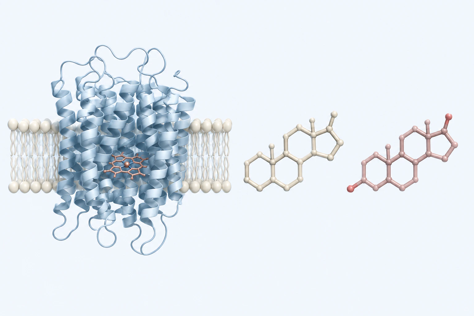
Human aromatase context. Protein and steroid forms are generic teaching abstractions, not measured structures, docking, catalytic conversion, expression, hormone concentration, mechanism, biomarker, or treatment evidence.
Scientific media record2 verified identifiers
- Subject
- Aromatasebiological-process
- Identifiers
- WikiBiome:aromataseUniProt:P11511
- Review
- Editorial review completeIdentifiers authority-verified · Accessibility validated · · aromatase|aromatase-mechanism-v1.webp
- Digital source
- Trained-algorithmic mediaCreated with a trained generative algorithm and reviewed by WikiBiome for subject identity, scientific framing, identifiers, provenance, and accessibility.
- Scientific basis
- Aromatase — UniProtKB P11511Aromatase
- License
- CC BY-SA 4.0Created
Aromatase (CYP19A1) is the cytochrome P450 enzyme that catalyzes the final and rate-limiting step of estrogen biosynthesis—the conversion of androgens (testosterone, androstenedione) to estrogens (estradiol, estrone).
It is expressed in the ovaries, placenta, adipose tissue, brain, bone, and—critically—in endometriotic lesions where local estrogen production drives disease progression.
Aromatase connects the metal-microbiome axis to estrogen-dependent disease through three pathways: metalloestrogen activation, adipose tissue Metal-Driven Inflammation, and estrobolome modulation.
Evidence map5 cited passagesInspect provenance +
cadmium and nickel are classified as metalloestrogens—metals that mimic estrogen by binding estrogen receptors (ERα) and activating estrogen-responsive gene transcription WITHOUT going through aromatase. This creates a paradox for aromatase inhibitor therapy:
zinc modulates aromatase activity—zinc deficiency is associated with altered androgen/estrogen ratios. Dietary zinc intake inversely associated with endometriosis risk, potentially through aromatase modulation.
Increased beta-glucuronidase activity → more estrogen recirculation → estrogen excess → endometriosis/breast cancer risk.
Sex differences in CRC: Higher estrogen from adipose aromatase in obese men may be protective or pathogenic depending on context.
Endometriotic lesions express aberrant aromatase—normally absent in eutopic endometrium but highly expressed in ectopic lesions, creating local estrogen production that feeds lesion growth independent of ovarian estrogen. Combined with dysbiotic estrobolome (excess beta-glucuronidase recirculating additional estrogen), this creates a dual-source estrogen e
Metal Connections#
Metalloestrogens#
Cadmium and Nickel are classified as metalloestrogens—metals that mimic estrogen by binding estrogen receptors (ERα) and activating estrogen-responsive gene transcription WITHOUT going through aromatase.[1]Role of Cadmium and Nickel in Estrogen Receptor Signaling and Breast Cancer: Metalloestrogens or Not?Aquino NB, Sevigny MB, Sabangan J et al. · 2012Open reference 1 ↓ This creates a paradox for aromatase inhibitor therapy.
Aromatase inhibitors (letrozole, anastrozole) block enzymatic estrogen production but cannot block metalloestrogen activation of ER. Environmental cadmium/nickel exposure may therefore undermine aromatase inhibitor efficacy in breast cancer and endometriosis.
Zinc and Aromatase#
- Zinc modulates aromatase activity—zinc deficiency is associated with altered androgen/estrogen ratios. Dietary zinc intake inversely associated with endometriosis risk, potentially through aromatase modulation.[2]Exploring the link between dietary zinc intake and endometriosis risk: insights from a cross-sectional analysis of American womenHuang Y, Wei Y, Liang F et al. · 2024Open reference 2 ↓
Microbiome Connection#
Estrobolome Circuit#
Aromatase produces estrogen → estrogen is conjugated in the liver → conjugated estrogen enters the gut via bile → gut bacterial Beta-Glucuronidase deconjugates it → free estrogen is reabsorbed (Estrobolome recirculation). Dysbiosis alters the estrobolome, disrupting this circuit.
Increased beta-glucuronidase activity → more estrogen recirculation → estrogen excess → endometriosis/breast cancer risk.[3]Kaliannan et al. 2018 — Estrogen-Mediated Gut Microbiome Alterations Influence Sexual Dimorphism in Metabolic Syndrome in MiceKanakaraju Kaliannan, Ruairi C. Robertson, Kiera Murphy et al. · 2018Open reference 3 ↓ Decreased beta-glucuronidase activity → less recirculation → estrogen depletion → metabolic syndrome, osteoporosis risk.
Aromatase and the estrobolome are two halves of estrogen homeostasis—one controls production, the other controls recycling. Both must be intact for normal estrogen levels.
Adipose Aromatase and Obesity-Microbiome Axis#
In obesity, adipose tissue becomes a major estrogen source via aromatase expression. The Gut Microbiome drives obesity-related inflammation → adipose expansion → increased aromatase → peripheral estrogen production. This explains.
Sex differences in CRC: Higher estrogen from adipose aromatase in obese men may be protective or pathogenic depending on context.[4]Sex differences in colorectal cancer: with a focus on sex hormone-gut microbiome axisZihong Wu, Yuqing Huang, Renyi Zhang et al. · 2024Open reference 4 ↓
Postmenopausal breast cancer: Adipose aromatase becomes the primary estrogen source after ovarian cessation; obesity-driven microbiome inflammation amplifies this.
Disease Relevance#
Endometriosis#
Endometriotic lesions express aberrant aromatase—normally absent in eutopic endometrium but highly expressed in ectopic lesions, creating local estrogen production that feeds lesion growth independent of ovarian estrogen.
Combined with dysbiotic estrobolome (excess beta-glucuronidase recirculating additional estrogen), this creates a dual-source estrogen excess that drives disease.[5]I Am the 1 in 10 -- What Should I Eat? A Research Review of Nutrition in EndometriosisPiecuch M, Garbicz J, Waliczek M et al. · 2022Open reference 5 ↓
Breast Cancer#
Aromatase inhibitors are first-line therapy for ER+ postmenopausal breast cancer. The microbiome modulates treatment response through. Estrobolome activity affecting circulating estrogen levels despite aromatase blockade.
Metalloestrogen exposure bypassing aromatase inhibition entirely.
Cross-References#
- Estrobolome—microbial estrogen recirculation (Primitive 7)
- Beta-Glucuronidase—the microbial enzyme controlling estrogen deconjugation
- Metalloestrogens—cadmium (Cd)/nickel (Ni) mimicking estrogen without aromatase
- Cadmium—metalloestrogen bypassing aromatase
- Nickel—metalloestrogen
- Zinc—modulates aromatase activity
- Endometriosis—aberrant aromatase in ectopic lesions
- Breast Cancer—aromatase inhibitor therapy
- Serotonin-Estrogen Axis—estrogen-serotonin neuroendocrine interaction
- Obesity—adipose aromatase as peripheral estrogen source
References 5
Numbered by first appearance in the article, then reconciled with its declared source list.
- 1
★Aquino NB, Sevigny MB, Sabangan J et al. (2012). Role of Cadmium and Nickel in Estrogen Receptor Signaling and Breast Cancer: Metalloestrogens or Not?. Journal of Environmental Science and Health Part C - Environmental Carcinogenesis and Ecotoxicology Reviews.
- 2
Huang Y, Wei Y, Liang F et al. (2024). Exploring the link between dietary zinc intake and endometriosis risk: insights from a cross-sectional analysis of American women. BMC Public Health.
- 3
Kanakaraju Kaliannan, Ruairi C. Robertson, Kiera Murphy et al. (2018). Kaliannan et al. 2018 — Estrogen-Mediated Gut Microbiome Alterations Influence Sexual Dimorphism in Metabolic Syndrome in Mice. Microbiome.
- 4
Zihong Wu, Yuqing Huang, Renyi Zhang et al. (2024). Sex differences in colorectal cancer: with a focus on sex hormone-gut microbiome axis. Cell Communication and Signaling.
- 5
Piecuch M, Garbicz J, Waliczek M et al. (2022). I Am the 1 in 10 -- What Should I Eat? A Research Review of Nutrition in Endometriosis. Nutrients.
Article network
Connect the evidence
Publicly readable discussion by ORCID-authenticated researchers. Questions, interpretation, methods, corrections, and new evidence stay attached to this record.
No discussion yet. Start with a precise question or a source-backed observation.
Activity and accepted changes
Accepted researcher context, editorial status, public discussion, and upstream Git revisions are shown together. Pending, declined, and withdrawn proposals remain private.
- published revision
Backfill gut microbiome concept links
Karen Pendergrass · +1 −1
Inspect exact Git diff ↗ - published revision
Backfill inflammation concept links
Karen Pendergrass · +1 −1
Inspect exact Git diff ↗ - published revision
Complete corpus-wide Dysbiosis linking
Karen Pendergrass · +1 −1
Inspect exact Git diff ↗ - published revision
massive wiki expansion: 149 stubs fixed, 100+ new pages, Rule 15 scan, keystone papers
WikiBiome Deploy Bot · +81 −0
Inspect exact Git diff ↗